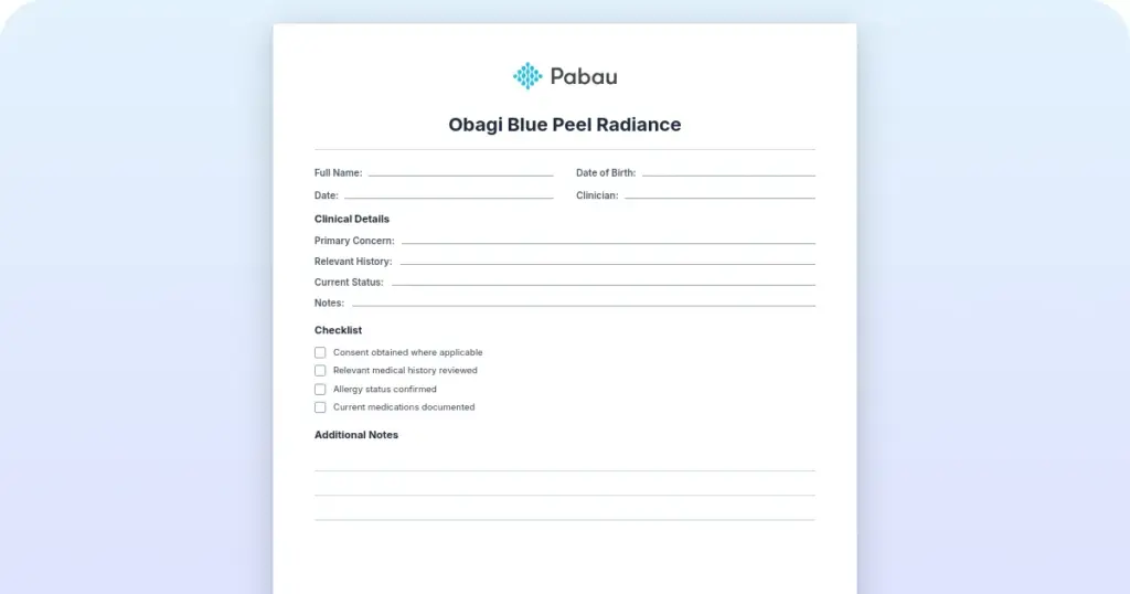
Obagi Blue Peel Radiance: Consent form and aftercare guide
An Obagi Blue Peel Radiance consent form records that your patient understood a ...

1
Last Updated: Sep 29, 2026

Ptosis surgery corrects drooping eyelids caused by weakened levator palpebrae superioris muscles, restoring both visual function and cosmetic appearance.
Pre-operative documentation must include levator function measurement, visual field testing demonstrating medical necessity, and margin reflex distance (MRD1) assessment.
Proper consent templates address the specific risks of blepharoptosis repair: lagophthalmos, dry eye syndrome, asymmetry, and revision surgery rates of roughly 10% (reported range 5-35% depending on technique and etiology).
Practice management software like Pabau offers digital forms and clinical documentation features that streamline ptosis surgery workflows, from pre-authorization letters to post-operative recovery tracking.
A ready-to-use clinical documentation template covering pre-operative ptosis assessment, levator function measurement, surgical planning, informed consent elements, and post-operative care instructions for blepharoptosis repair procedures.
Download templatePtosis surgery — also called blepharoptosis repair — is a specialized eyelid procedure that corrects a drooping or sagging upper eyelid (blepharoptosis), which can impair visual function and affect appearance. This guide provides ready-to-use documentation templates for oculoplastic surgeons, aesthetic practitioners, and eye care practices.
These templates streamline pre-operative assessment, surgical planning, informed consent, and post-operative recovery workflows. They cover essential clinical documentation, insurance pre-authorization requirements, and patient education materials needed for safe, compliant ptosis repair procedures.
Ptosis (blepharoptosis) occurs when the levator palpebrae superioris muscle becomes weakened, stretched, or damaged. This muscle is primarily responsible for lifting the upper eyelid. Causes include age-related degeneration, neurological conditions, myasthenia gravis, trauma, or post-surgical changes.
When the eyelid droops below its normal position, it can obstruct the visual axis. This impairs superior visual field perception and creates the appearance of fatigue or heaviness.
Ptosis surgery addresses this by tightening, resecting, or repositioning the levator muscle. In cases of severe levator dysfunction, surgeons use alternative techniques such as frontalis sling suspension or Müller’s muscle resection. The goal is to restore the upper eyelid to its normal anatomical position — typically a margin reflex distance of 4–4.5mm.
This improves visual function and restores a more alert appearance. Procedures fall under CPT codes 67901.
Medicare covers the procedure when medical necessity criteria are met, including a documented superior visual field deficit (typically ≥12 degrees by Humphrey visual field testing) or margin reflex distance ≤2mm, supported by clinical documentation and imaging.
The ptosis surgery template packet includes five interconnected clinical documentation forms designed to support the complete surgical workflow:
Each section can be customized to reflect your practice’s protocols, surgical approach preferences, and regional regulatory requirements. Use digital intake forms to capture pre-operative assessment data electronically, reducing manual data entry and streamlining chart review. Pair the template with AI-powered clinical documentation to generate post-operative progress notes from dictation. This captures levator function measurements and wound healing observations without delay.
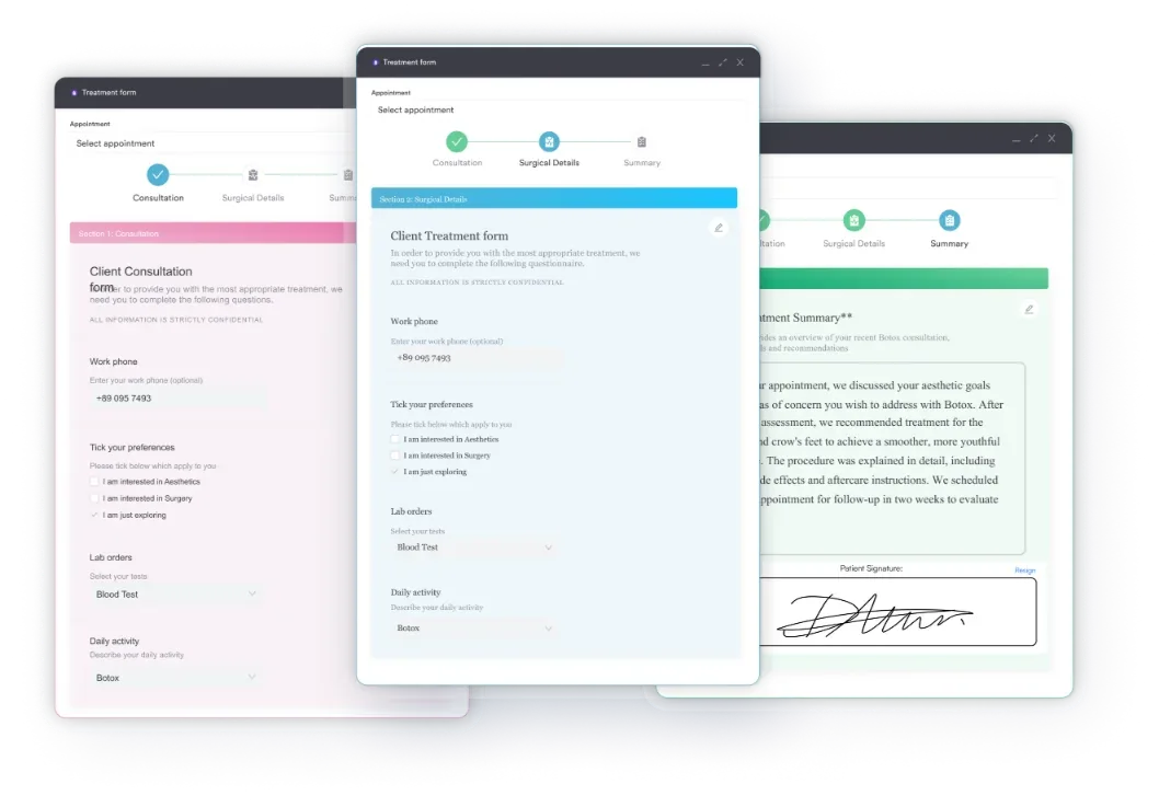
This template packet is designed for healthcare practitioners and practices involved in oculoplastic surgery and aesthetic eyelid procedures:
Whether you operate in a single-location practice or manage multi-location surgical centers, the template ensures consistency across all providers and locations, reducing missing or inconsistent documentation and supporting regulatory compliance.
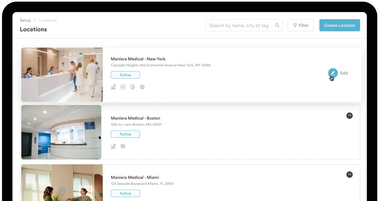
If you are new to digital tooling, start with what practice management software does before digitizing forms like this.
Regulatory compliance. Pre-operative assessment templates document the clinical justification for surgery. Specifically, they capture levator function grading and visual field defect severity required by your Medicare Administrative Contractor’s Blepharoplasty, Blepharoptosis Repair and Brow LCD (for example, L34028), reducing claim denials and audit risk.
Informed consent standardization. Ready-made consent templates ensure consistent, comprehensive risk disclosure before surgery. As a result, your practice is protected from consent-related liability claims and patients can make truly informed decisions.
Workflow efficiency. Standardized forms eliminate the need to create templates from scratch for each patient. In turn, this reduces administrative burden and allows clinical staff to focus on pre-operative education and surgical scheduling.
Documentation completeness. Structured assessment forms ensure that no critical clinical measurements are missed during pre-operative evaluation. For example, they capture levator function, MRD1, and visual field results consistently, supporting medical necessity documentation and outcome tracking.
Insurance pre-authorization support. The template includes a pre-authorization letter aligned to Medicare and commercial payer criteria. Furthermore, this speeds approval timelines and reduces claim rejections.
Patient education and recovery compliance. Clear, written post-operative instructions improve patient understanding of the recovery timeline and warning signs. Consequently, unplanned follow-up visits are reduced and complications can be detected earlier, supporting better surgical outcomes.
Flag patients with myasthenia gravis or third nerve palsy for extended pre-operative observation (3-6 months for neurogenic causes) before scheduling definitive surgery. Some neurogenic ptosis resolves spontaneously without intervention, and myasthenic ptosis requires medical optimization before surgical planning. Document this clinical reasoning in the assessment template to support payer review and demonstrate surgical appropriateness.
The foundation of safe ptosis surgery is accurate pre-operative assessment. Surgeons grade levator function in millimeters of superior eyelid excursion — how much the upper lid moves when the patient looks up, with the brow held fixed to eliminate frontalis compensation — from poor (≤4mm) to good (≥8mm):
Your template should include dedicated measurement fields for:
This standardized data supports both surgical planning and insurance pre-authorization. Medicare explicitly requires documented ptosis with levator function grading and visual field impact assessment, per your MAC’s Blepharoplasty, Blepharoptosis Repair and Brow LCD (for example, L34028).
Store these measurements in your secure patient portal for pre-operative review by your surgical team. This also enables compliance management audits to verify documentation completeness across all pre-operative charts.
Post-operative success depends on clear patient education and structured follow-up. Your post-operative instructions template should address four key areas: (1) swelling progression (peaks day 2–3, resolves by week 4–6), (2) suture removal timing (typically day 7–14), (3) activity restrictions (avoid heavy lifting for 2 weeks), and (4) symptom monitoring.
In addition, key post-operative complications to address in patient education materials and clinical follow-up protocols include:
Schedule post-operative visits at day 7–10 (suture removal and initial healing assessment), week 4 (early outcome review), and week 12 (final outcome assessment). Eyelid position stabilizes by the 12-week mark. Use your template’s follow-up section to document levator function preservation, eyelid symmetry, and patient satisfaction with functional and cosmetic outcomes.

From pre-operative assessment to post-operative follow-up, Pabau’s digital forms and clinical notes management keep all ptosis surgery documentation organized, compliant, and accessible. Book a demo to see how oculoplastic surgeons use Pabau to manage complex eyelid procedures.

Medicare and most commercial payers require pre-authorization for ptosis repair. Your template should include a pre-authorization letter covering four elements:
Reference the applicable CPT codes in your pre-authorization template: CPT 67901 (frontalis suspension with suture or banked fascia), 67902 (frontalis sling with autologous fascia), 67903/67904 (levator resection or advancement, internal/external approach), 67906 (superior rectus sling, e.g. Whitnall’s sling), or 67908 (conjunctival-Müller’s resection). Also include codes for any concurrent procedures — blepharoplasty CPT 15820–15823 and brow ptosis repair CPT 67900.
Many payers reference the NCBI StatPearls literature on blepharoptosis when evaluating medical necessity. Citing this in your pre-authorization request strengthens your case and demonstrates alignment with peer-reviewed surgical standards.
Ptosis surgery templates provide a standardized, evidence-based framework for managing blepharoptosis repair from initial evaluation through post-operative recovery. Structured pre-operative assessment, comprehensive informed consent, and clear recovery protocols protect both your patients and your practice.
Download the template packet above to establish consistent, compliant documentation across your surgical practice. Then book a demo with Pabau to see how digital forms and clinical documentation tools integrate these templates directly into your workflow — eliminating paper-based charting delays.

Need guidance on cosmetic surgery consent and intake? Cosmetic surgery intake form covers informed consent, risk disclosure, and pre-operative intake documentation for aesthetic procedures, including eyelid surgery.
Want to understand CPT codes for oculoplastic procedures? plastic surgery software guide discusses surgical coding, billing workflows, and practice management integration for cosmetic surgery centers.
Performing breast augmentation procedures too? Breast augmentation aftercare template standardizes post-operative guidance on wound care, compression garments, and activity restrictions.
Ptosis surgery lifts a drooping upper eyelid by tightening or repositioning the levator palpebrae superioris muscle, or using frontalis sling suspension in severe cases, to restore normal eyelid position and improve visual function.
Medicare and most commercial payers cover ptosis repair when medical necessity is documented — typically a superior visual field defect of ≥12 degrees or margin reflex distance ≤2mm, supported by levator function grading.
Risks include lagophthalmos, dry eye syndrome, swelling, bruising, eyelid asymmetry, and a revision rate of roughly 10% (reported range 5-35% depending on technique and etiology). Rarely, infection or hematoma may occur.
Swelling peaks at days 2–3 and resolves over 4–6 weeks. Sutures are removed at 7–14 days, and final eyelid position stabilizes by 12 weeks. Most patients resume light activities within one week.
Common codes include CPT 67901 (frontalis suspension with suture/fascia), 67902 (frontalis sling), 67903/67904 (levator resection/advancement, internal/external), 67906 (superior rectus sling), and 67908 (conjunctival-Müller’s resection). Concurrent blepharoplasty uses 15820–15823.
MRD1 measures the distance from the pupillary light reflex to the upper eyelid margin (normal 4–4.5mm). An MRD1 ≤2mm signals ptosis and is a Medicare medical necessity criterion for surgical coverage.