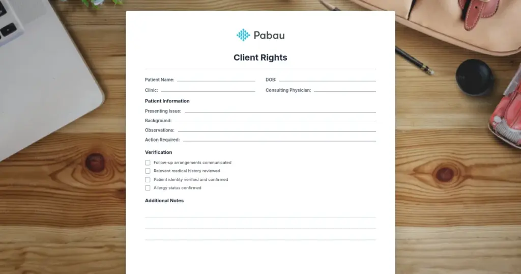


Client rights policy template: Free PDF download
The 7 core client rights: confidentiality, informed consent, refusing treatment,...
July 23, 2026
An eye movement test (H-test) evaluates extraocular muscle function across six cardinal gaze positions using systematic directional prompts.
Abnormal findings such as nystagmus, restricted gaze, or pursuit deficits can signal neurological conditions, central nervous system pathology, or cranial nerve involvement.
Documented eye movement assessment supports concussion screening, neurological workup, and tracking treatment response in clinical practice.
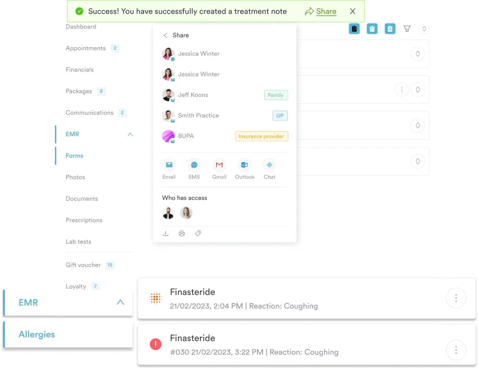
Pabau’s digital forms and client records enable structured documentation of eye movement findings with automated clinical note generation via Echo AI.
A structured clinical assessment form for evaluating extraocular muscle function, saccadic eye movements, smooth pursuit tracking, and ocular alignment. Includes H-test protocol, cover test documentation, and findings interpretation guide.
Download templateAssessing eye movements is a fundamental skill in clinical practice, yet many practitioners lack a structured approach to documenting findings. The eye movement test is a standardized clinical assessment tool that helps you systematically evaluate ocular motility, detect neurological deficits, and support diagnosis across specialties. Whether you’re screening for concussion, investigating dizziness, or monitoring neurological status, this guide walks you through the steps and provides a downloadable template for your clinic.
An eye movement test is a clinical examination that evaluates how the eyes move and track. The test assesses the function of the six extraocular muscles-the muscles surrounding each eye that control its position and motion.
The most common eye movement test is the H-test (sometimes called the cardinal gaze test), which moves the eyes through six cardinal gaze positions to assess muscle function. The test takes its name from the visual path created by these six points, which resembles the letter H.
During an eye movement test, you observe how smoothly the patient’s eyes follow your finger or a fixation target, whether they move together (conjugate gaze), and whether any involuntary movements (nystagmus) are present. This simple assessment provides crucial information about ocular motility and potential neurological involvement.
Eye movement testing is performed across multiple healthcare specialties including ophthalmology, optometry, neurology, physical therapy, and sports medicine. It’s particularly valuable in concussion assessment, stroke evaluation, and monitoring patients with vestibular or neurological conditions.
The eye movement test follows a standardized five-step clinical procedure. Each step builds on the previous one, creating a complete assessment of oculomotor function.
Throughout the assessment, note any nystagmus (involuntary eye movements), diplopia (double vision) reported by the patient, or complaints of difficulty tracking. Document the findings using a structured clinical record that includes position-by-position observations and any abnormalities detected.
Normal eye movement findings include smooth, conjugate eye movement through all six cardinal positions without nystagmus, strabismus, or restriction. The eyes should track smoothly during pursuit tasks and make quick, accurate saccades.
Abnormal findings warrant further investigation. Restricted eye movement in a specific direction may indicate cranial nerve involvement (CN III controls most extraocular movements, CN IV controls the superior oblique, CN VI controls lateral rectus). Nystagmus-involuntary rhythmic eye oscillations-can signal vestibular dysfunction, neurological disease, or central nervous system pathology.
Smooth pursuit deficits (jerky or broken tracking) often indicate central pathology affecting brainstem or cerebellar circuits. Saccadic abnormalities such as hypometric saccades (undershoot) or hypermetric saccades (overshoot) suggest cerebellar dysfunction or oculomotor nerve lesions.
Always correlate eye movement findings with the patient’s clinical presentation, history, and other examination findings. An abnormal eye movement test alone does not diagnose a condition but prompts further investigation and specialist referral when indicated.
Structured digital forms, automated clinical notes, and secure patient records in one integrated system.

Eye movement assessment is essential for practitioners across multiple healthcare settings and specialties.
Standardized eye movement testing delivers measurable clinical and operational benefits. A structured assessment reduces the risk of missing subtle findings and improves documentation quality.
Clinical benefit: Systematic eye movement assessment improves diagnostic accuracy and supports early detection of neurological conditions. The test is non-invasive, requires no equipment beyond a fixation target, and can be performed in any clinical setting in under five minutes.
Documentation clarity: A structured template ensures consistent, complete recording of findings across all patients and clinicians in your practice. This improves continuity of care and provides clear baseline documentation for tracking changes over time.
Compliance: Standardized assessment supports documentation standards expected by regulators and insurers, reducing audit risk and improving claim defensibility.
Patient safety: A checklist-based approach reduces the likelihood of skipping assessment steps, improving consistency and reducing liability exposure.
Establish a baseline eye movement assessment early in your patient relationship. This baseline becomes invaluable if a patient reports new visual symptoms or neurological changes-you can quickly detect departures from normal and track deterioration or recovery over time.
Eye movement abnormalities are sensitive markers of concussion and neurological injury. Impaired smooth pursuit, slowed saccades, and nystagmus often emerge after head trauma even when other neurological findings are subtle.
Sports medicine and physical therapy practices routinely incorporate eye movement assessment into concussion baseline testing and return-to-play protocols. Baseline eye movement data collected during the pre-season allows clinicians to detect objective changes post-injury, supporting safe return-to-activity decisions.
In neurological screening contexts, eye movement assessment helps differentiate peripheral from central causes of vision and balance complaints. A patient with peripheral vestibular dysfunction typically shows normal eye movements at rest but abnormal vestibulo-ocular reflex (VOR) on head thrust testing. Central causes often produce overt eye movement abnormalities during routine H-testing.
Effective eye movement assessment requires clear, actionable documentation. When you use a structured assessment template, findings are recorded consistently, making them easy to retrieve and compare over time.
Modern practice management systems allow you to embed eye movement assessment forms directly into patient records. This integration means assessment data flows seamlessly into the clinical note, reducing transcription errors and saving time. Features like automated clinical documentation can further streamline your workflow by converting structured findings into narrative note text automatically.
Recording eye movement findings alongside other neurological assessments creates a comprehensive baseline against which future assessments can be compared. This longitudinal tracking is especially valuable in conditions like multiple sclerosis, Parkinson’s disease, or post-stroke rehabilitation, where subtle progression matters for treatment decisions.
The eye movement test is a quick, non-invasive assessment that yields valuable clinical information across specialties. By using a standardized template and integrating it into your clinical workflow, you improve assessment consistency, enhance documentation quality, and support better patient outcomes.
Download the free template above to establish a structured approach to ocular motility assessment in your practice. For clinics looking to streamline documentation further, book a demo to see how Pabau’s digital forms and automated clinical note generation can simplify assessment administration and improve care coordination.
Need structured neurological assessment across your clinic? Clinical record management provides templates and organization tools for comprehensive patient assessment documentation.
Looking to automate clinical note writing? Pabau’s AI medical scribe converts structured assessment findings into clinical notes automatically, reducing documentation time without sacrificing detail.
Want to improve patient intake workflows? Mental health practice software features digital intake forms that capture detailed history and baseline assessments upfront, supporting more efficient clinical encounters.
An eye movement test is a clinical examination that assesses how the extraocular muscles control eye position and motion across all directions of gaze. The H-test (cardinal gaze test) is the most common format, moving the eyes through six positions to detect restriction, nystagmus, or other abnormalities.
A basic eye movement test typically takes 3-5 minutes to perform and document. It requires no special equipment and can be completed in any clinical setting.
Nystagmus (involuntary eye oscillations) during eye movement testing can indicate vestibular dysfunction, neurological disease, or central nervous system pathology. Horizontal nystagmus often suggests vestibular involvement; vertical nystagmus typically indicates central (brainstem or cerebellar) pathology.
Eye movement abnormalities support concussion assessment but do not diagnose concussion alone. Impaired smooth pursuit, slowed saccades, and convergence insufficiency are objective markers of oculomotor dysfunction that often appear after head trauma. A positive eye movement test should be interpreted alongside other baseline and post-injury metrics.
Restricted eye movement in a specific direction typically suggests cranial nerve involvement (CN III, CN IV, or CN VI) or orbital restriction (thyroid eye disease, mechanical obstruction). The specific direction of restriction helps localize the affected muscle or nerve.
Repeat testing frequency depends on clinical context. Baseline testing for concussion is typically done once pre-season; post-injury testing occurs immediately and during return-to-play progression. For chronic conditions, repeat testing is guided by clinical changes or treatment milestones rather than a fixed schedule.