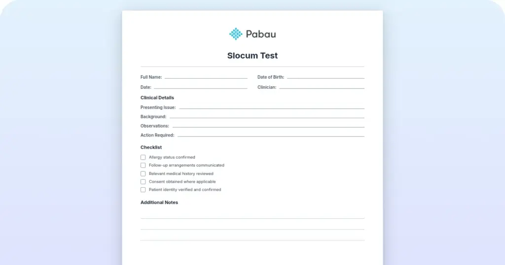
Slocum test: How to perform, interpret and record it
The Slocum test is a knee drawer test performed with the tibia rotated. It check...

3
Last Updated: Oct 21, 2026

The plantar fascia rupture test is a structured clinical assessment used to confirm or rule out a tear of the plantar fascia.
Sudden sharp pain, an audible pop, bruising, swelling and loss of weight-bearing point to rupture rather than plantar fasciitis.
MRI is the gold standard for confirming a rupture, while ultrasound adds real-time assessment under load.
Avoid corticosteroid injection in a confirmed or suspected rupture, because steroid injection is a documented risk factor for the tear.
Practice management software like Pabau stores the completed assessment in the patient record, so the whole team can review it at the next visit.
A one-page clinical form covering patient and clinician details, a medical history prompt, Windlass test results, and four yes/no symptom checks with comment boxes. It closes with a free-text field for the imaging test you conducted, its results, and what you plan next.
Download templateA plantar fascia rupture test is a structured examination that separates a torn fascia from plantar fasciitis. Onset, bruising and weight-bearing tolerance do most of that work, and imaging confirms it. Treat a tear as fasciitis and the patient loses weeks to the wrong protocol.
The template above records what you found and what you plan next, on a single page. The sections below cover the examination sequence, the imaging thresholds, and the one injection to avoid once a rupture is confirmed.
A plantar fascia rupture test is a clinical assessment protocol for evaluating a suspected tear of the plantar fascia. The plantar fascia is the thick band of connective tissue running along the sole from the calcaneus to the toes. It provides arch support and shock absorption during weight-bearing activity.
The test is a structured examination rather than a single maneuver. It combines patient history, palpation, loaded movement testing, and imaging referral criteria. Rupture presents acutely, with sudden sharp pain, an audible pop, bruising and swelling. Plantar fasciitis develops gradually, with inflammation and no structural tearing.
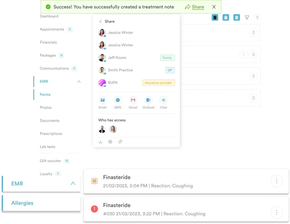
Accuracy is highest when the examination and the imaging are read together. Palpation and the Windlass test narrow the diagnosis, and MRI or ultrasound settles it. Documenting each step in the same place keeps the findings usable at the follow-up visit.
Separating rupture from fasciitis drives the whole treatment decision. Plantar fasciitis is a degenerative overuse condition at the origin of the fascia. A rupture is a structural tear, and the two carry different prognoses and pathways.
A structured form captures these differentiating features at the first assessment and routes the patient correctly. Recording them in safer clinical notes also keeps the distinction defensible if the diagnosis is questioned later.
Document these cardinal symptoms during the plantar fascia rupture test:
Acute presentations are usually unmistakable. Subacute and chronic ruptures develop in degenerative tissue and present more quietly, with progressive pain and functional loss. Record the pattern in the comments boxes, or mark the tender area on a pain locator chart. That record is what justifies the imaging decision later.
The plantar fascia rupture test follows a systematic clinical protocol. These are the five key steps:
You can also run this protocol on screen instead of on paper. Pabau’s digital forms feature lets you build the same assessment as a custom form, with your own checkboxes and rating fields. Completed forms save straight into the patient record.

Clinical examination alone cannot definitively confirm a rupture. Imaging is what grades the tear and settles the diagnosis.
Record the imaging test you conducted and its results in the final field of the template. Add what you are planning for the patient in the same field, so the next clinician reads the finding and the plan together.
Once the plantar fascia rupture test confirms or strongly suggests rupture, treatment follows a stepped approach:
Once the plan is agreed, Pabau’s automated workflows can trigger the recall appointment and the reminders that go with it. That keeps a patient waiting on imaging results from dropping out of the pathway.
The template above is a one-page form you print or complete on screen during the appointment. Here is how each field works:
The form deliberately leans on comments boxes rather than fixed scores. That keeps it usable across podiatry, physical therapy and sports medicine, where the same findings get described in different terms. Save the completed page to the patient’s record in your practice management system. The assessment is then available at every later visit.
The plantar fascia rupture test template is written for:
Sports medicine practices and physical therapy practices benefit most. A standardized assessment improves handover between the treating clinician, the referring physician and the rest of the care team.
These six come up repeatedly, and every one of them costs the patient time:
The plantar fascia rupture test reduces these risks by enforcing the same sequence every time and leaving a written record behind.
A paper form usually gets scanned into a folder days after the appointment. The scan cannot be searched, and the next clinician rarely opens it before the follow-up visit. Practices that go fully paper-free, like Kingswood Aesthetics, avoid that delay altogether.
In Pabau, the same assessment lives as a digital form attached to the patient record. You complete the Windlass test result and the symptom checks on screen during the appointment. The imaging note and treatment plan sit in the same place, timestamped and searchable at the next visit.
Follow-up is where that pays off. Pabau books the recall appointment from the plan you recorded and sends the reminders automatically. Your team opens the record at the next visit and sees exactly what you found. Nobody repeats the examination from scratch.

Pabau’s digital forms let you complete assessments like the plantar fascia rupture test on screen and store them in the patient record. The follow-up books itself, and your team sees the findings at every later visit.

A plantar fascia rupture is rarely subtle, but a quick history can still mislabel it as fasciitis. Onset, bruising and weight-bearing tolerance separate the two, and the Windlass test shows whether the loaded fascia hurts. If a steroid injection sits in the record, weight the diagnosis toward a tear.
The trade-off worth remembering is imaging. Examination narrows the diagnosis but cannot grade the tear. An acute injury with a pop and heavy bruising warrants imaging rather than a watch-and-wait plan. That single decision sets the recovery timeline you give the patient.
Download the template above, use it at the next acute heel injury, and store the completed form where the whole team can find it. Book a demo to see how Pabau keeps clinical assessments and their follow-up appointments in one patient record.

Ruling out an occult fracture? Patellar pubic percussion test walks through a bedside test for suspected fracture and the form that records it.
Building out the neuro screen? Deep tendon reflex exam covers the 0-4+ grading scale and how to chart each reflex consistently.
Measuring activity participation after an injury? Activity Card Sort uses 89 photographic cards across instrumental, leisure, and social domains to track participation changes.
Tracking pain through recovery? Pain journal gives patients a structured way to log symptoms between appointments.
Need a quick severity score? Pain scale 1-10 explains how to use the scale and what a meaningful change looks like.
Most uncomplicated partial ruptures heal in six to 12 weeks with conservative management, such as a boot, rest and physical therapy. Full-thickness ruptures usually need 12 to 16 weeks or longer, and surgical repair can add another four to eight weeks. Recovery varies with the severity of the rupture, the patient’s age, other conditions and adherence to rehabilitation. Note the expected recovery window in the template’s final field so the follow-up appointment matches the healing timeline.
Plantar fasciitis is inflammation of the fascia, with gradual onset, aching pain and minimal swelling. A rupture is a structural tear, with sudden onset, sharp pain, significant bruising and difficulty bearing weight. Fasciitis responds to stretching and physical therapy, while a rupture needs immobilization and imaging. The plantar fascia rupture test separates the two by checking onset, tenderness, bruising, swelling and the Windlass test result.
Yes. Most partial ruptures and many complete ruptures heal with conservative management, including rest, ice, a protective boot, physical therapy and NSAIDs. Surgery is reserved for complete full-thickness tears, failed conservative care after three to six months, or patients who need a rapid return to activity. MRI findings guide that decision. If imaging shows a full-thickness gap and progress stalls at six to eight weeks, seek a surgical opinion.
MRI is the gold standard because it gives high-resolution soft tissue images and shows full or partial tears clearly. Ultrasound is cheaper and immediate, and it is increasingly used first in sports medicine because it allows dynamic assessment under load. Operator skill matters more with ultrasound. Either modality can confirm a rupture, so choose on local availability, cost and patient factors such as MRI contraindications.