Sulcus Test: Shoulder Instability Assessment
The sulcus test is an orthopedic special test that checks for inferior glenohume...

2
Last Updated: Oct 22, 2026

The deep tendon reflex exam checks the reflex arc, testing sensory nerves, the spinal cord synapse, and motor fibers in seconds.
The 0-4+ grading scale runs from absent reflexes (0) to clonus (4+), with grade 2+ recorded as normal.
Five reflexes map to set nerve roots: biceps and brachioradialis (C5-C6), triceps (C7-C8), patellar (L3-L4), and Achilles (S1-S2).
The downloadable template is a two-page recording form, so use the grading scale and nerve root tables on this page as your reference.
Pabau’s digital intake forms capture every reflex grade in the patient record, so you can compare findings visit to visit.
A printable two-page form for recording deep tendon reflex findings at the bedside. It covers patient details, vitals, left and right responses for biceps, triceps, brachioradialis, patellar, ankle, and plantar reflexes, plus a description box for each.
Download templateThe deep tendon reflex exam is the most common neurological test in clinical practice. A single tap with a reflex hammer produces an involuntary muscle contraction. That contraction tells you whether the sensory nerve, the spinal cord synapse, and the motor nerve are all working.
The appeal is speed. You need a reflex hammer, a quiet room, and a patient who can relax. In under two minutes you can check several nerve root levels at once. What you find ranges from mild peripheral neuropathy to early signs of spinal cord compression.
This guide covers patient positioning, the five reflexes and their nerve roots, the 0-4+ grading scale, and what abnormal results point to. The downloadable form above gives you somewhere to write the findings down while you work.
The template is a recording form rather than a reference chart. Across two pages it gives you blank fields to complete during the exam, and nothing else.
The form does not print the 0-4+ scale, the nerve root map, a technique checklist, or reinforcement maneuver prompts. Those references sit in this article instead. Keep the page open beside you while the scale is still new, or add your own header to the form before printing it.
The form labels the ankle jerk as Ankle, while most textbooks call it the Achilles reflex. Both names describe the same S1-S2 response.
A deep tendon reflex exam tests a three-link chain. Sensory neurons carry a signal from the stretched tendon to the spinal cord. The spinal cord synapse bridges the gap. Motor neurons then fire back and contract the muscle.
This reflex arc bypasses the brain entirely. Tapping the patellar tendon makes the quadriceps contract before the patient has thought about it. Testing the arc at five sites maps the spinal cord and peripheral nerves from the neck down to the ankles.
The exam is non-invasive and gives you an answer immediately. It needs no equipment beyond a reflex hammer, and it fits inside a wider neurological exam checklist.
The 0-4+ scale is the clinical standard for grading deep tendon reflexes. It works the way the numeric pain rating scale does, with every clinician using the same five categories. A grade 2+ recorded today reads the same way in five years, or when another practitioner opens the note.
Grade 2+ is the reference point for normal. Grades 0 and 1+ suggest a problem inside the reflex arc itself. Grades 3+ and 4+ point the other way, toward lost inhibition from above. Grading all five reflexes the same way is what lets you localize the level involved.
The upper extremity reflexes test the cervical spine, from C5 through C8. Each one has its own tendon, technique, and nerve root.
Diminished biceps and brachioradialis reflexes both point to C5-C6. That pattern suggests cervical radiculopathy or nerve root compression, a frequent finding in sports medicine caseloads. An absent triceps reflex localizes the problem lower, at C7-C8.
The lower extremity reflexes test the lumbar and sacral spine, from L3 through S2. These two are the most commonly examined DTRs, because they are easy to elicit and quick to interpret.
Absent patellar reflexes (L3-L4) are common in older patients and in peripheral neuropathy. Absent Achilles reflexes (S1-S2) often signal lower lumbar or sacral involvement. Disc herniation is a frequent cause, and a familiar pattern in physical therapy practices.
The plantar response is not a deep tendon reflex, even though the template records it alongside them. Stroking the sole of the foot tests a superficial cutaneous pathway rather than a muscle stretch reflex. It is graded by direction, not on the 0-4+ scale.
A normal adult response is downward flexion of the big toe. Upward extension of the big toe with fanning of the others is the Babinski sign, which indicates upper motor neuron dysfunction. Record it as flexor or extensor, never as a number.
The Hoffmann sign is the upper limb equivalent. Flicking the middle fingernail produces thumb and index finger flexion when corticospinal pathways are affected. Abdominal and cremasteric reflexes are superficial too, and they are documented less often but still worth knowing.
Technique decides whether a borderline finding shows up at all. Small changes in positioning separate a detected abnormality from a missed one.
A reinforcement maneuver amplifies a reflex that looks absent or borderline. The Jendrassik maneuver is the standard choice for the lower limbs. Ask the patient to interlace their fingers and pull their hands apart hard while you tap.
For the upper limb reflexes, ask the patient to clench their teeth or press their knees together instead. Either action lifts spinal cord excitability for a moment, which is often enough to bring out a weak response.
Time the tap to land during the effort rather than before or after it. If the reflex is still absent with reinforcement, document it as truly absent. Note that you used reinforcement, so the next clinician knows the zero is real.
Hyperreflexia (grade 3+ or 4+) suggests an upper motor neuron lesion. Damage to the brain or spinal cord removes the inhibition that normally damps the reflex arc. Stroke, multiple sclerosis, and spinal cord compression all produce this pattern. In acute injury, reflex testing sits in the disability step of the primary trauma survey.
Hyporeflexia or areflexia (grade 0 or 1+) points to the lower motor neuron side. Peripheral nerve damage, radiculopathy, and nerve root compression all reduce the response. Diabetic neuropathy classically blunts the Achilles reflex first, and absent reflexes at all five sites suggest a systemic neuropathy.
Asymmetry matters more than the absolute grade. One side normal and the other abnormal points to localized pathology on the affected side. Compare sides at every site, every time.
A single absent Achilles reflex in an older patient is often normal aging. Multiple absent reflexes, new asymmetry, or a grade that has shifted since the last visit are not. Any of those three warrants imaging or a specialist referral.
Reflex grades are most useful as a trend. Tracking them in a practice dashboard alongside other neurological findings makes a slow change visible. Treat DTR results as one input into the wider assessment, since they are not diagnostic on their own.
Documentation is what turns a two-minute exam into something you can compare later. Reflex grades belong in the objective section of your progress notes. Standardized workflow templates keep the format identical across everyone on your team.

This guide follows established clinical references. For technique detail, see Merck Manual’s reflex guidance. For hammers and elicitation methods, see this review of reflex tools. The Cleveland Clinic DTR overview is a useful patient-facing summary.
The American Academy of Neurology and the American Association of Neuromuscular and Electrodiagnostic Medicine both publish examination standards. Check their current guidance for your own setting and patient population.
A paper form solves the bedside problem and creates a filing one. The sheet gets scanned, emailed, or left in a folder. Six months later nobody can answer whether the left patellar reflex has actually changed.
Practice management software like Pabau removes that step. Build the reflex exam as a digital form once, and it opens inside the patient’s record at every visit. Grades, side comparisons, and your free-text description all save to the same timeline.
Because the findings live in the record rather than in a scan, you can pull up the last three exams during the consultation. Follow-up appointments book from the same screen, and the referral letter carries the notes with it.
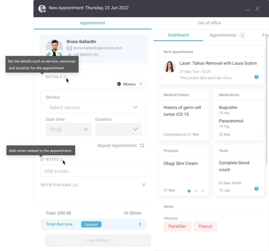

Pabau’s digital forms capture DTR grades, side comparisons, and your written description inside the patient’s chart. Findings from every visit sit on one timeline, so change over time is easy to spot.

The reflex hammer is cheap and the exam is quick. What limits the value of a reflex finding is whether it was graded consistently and written down where somebody can compare it next time.
Print the form, grade against the 0-4+ scale on this page, and keep the plantar response separate from the deep tendon reflexes. Those three habits are what make a reflex grade worth revisiting.
The trade-off with paper is that it only helps once. Book a demo to see how Pabau turns the same form into a record you can compare visit by visit.

Working through the rest of the neuro exam? Cranial nerve examination checklist walks the twelve nerves in the order you test them.
Testing shoulder function in the same exam? Belly press test The technique, its positive signs, and what the accuracy numbers mean for the next shoulder exam.
Need a grading scale for muscle power? MRC muscle strength scale gives you the 0-5 equivalent of the reflex scale.
Assessing joints alongside the reflex exam? Range of motion assessment explains how to measure and record joint movement.
Choosing software for your practice? Physical therapy practice management software The category has exploded with options, but most choices affect billing, prior authorizations, and Medicare audit risk.
A deep tendon reflex exam tests the three-link reflex arc: sensory nerve → spinal cord → motor nerve. Tapping a tendon with a reflex hammer triggers an involuntary muscle contraction, revealing whether the nervous system is functioning normally. It takes less than two minutes and requires only a reflex hammer.
The standardized 0-4+ scale ranges from absent (0) to clonus present (4+), with grade 2+ representing normal. Grade 1+ is diminished, 3+ is increased or brisk, and 4+ indicates rhythmic repeating contractions typical of upper motor neuron hyperexcitability.
Absent reflexes (areflexia) typically indicate lower motor neuron disease, peripheral nerve damage, or spinal nerve root compression. Common causes include diabetic neuropathy, sciatica, Guillain-Barré syndrome, or spinal cord lesions. Age-related diminished reflexes in elderly patients are also normal, particularly Achilles reflexes.
Biceps and brachioradialis reflexes correspond to C5-C6, triceps to C7-C8, patellar to L3-L4, and Achilles to S1-S2. This nerve root mapping is essential for localizing spinal cord and nerve root pathology when reflexes are abnormal.
No. The plantar reflex is a superficial cutaneous reflex elicited by stroking the sole of the foot, not a muscle stretch reflex. Record it as flexor or extensor rather than on the 0-4+ scale, even though the template lists it beside the deep tendon reflexes.