


Dental examination form template: Complete clinical guide
A dental examination form documents patient history, clinical findings, tooth ch...
July 23, 2026
An anemia chart is a diagnostic reference tool that classifies anemia into microcytic, normocytic, and macrocytic categories based on MCV, RDW, and hemoglobin levels.
WHO defines anemia as hemoglobin <13 g/dL in adult males and <12 g/dL in non-pregnant adult females; normal RDW ranges from 11.5% to 14.5%.
Key differentiators include serum ferritin, TIBC, reticulocyte count, and peripheral blood smear findings that help distinguish iron deficiency from B12/folate deficiency and chronic disease anemia.
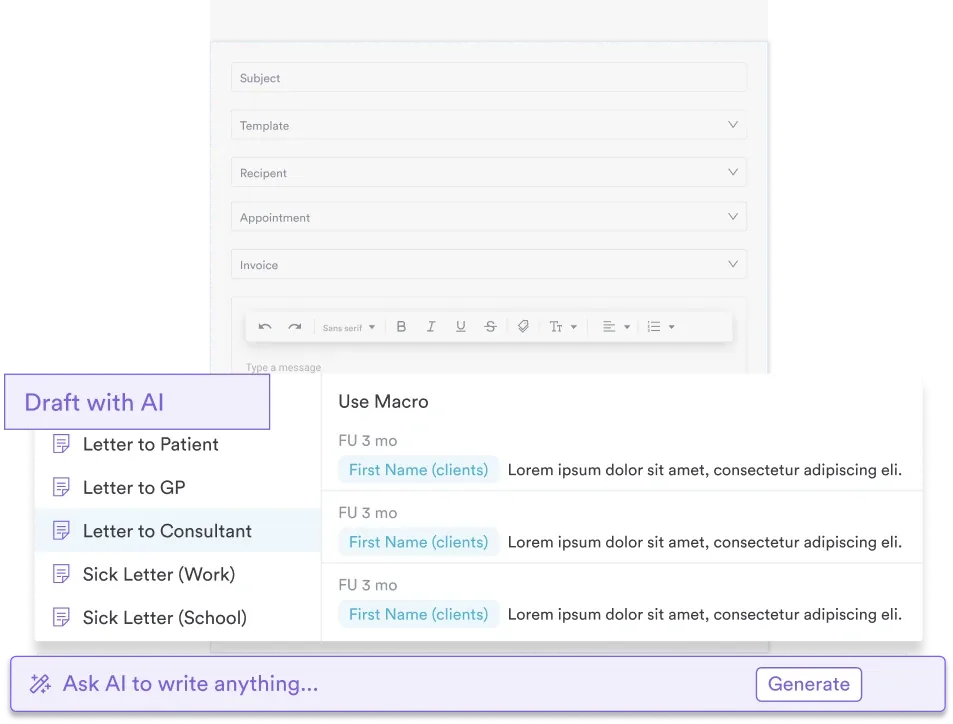
Practice management software like Pabau captures anemia workup findings — MCV category, hemoglobin severity, and next-step tests — in organized patient records.
Severity can be graded on WHO’s three-tier scale (mild, moderate, severe) for general practice, or CTCAE’s four-point scale for oncology settings — matching the system to your setting avoids miscommunication.
A comprehensive diagnostic reference covering morphologic classification (microcytic, normocytic, macrocytic), hemoglobin severity thresholds, lab value interpretation (MCV, RDW, ferritin, TIBC, reticulocyte count), WHO diagnostic criteria, and stepwise differential diagnosis pathways for clinical practice.
Download templateAn anemia chart is a clinician-ready reference tool for classifying, evaluating, and tracking patients with suspected or confirmed anemia. It pulls the classification of anemia by morphology (microcytic, normocytic, macrocytic), WHO hemoglobin severity thresholds, and the key lab markers — MCV, RDW, serum ferritin, TIBC, reticulocyte count, and peripheral smear findings — into a single visual guide.
A practice-focused chart connects each lab result to the next clinical decision, so you can reach a differential diagnosis and prioritize the workup in seconds instead of flipping between references.
Anemia affects an estimated 9.3% of the US population ages 2 and older, and is more common in females (13.0%) than males (5.5%), according to CDC NCHS data from August 2021 to August 2023. A chart that ties hemoglobin levels to severity grading and clinical features gives you that read at a glance and supports consistent biomarker interpretation.
The chart serves three immediate purposes: first, it confirms WHO diagnostic thresholds for anemia (hemoglobin <13 g/dL in adult males, <12 g/dL in non-pregnant females); second, it maps morphologic findings (low MCV, normal MCV, high MCV) to probable etiologies; and third, it guides next-step testing based on reticulocyte count and iron metabolism markers.
A well-designed anemia diagnosis chart follows a stepwise diagnostic logic that mirrors real practice workflows. The five-step pathway below guides practitioners from initial presentation through differential diagnosis confirmation.
An effective anemia chart consolidates these five steps into a single reference — eliminating back-and-forth flipping through multiple resources and reducing the risk of missed differential diagnoses.
Discover how practitioners use Pabau's structured forms and clinical documentation to capture comprehensive anemia workup findings, enabling faster diagnosis and better patient outcomes.

An anemia chart is essential for any healthcare setting where practitioners evaluate patients with suspected blood disorders or chronic conditions that commonly present with anemia.
A structured anemia chart delivers measurable operational and clinical benefits:
Flag patients with elevated RDW (>14.5%) alongside normal MCV for follow-up: mixed causes of anemia (e.g., early iron deficiency plus B12 deficiency) may present with normocytic indices but abnormal RDW. Repeat testing in 4-6 weeks if the initial workup is inconclusive.
The morphologic approach to anemia — classifying cases as microcytic, normocytic, or macrocytic based on MCV — remains the gold standard for differential diagnosis. As an anemia type chart, it maps each category to a distinct set of etiologies; as an anemia investigation chart, it also flags the next-step tests that narrow the differential fastest.
Microcytic anemia (MCV <80 fL) most often points to iron deficiency, thalassemia trait, or anemia of chronic disease (AOCD). Low ferritin confirms iron deficiency, and elevated TIBC supports it; normal-to-high ferritin with low TIBC instead suggests AOCD or chronic inflammation. Key lab markers: serum ferritin, TIBC, serum iron, and a peripheral smear for target cells. Many practices keep this section open as a standalone iron deficiency anemia chart, since low-ferritin, microcytic presentations account for the majority of anemia referrals.
Normocytic anemia (MCV 80-100 fL) covers a wider range — from acute blood loss and hemolytic anemia to bone marrow disorders and renal disease. Check the reticulocyte count: elevated values suggest hemolysis or acute bleeding, while low values point to bone marrow failure or renal insufficiency. Follow-up tests: haptoglobin, LDH, Coombs test, renal function (creatinine, BUN), and EPO level.
Macrocytic anemia (MCV >100 fL) is most commonly caused by vitamin B12 or folate deficiency (megaloblastic) or alcohol-related bone marrow suppression (non-megaloblastic). Order serum B12 and folate levels; if both are low, investigate the underlying cause — pernicious anemia, dietary insufficiency, or malabsorption. A peripheral smear showing hypersegmented neutrophils confirms megaloblastic anemia.
WHO severity grading overlays hemoglobin thresholds onto this morphologic framework:
An integrated anemia chart shows all three dimensions — morphology, severity, and etiology — in one reference.
The anemia chart lab values below cover the constellation of laboratory markers used in a routine anemia workup. Understanding normal ranges and clinical significance prevents misinterpretation and guides targeted follow-up.
| Lab Marker | Normal Range | Clinical Significance in Anemia |
|---|---|---|
| Hemoglobin (Hgb) | Adult M: 13-16 g/dL; Adult F: 12-15 g/dL | Primary diagnostic criterion; values below WHO thresholds confirm anemia presence and severity grade. |
| Mean Corpuscular Volume (MCV) | 80-100 fL | Classifies anemia morphologically; directs differential diagnosis pathway (microcytic, normocytic, macrocytic). |
| Red Cell Distribution Width (RDW) | 11.5-14.5% | Measures RBC size variation; elevated RDW suggests mixed causes or early deficiency; helps differentiate iron deficiency from thalassemia. |
| Reticulocyte Count | 0.5-2% | Indicates bone marrow response; elevated suggests hemolysis or acute bleeding; low suggests production defect (iron, B12, folate, bone marrow disorder). |
| Serum Ferritin | 12-150 ng/mL (F); 24-300 ng/mL (M) | Reflects iron stores; low (<15 ng/mL) confirms iron deficiency; elevated may indicate chronic disease or hemochromatosis. |
| Total Iron-Binding Capacity (TIBC) | 250-425 mcg/dL | Measures transferrin availability; elevated TIBC with low ferritin confirms iron deficiency; low TIBC with normal-to-high ferritin suggests AOCD. |
| Serum B12 | 200-900 pg/mL | Confirms B12 deficiency in macrocytic anemia; levels 200-400 may indicate early deficiency requiring methylmalonic acid and homocysteine testing. |
| Serum Folate | >5.4 ng/mL | Assesses folate status; low folate (along with elevated homocysteine) confirms folate deficiency as cause of macrocytic anemia. |
Note: Reference ranges vary slightly between laboratories. Always reference your facility’s specific range when interpreting results. For functional medicine practitioners, optimizing these ranges (e.g., ferritin 30-100 ng/mL for symptom resolution) may extend beyond standard clinical thresholds.
Implementing an anemia workup chart in routine practice requires more than printing a reference — it requires integration into practice workflows, staff training, and patient communication protocols.
Distribute the chart to all clinical staff and include it in new-hire orientation. Conduct a brief training session explaining the morphologic classification logic and how to use the chart to guide test ordering and clinical documentation. Make the chart visible in consultation rooms or break rooms so practitioners reference it during patient visits. Link the chart to your WHO guidelines and ARUP’s anemia testing algorithm as authoritative references for your team.
For multi-location teams, keeping the chart alongside your clinical records keeps interpretation consistent across practitioners. Practice management software like Pabau lets clinicians record each anemia workup in structured clinical documentation — MCV category, hemoglobin severity, and next-step tests — tied directly to the patient record. Every result and the reasoning behind it stays in one place, so any clinician picking up the case sees the full picture.
An anemia chart turns blood work interpretation into a fast, repeatable process. Consolidating morphologic classification, WHO severity thresholds, key lab markers, and diagnostic pathways into one reference lets clinicians move from lab results to treatment decisions with confidence.
Whether you run a medical spa screening for anemia before IV therapy or a primary care practice managing chronic disease anemia, a well-built chart cuts diagnostic variability and speeds up care. Download the template above and add it to your team’s standard protocols, then see how Pabau keeps every result tied to documented clinical reasoning.
Need a framework for blood work interpretation? Interpreting Biomarkers Without Overpromising provides practical guidance on translating lab results into clinically meaningful patient conversations.
Looking to standardize clinical documentation? Safer Clinical Notes outlines best practices for documenting anemia findings and diagnostic reasoning in patient records.
Want to scale anemia protocols across your team? Paperless Clinical Workflows shows how integrated clinical documentation systems support consistent diagnostic protocols across multiple practitioners and locations.
An anemia chart is a diagnostic reference tool that classifies anemia by morphology (microcytic, normocytic, macrocytic), hemoglobin severity, and key lab values (MCV, RDW, ferritin, TIBC, reticulocyte count) to guide differential diagnosis and treatment decisions in clinical practice.
Microcytic anemia (MCV <80 fL, common causes: iron deficiency, thalassemia), normocytic anemia (MCV 80-100 fL, causes: hemolysis, acute bleeding, renal disease), and macrocytic anemia (MCV >100 fL, causes: B12 or folate deficiency, alcohol-related bone marrow suppression).
Hemoglobin below WHO thresholds (13 g/dL in adult males, 12 g/dL in non-pregnant adult females) confirms anemia. MCV determines morphologic classification; RDW (normal 11.5-14.5%) and reticulocyte count (normal 0.5-2%) help differentiate causes and assess bone marrow response.
Microcytic anemia shows small, hypochromic RBCs; target cells suggest thalassemia. Normocytic anemia with spherocytes and elevated bilirubin suggests hemolytic anemia. Macrocytic anemia displays hypersegmented neutrophils (B12 deficiency) or macroovalocytes. Peripheral smear results confirm lab-based classification and narrow differential diagnosis.
Normal hemoglobin is 13-16 g/dL in adult males and 12-15 g/dL in non-pregnant adult females. WHO defines anemia as hemoglobin below 13 g/dL in males and below 12 g/dL in non-pregnant females — low hemoglobin levels below these cutoffs confirm the diagnosis and set the severity grade. Pediatric and pregnant thresholds differ and should be referenced separately.
After initial diagnosis and treatment initiation, recheck hemoglobin in 4-6 weeks for iron supplementation, 8-12 weeks for B12 or folate replacement. For chronic disease anemia, monitor every 3 months. Acute bleeding or hemolytic episodes warrant urgent rechecking. Always reference your practice’s protocols and the patient’s clinical response to guide timing.
By cause, the common types are iron deficiency anemia, vitamin deficiency anemia (B12 or folate), anemia of chronic disease, and aplastic or hemolytic anemia. By red cell size (MCV), the three morphologic categories on an anemia chart are microcytic, normocytic, and macrocytic.
Download the free anemia chart PDF from the box at the top of this page. It covers morphologic classification, WHO severity thresholds, and lab value interpretation on a single printable reference sheet.
Beyond the three morphologic categories on this chart, clinicians also describe anemia by cause: iron deficiency, vitamin deficiency (B12 or folate), anemia of chronic disease, aplastic, hemolytic, sideroblastic, and thalassemia-related anemia. This etiological classification of anemia sits alongside the morphologic one — a single case can appear in both, for example iron deficiency anemia is also microcytic.
WHO’s three-tier grading (mild, moderate, severe) is the most common in general practice, but oncology settings often use the CTCAE scale, which grades anemia on a four-point scale: Grade 1 (hemoglobin just below normal down to 10 g/dL), Grade 2 (10 to 8 g/dL), Grade 3 (below 8 g/dL, transfusion indicated), and Grade 4 (life-threatening, requiring urgent intervention). Match the grading system to the clinical context you’re documenting.
A borderline result usually sits just above the WHO cutoff — roughly 12-12.9 g/dL in non-pregnant women or 13-13.9 g/dL in men — before hemoglobin drops into the mild anemia band. These readings are often picked up incidentally and warrant a recheck alongside ferritin and reticulocyte count rather than immediate treatment.