







Breastfeeding weaning plan: Free template and 6-week schedule
A breastfeeding weaning plan is a structured way to move your baby from breast m...

1
Last Updated: Sep 16, 2026

The Roos stress test is a provocative test for thoracic outlet syndrome (TOS).
The patient holds both arms at 90° abduction and external rotation. They then open and close their hands for up to three minutes. Symptom reproduction inside that window, or an inability to finish it, is a positive finding.
A 2019 study of microvascular responses to the test reported test-retest reliability that was good but not perfect. The authors attributed part of that to inconsistent symptom reproduction between the two trials. They recommend repeating the maneuver at least twice.
Physical therapists, osteopaths, chiropractors, and sports medicine clinicians meet this presentation often. Upper limb pain, numbness, and heaviness resist an obvious diagnosis, and TOS is easy to miss. This guide covers the procedure, how to read the findings, how it compares with the other TOS tests, and what to document.

The Roos stress test, also called the elevated arm stress test (EAST), screens for thoracic outlet syndrome.
The patient holds both arms at 90° abduction and external rotation, opening and closing the hands for three minutes.
Symptom reproduction within three minutes, or failure to complete the window, is a positive finding.
Reported sensitivity and specificity vary widely, so the result belongs in a cluster with the history and other provocative tests.
Onset timing, symptom type and laterality are what make the record useful later, so document all three.
The Roos stress test puts the neurovascular structures of the thoracic outlet under sustained mechanical stress. It is also called the elevated arm stress test, or EAST, and the two names are used interchangeably. D.B. Roos described it in the 1960s, and it remains the most widely used provocative test for TOS.
Thoracic outlet syndrome is not one pathology. Clinicians recognize three subtypes:
The test is sensitive across all three subtypes, though a positive presentation looks different depending on whether the compression is neural or vascular. Which subtype you suspect changes how you read the findings.
The thoracic outlet is a triangular space bounded by the first rib below, the clavicle in front, and the scalene muscles behind. The brachial plexus and subclavian vessels pass through it before running under the pectoralis minor toward the axilla. In TOS, one or more of those structures is compressed.
Compression comes from a cervical rib, a hypertrophied scalene, or postural narrowing of the costoclavicular space. Elevating the arms to 90° abduction and external rotation tightens that space and stretches the plexus across the first rib.
Repeated hand opening and closing raises metabolic demand in the distal muscles. Where flow is already compromised, symptoms come on faster than positioning alone would produce them.
The pectoralis minor adds a second compression point where the bundle passes under the coracoid process. Patients with a hypertrophied pectoralis minor from overhead sport often show earlier onset. Swimmers, baseball pitchers, and volleyball players are the usual examples.
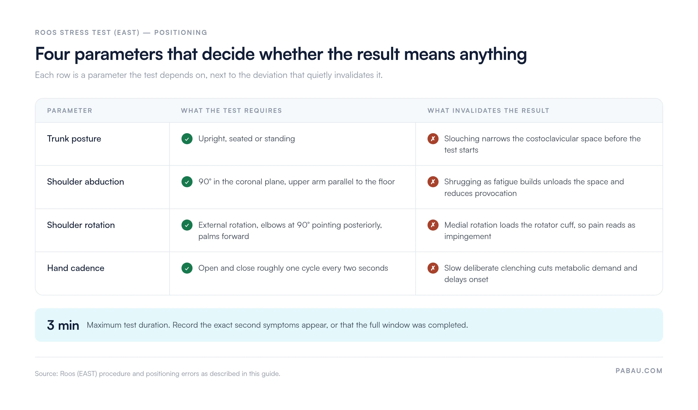
Positioning decides the result. Small deviations in abduction angle or forearm rotation can fail to provoke TOS at all, or produce a false positive from rotator cuff impingement. A purpose-built physical therapy EMR lets you store the procedure as a checklist, so technique stays consistent across practitioners.
Shoulder elevation is the most frequent error. Patients instinctively shrug as fatigue builds, which unloads the costoclavicular space and reduces provocation. Remind them to keep the shoulders relaxed and level throughout.
Insufficient external rotation comes second. If the forearms rotate inward, the test loads the rotator cuff instead of the thoracic outlet. Any pain that follows points at the subacromial space, and the test result carries no TOS information. For a quick visual check, the elbows should point posteriorly instead of laterally.
Cadence matters too. Some patients perform slow, deliberate fist clenches instead of rapid alternating cycles. That lowers metabolic demand and delays or prevents provocation in borderline cases. A two-second cadence is the clinical standard.
Taken together, the four parameters below are what a reader of the note is trusting when they see a positive or negative result.
A positive result reproduces the patient’s characteristic symptoms within the three-minute window. It also counts as positive when symptom severity stops the patient short of three minutes.
New pain on its own, such as localized shoulder fatigue, does not qualify. The reproduced symptoms have to match what the patient reports in daily life.
Positive indicators vary by TOS subtype:
A negative result does not exclude TOS. Mild or intermittent compression can present negatively if provocation is insufficient. Recent corticosteroid treatment can also reduce inflammatory compression enough to mask the finding.
No established guideline maps onset timing to severity, so treat the table below as clinical reasoning rather than a validated rule. Earlier onset generally suggests more significant compression. That relationship is not linear, and the timing belongs alongside the rest of the examination.
Reported accuracy for the Roos stress test varies widely across studies, which reflects how loosely TOS itself is defined. Physiotutors cites a 2001 study by Gillard and colleagues, reporting 84% sensitivity and 30% specificity. Other studies report lower sensitivity, and no universally accepted reference standard exists to compare them against.
Key points from the evidence base:
The Physiopedia clinical reference recommends using the test as part of a cluster. A decision to investigate further should rest on several positive provocative tests, the history, and imaging.
The Roos stress test is the most widely used of the TOS provocative tests, and most clinicians run a battery. Each one stresses the thoracic outlet by a different mechanism, which changes what it detects best.
The Adson test has relatively low specificity on its own, because pulse diminution occurs in many asymptomatic people. The Roos test is generally more useful for neurogenic TOS, since it targets functional limitation and not vascular occlusion alone. If you run the Adson test alongside it, record both results in the same note.
The Wright test is the one to add when hyperabduction reproduces the symptoms, because it points at the subcoracoid space instead of the first rib.
Reach for the test when upper limb symptoms are positional, activity-related, or provoked by overhead work. Three presentations come up most often:
Screen for red flags before you test. Acute hand pallor, pulselessness, or signs of arterial thrombosis make provocative testing inappropriate. Refer urgently instead.
The test earns its keep inside a cluster. One positive provocative test raises suspicion. Two or more, in a clinically consistent presentation, move TOS up the differential substantially. The Morley test adds a direct palpation check over the brachial plexus in the supraclavicular fossa.
Document the Roos, Adson, and Wright results in the same appointment note when you use a cluster approach. Recording onset timing and symptom description for each gives a far richer picture than three separate notes. It also makes the referral letter quicker to write.
The test has limitations worth understanding before you lean on it as a primary diagnostic tool.
False positives are the known concern. Rotator cuff pathology, subacromial impingement, and C8-T1 cervical radiculopathy all produce arm pain and paresthesia during sustained elevation. A cervical screen and a rotator cuff assessment beforehand reduce the misclassification risk. The arm squeeze test is one quick way to separate cervical radiculopathy from shoulder pathology.
False negatives turn up in mild or intermittent compression. A recent corticosteroid injection or manual therapy can temporarily reduce tissue irritability enough to suppress the response. A negative result in a patient with a suggestive history is a reason for combination testing, and it does not exclude TOS.
Patient compliance limits the test too. Significant shoulder pain from any cause, or restricted mobility after surgery, can stop a patient holding 90° abduction for three minutes. In those cases the test is inconclusive, and recording it as negative is a documentation error.
Contraindications include:
None of these are unique to the Roos test. They follow from the same safety reasoning that governs any sustained provocative position.
A vague note reading “Roos test positive” throws away the signal that onset timing, laterality and symptom character carry. Write down the procedure parameters and the patient’s exact response alongside the verdict.
A complete entry covers:
Free-text notes are the default for this, so every practitioner records it slightly differently. Six months later, comparing the first Roos test with the third means reading three paragraphs of prose.
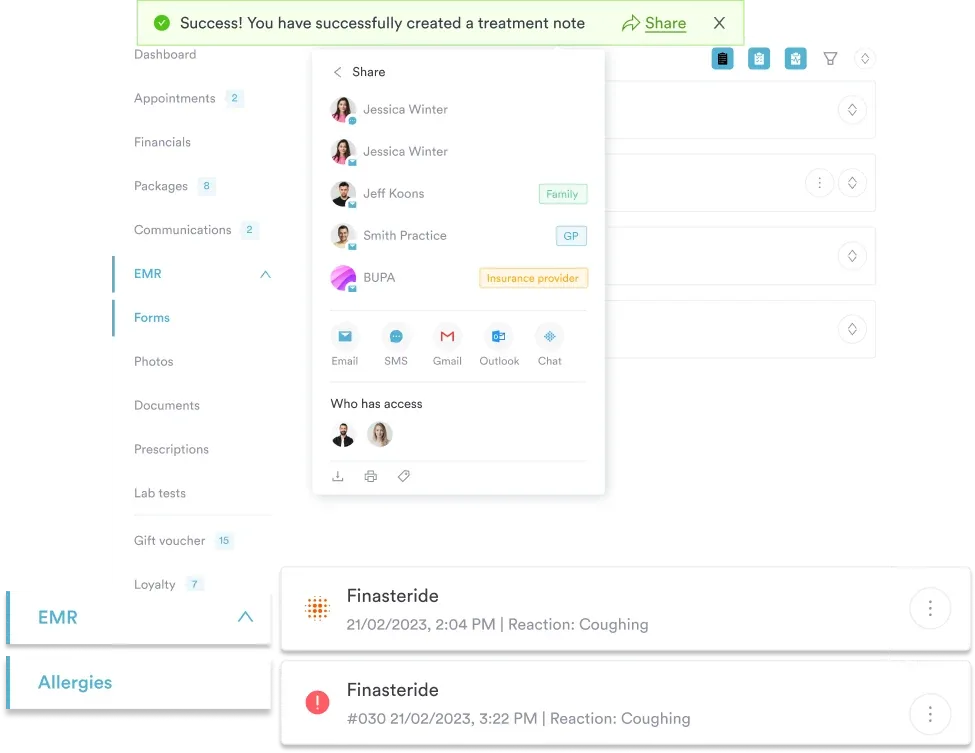
Practice management software like Pabau lets you build the entry once as a custom clinical form. You define the fields for onset in seconds, symptom type, laterality and interpretation. Every practitioner then fills the same ones.

The same template can carry the Adson and Wright fields, so a cluster assessment lands in one note instead of three. That note attaches to the client record the appointment already sits in, which is what makes the referral letter quick to write.
Pabau Scribe, our AI scribe, can draft the narrative part of the note during the appointment, so it is finished before the patient leaves.

Pabau’s custom clinical forms capture Roos test onset timing, symptom type and laterality as structured fields. The note lands in the client record with no transcription step.

The Roos stress test is quick, cheap and easy to perform badly. Getting the position right, then writing down what happened and when, is what separates a useful record from a line that says positive.
Low specificity keeps a positive result in the category of suspicion. Pair it with the Adson or Wright test, weigh the history, and refer when the vascular picture demands it.
If your TOS assessments live in free text today, a structured template is the cheapest change you can make. Book a demo to see how Pabau structures musculoskeletal assessment notes.

List Item #1
List Item #2
The Roos stress test screens for thoracic outlet syndrome. Sustained arm elevation and repeated hand exercises reproduce neurovascular compression symptoms. It covers all three TOS subtypes, neurogenic, arterial and venous, but is most sensitive for the neurogenic form. A positive result supports further investigation without confirming a diagnosis.
The patient sits or stands upright and abducts both arms to 90° in the coronal plane. The shoulders externally rotate so the elbows bend to 90° with the palms facing forward. The patient then opens and closes their hands at a steady pace for up to three minutes. Record when symptoms appear, what type they are, and which side is affected.
A positive result means the patient’s characteristic symptoms return within the three-minute window. Arm pain, numbness, tingling or heaviness all count, as does stopping early because symptoms became severe. It raises suspicion for thoracic outlet syndrome, and confirmation usually needs clinical correlation plus nerve conduction studies or imaging.
Accuracy varies widely by study. Physiotutors cites a 2001 study by Gillard and colleagues reporting 84% sensitivity and 30% specificity. Low specificity is the practical limitation, so use the test inside a cluster with the Adson test, the Wright test and the patient’s history.
The Roos test provokes symptoms through sustained arm elevation and the metabolic demand of repeated hand exercises. The Adson test targets arterial compression. The patient rotates the head to the affected side, extends the neck and holds a breath while you monitor the radial pulse. Adson is more specific for arterial TOS and anterior scalene compression, and Roos is more sensitive for the neurogenic form. Running both improves accuracy.
False positives are the biggest limitation. Rotator cuff pathology and C8-T1 cervical radiculopathy both mimic TOS symptoms during sustained elevation. False negatives occur in mild or intermittent compression. Patients with shoulder surgery restrictions or acute glenohumeral instability cannot hold the position, which makes the result inconclusive. The test is contraindicated when acute arterial or venous occlusion is suspected.