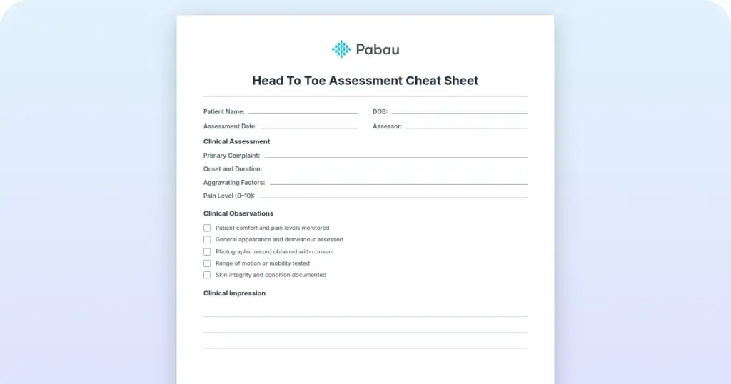
Head to toe assessment cheat sheet: Printable nursing guide
A head to toe assessment cheat sheet gives you one fixed order at the bedside, s...

3
Last Updated: Oct 23, 2026

A vision screening test checks visual acuity, color vision, eye alignment, and depth perception. It never replaces a comprehensive eye exam.
Most settings pass an adult at 20/40 or better, but the criteria vary by state, age, and screening setting.
The AAP recommends screening children at ages 3 to 5, then annually through the school years.
Any abnormal color vision, alignment, or depth perception finding fails the screening, even when acuity is normal.
Practice management software like Pabau stores each screening result and referral decision in the patient’s chart.
A printable clinical form covering patient details, visual acuity for each eye, color and depth perception, eye alignment, and the referral decision. Ready to print, or to import into your practice management system.
Download templateA vision screening test takes 5 to 15 minutes, and it answers one question. Does this patient need to see an eye care professional? Getting that call right depends on the pass threshold for the person in front of you. Those thresholds move with age, state, and setting.
This page gives you the printable form, the acuity thresholds by population, and the referral route for each kind of failure. It also covers what happens once the paper is filled in, because a screening only helps the patient if the referral gets followed up.
A vision screening test is a short, focused eye examination that checks for possible vision problems or eye disease. It measures visual acuity, tests color vision and eye alignment, and can pick up signs of amblyopia (lazy eye) and strabismus (eye misalignment).
A comprehensive eye exam goes further. Performed by an optometrist or ophthalmologist, it uses dilating drops and evaluates the internal and external structures of the eye. A screening does neither. If it identifies a possible problem, the patient is referred on for that full examination.
Vision screening tests are systematic and reproducible, which is what makes them usable at scale. Primary care doctors, pediatricians, school nurses, occupational health providers, and trained screeners can all administer one. They run in practices, schools, workplaces, and community settings, and they give you a quick risk assessment rather than a diagnosis.
A screening cannot replace a comprehensive exam. It is a first-line tool that identifies people at risk and routes them for proper care. That is what keeps it affordable across a large population. Our guide to the eye physical examination covers what the full workup adds once the patient arrives.
Vision screenings combine several methods, each aimed at a different part of visual function. Knowing what each one covers helps you choose the right tools for your patient population. Depth perception has its own scoring conventions, which the stereopsis test template sets out in full.
The Snellen chart remains the standard for measuring visual acuity. Each row of letters has a designated distance at which a person with 20/20 vision should read it clearly. In a 20/40 result, the numerator is the patient’s distance from the chart. The denominator is the distance at which normal vision reads that same line. Because the notation is standardized, you can track change over time and hand a specialist a figure they recognize.
Screening intervals vary by age, risk factors, and setting. Agreeing one schedule across the practice is what keeps those intervals from drifting.
Commercial drivers sit under a separate federal standard rather than a state one. The DOT eye test form records the acuity, field of vision, and color recognition findings the examiner has to certify.
Thresholds depend on the patient’s age and the screening setting. No single universal standard exists, so criteria vary by state, school district, and occupational body.
The card below sets each threshold beside the referral route it triggers, so a screener can make both calls from one place.
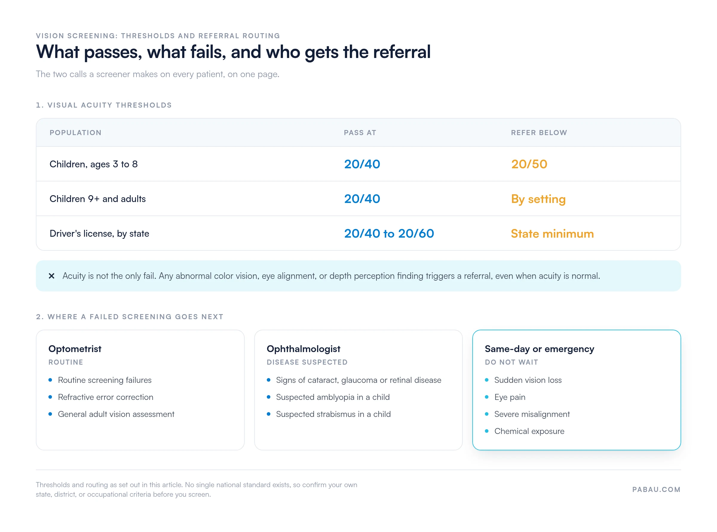
Important: These are general guidelines. Always confirm your state or local screening standards before you set a pass threshold. Then document the test method, the result, and the referral decision for every patient.
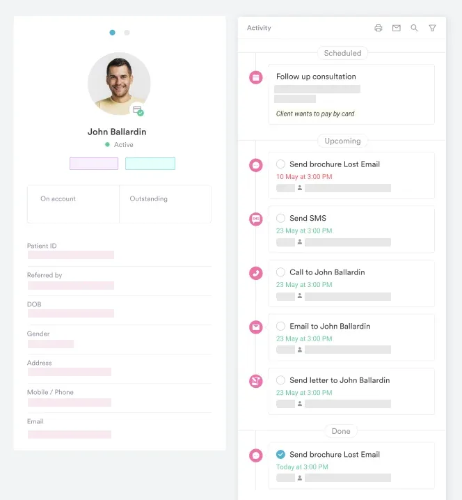
The downloadable form gives you a set order for running and recording a screening. Follow these steps to use it in practice.
Each component of a screening has its own section on the printable form. Here is what you get when you open it.
Once the paper screening is complete, the results need to come off paper. Digital intake forms give you long-term storage, referral tracking, and a record that survives a compliance audit.
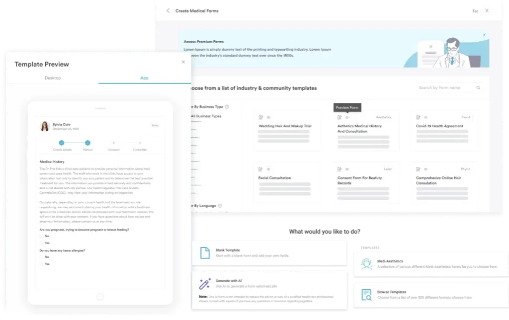
Store the result and the referral decision in the patient’s permanent chart, so the next clinician sees the assessment immediately. Where a referral was issued, track its status through scheduled, completed, and result received.
Digital patient records also keep the file inside a HIPAA-compliant system, which matters because screening records hold sensitive health data.
Linking the result to the appointment history pays off later. It lets you recall a school-age child at the right interval without anyone reading back through a folder of paper forms.
Several organizations publish vision screening guidelines, and they do not agree on every detail. Pick one standard for your patient population and clinical setting, then apply it consistently.
The referral is the outcome that matters most. Any abnormal finding warrants one, whether that is acuity below your threshold, abnormal color vision, eye misalignment, or reduced depth perception. Write down the specific reason, so the specialist reads the result in the context you saw it.
Paper screening forms create their own admin. The form goes into a folder and the referral gets written on a note. A week later nobody can say whether the patient booked the exam.
Pabau keeps the whole screening in one record. You build the form once as a digital intake form, and the acuity, color, alignment, and depth results save straight to the patient’s chart. The pass or fail decision sits in the same place as the appointment that produced it.
From there the follow-up runs itself. Automated recalls bring school-age children back at the right interval, and the referral stays on the chart until someone marks it complete. Your team stops rebuilding the picture from paper, and every result stays inside a HIPAA-compliant record.

Run vision screenings as digital forms, store the pass or fail decision against the appointment, and track referrals until they close. Your team spends the time on patients instead of paperwork.

A screening is only as good as the referral that follows it. Print the form, agree one pass threshold across the team, and write the reason on every fail. That alone removes most of the variation between screeners.
The trade-off worth remembering is scope. A screening buys you speed and reach, and it gives up depth in exchange, so it never carries a diagnosis. Treat every fail as a handover rather than a verdict. Book a demo to see how Pabau stores each screening result and keeps the referral visible until it closes.

Documenting pupil findings alongside acuity? The PERRLA eye exam form template gives you a structured place to record pupil size, reaction, and accommodation.
Need to monitor central vision between exams? The Amsler grid chart is the printable tool for tracking distortion in patients at risk of macular change.
Screening patients with diabetes? The diabetes eye exam form covers the retinal findings and recall intervals that group needs.
Wondering whether a fail is refractive or something worse? The pinhole test is the quick check that separates blurred vision from disease.
Following up an alignment finding? Our guide to the eye movement test walks through tracking, saccades, and what each result points to.
A vision screening test is a brief eye examination that checks visual acuity, color vision, eye alignment, and depth perception. It flags people who may have a vision problem. It is not a comprehensive eye exam and cannot detect every eye condition. Anyone with an abnormal result is referred to an eye care professional for full evaluation.
A vision screening is a quick 5 to 15 minute test that checks for obvious vision problems. An eye exam is a 30 to 60 minute evaluation by an optometrist or ophthalmologist. It adds a dilated examination, visual fields, intraocular pressure, and a diagnosis. Screening identifies the people who need that exam, and does not replace it.
Vision screenings test for visual acuity, color blindness, eye misalignment (strabismus), and depth perception abnormalities. They can flag amblyopia (lazy eye), refractive errors, and other conditions that warrant a specialist referral. They do not diagnose disease or set a treatment plan.
Children aged 3 to 5 and all school-age children should be screened annually, per AAP and AOA guidelines. Adults should be screened every one to three years, depending on risk factors. Those 65 and older benefit from annual screening. Driver’s license renewal, occupational health assessments, and pre-treatment clearance often require one too.
A failed screening means a referral to an optometrist or ophthalmologist for a comprehensive eye exam. A fail can come from acuity below the threshold, or from an abnormal color or alignment finding. The specialist diagnoses the cause and prescribes treatment if needed, such as glasses or eye exercises. A fail does not mean disease, and most failures come down to refractive error.
No. A screening identifies people at risk and routes them for further evaluation. It cannot detect internal eye disease, measure eye pressure, or assess visual fields. Only a comprehensive exam by an eye care professional does that. Screening is the gateway to care, and the exam is where a diagnosis happens.