Sulcus Test: Shoulder Instability Assessment
The sulcus test is an orthopedic special test that checks for inferior glenohume...

3
Last Updated: Oct 22, 2026

An eye physical examination covers eight components: visual acuity, pupils, eye movements, visual fields, intraocular pressure, external inspection, anterior segment, and posterior segment.
The American Academy of Ophthalmology sets adult exam frequency by age band, and high-risk patients need an annual exam whatever their age.
The AAO and the American Optometric Association publish different intervals, so every practice needs one written recall policy.
Eye exams pick up systemic disease, including hypertension and diabetic retinopathy, so they matter well beyond vision correction.
Practice management software like Pabau handles digital intake, automated recall, and clinical notes across the whole exam workflow.
An eye physical examination is a structured assessment of the visual system, covering eight components in one appointment. Each component checks a different part of the eye. Each one can surface disease the others would miss.
In 2017, about 93 million US adults were at high risk for vision loss. Around 40% of them had not seen an eye doctor in the previous year, according to CDC data. Most of that shortfall comes down to access and workflow rather than clinical judgment.
This guide covers what the examination includes, who performs it, how often patients need one, and how to prepare them. It is written for optometrists, ophthalmologists, and the practice managers who support them.
An eye physical examination assesses function and structure together. Function covers acuity, pupil responses, eye movements, and visual fields. Structure covers the eyelids and cornea at the front, then the lens, retina, and optic nerve behind them.
That breadth is what separates the examination from a vision screening. A screening checks acuity and flags who needs a referral. A home nearsighted test with a printed Snellen chart is one such screening, since it measures distance acuity alone. The full examination looks for disease, including disease the patient cannot feel yet.
A routine examination takes 30 to 45 minutes. Adding dilation pushes it to 60 to 90 minutes, because mydriatic drops need 20 to 30 minutes to work. Build both lengths into your appointment types so the schedule matches what actually happens in the room.
Optometrists and ophthalmologists both perform eye physical examinations, and scope of practice varies by state. Optometrists typically manage refractive errors, ocular surface disease, and screening examinations.
Ophthalmologists handle surgical conditions, complex retinal disease, and specialist referrals. Some patients see both within a single episode of care.
Family physicians sit upstream of both. A routine physical often picks up the visual complaint first, and primary care software is where that referral gets raised and tracked. Vision checks are also a standard part of the preparticipation physical for young athletes.
Frequency depends on age and risk. The American Optometric Association recommends an exam at least every two years for low-risk adults aged 18 to 64.
From 65 onward, the recommendation is annual. Anyone with diabetes, a family history of glaucoma, or existing eye disease should be seen at least annually.
The American Academy of Ophthalmology bands its adult advice differently. It suggests every two to four years from 40 to 54, then every one to three years from 55 to 64. From 65 the interval is one to two years. High-risk patients are seen annually at any age.
Two respected bodies, two different intervals. Pick one as your practice policy, write it down, and encode it in your recall rules. Leave it unwritten and each clinician sets their own interval, which is how a recall list quietly drifts out of date.
Children are examined more often than adults. The AOA recommends a first exam at 6 to 12 months, and at least one more between ages 3 and 5. After that comes an exam before first grade, then annually through age 17. Vision screening usually runs alongside a broader developmental assessment at those early visits.
Efficient appointment scheduling and automated recall messaging are what turn a frequency policy into booked appointments. Without a system tracking when each patient is due, the recommendation stays on paper.
Patients arrive under-prepared, and the examination loses five to ten minutes catching up. A short pre-visit checklist, sent digitally before the appointment, removes most of that friction. Nothing else in the appointment gives back that much time for so little effort.
Good patient compliance with pre-visit preparation starts with clear instructions. Here is what to bring and what to do before arrival.
Sending digital intake forms ahead of the appointment captures medical history, symptoms, and medication lists before the patient arrives. The checklist stops being a verbal prompt and becomes a structured clinical record, pulling straight into the patient file instead of onto a paper clipboard.
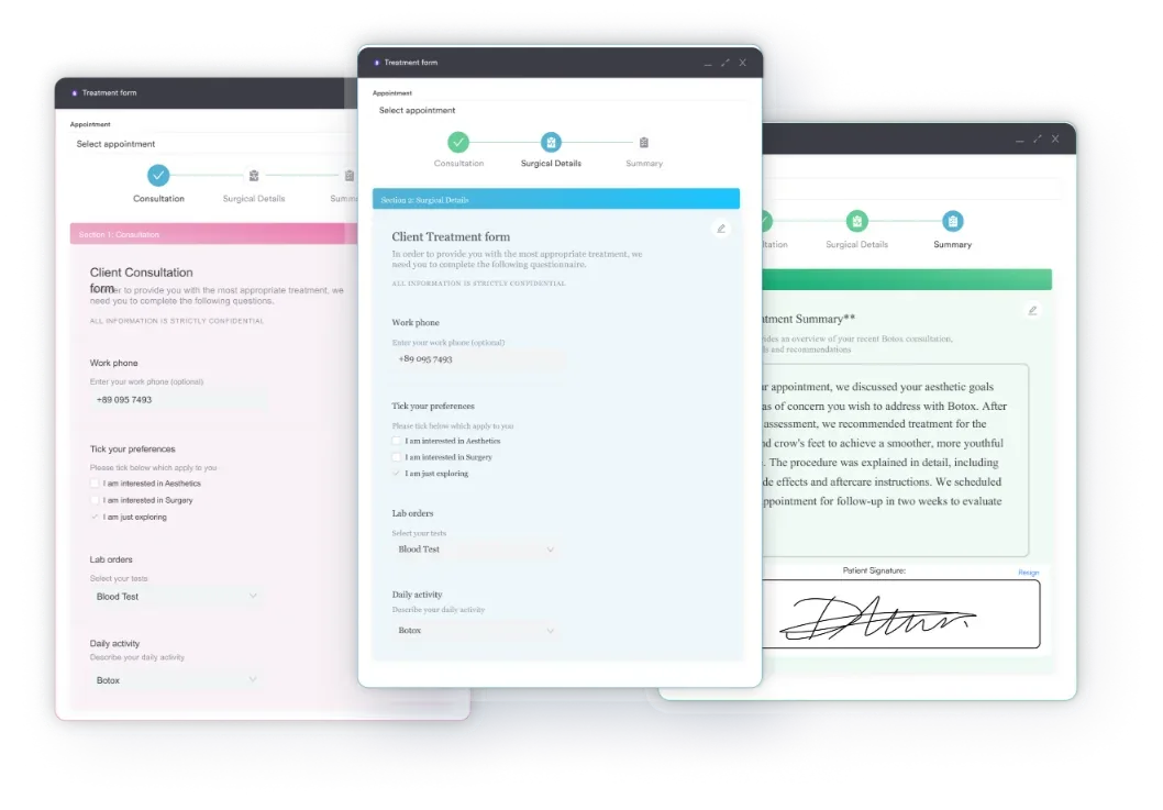
Complete medical intake forms also help clinicians decide how deep to go. A patient reporting floaters and flashes needs fundoscopy prioritized. A patient booked for a routine refractive check needs a different flow. Pre-visit data shapes the appointment before the patient walks in.
The American Academy of Ophthalmology’s 8-point examination framework sets the clinical standard for a comprehensive eye exam. The sequence below follows that structure. It adds the diagnostic context that helps practice teams document findings accurately and understand what each step is looking for.
Visual acuity is measured with a Snellen chart at 20 feet (6 meters), or a LogMAR chart where greater precision is needed. Each eye is tested separately, first without correction, then with habitual correction in place. Near vision is assessed with a near card when indicated.
The result is recorded as a fraction. Normal resolution is 20/20, while 20/40 means the patient sees at 20 feet what a normally sighted person resolves at 40. Results below 20/30 typically prompt refraction testing.
Pupil assessment looks for symmetry, reactivity, and the relative afferent pupillary defect (RAPD). The swinging flashlight test alternates a bright light between the eyes. An RAPD, also called a Marcus Gunn pupil, points to asymmetric optic nerve or retinal disease. Both direct and consensual responses are recorded.
Extraocular movements are assessed by asking the patient to follow a target through the diagnostic H pattern. Limitations, diplopia, or nystagmus at any position of gaze indicate muscle, nerve, or central pathology that needs further investigation. Where reduced binocular vision is suspected, a randot test measures depth perception in seconds of arc.
Confrontation visual fields provide a rapid bedside screen. The clinician presents targets in all four quadrants while the patient fixates centrally. Field defects suggest glaucomatous damage, a neurological lesion such as hemianopia from stroke or tumor, or retinal pathology. When confrontation suggests a defect, formal automated perimetry gives quantified thresholds for documentation and monitoring.
The slit lamp is a specialized binocular microscope that illuminates and magnifies anterior ocular structures in cross-section.
The examination proceeds systematically through the eyelids and margins, conjunctiva and sclera, cornea, anterior chamber depth and cells, iris, and lens. The slit lamp identifies corneal abrasions, uveitis, early cataract, pterygium, and blepharitis, among other anterior segment pathologies.
Normal intraocular pressure (IOP) ranges from 10 to 21 mmHg. Values above 21 mmHg are classified as ocular hypertension and carry elevated glaucoma risk. Elevated IOP alone does not confirm a glaucoma diagnosis, because individual optic nerve susceptibility varies.
Goldmann applanation tonometry, performed at the slit lamp after fluorescein staining, is the clinical gold standard. Non-contact tonometry, the air puff test, is widely used for screening but is less precise for definitive measurement.
Mydriatic drops, typically tropicamide 0.5-1% or phenylephrine 2.5%, dilate the pupil to allow a wider view of the posterior segment. Dilation takes 20 to 30 minutes to achieve and the effects last two to four hours. Direct ophthalmoscopy gives a magnified but narrow view, while indirect ophthalmoscopy shows more of the peripheral retina.
The examination assesses the optic disc, the macula, the retinal vasculature, and the peripheral retina. On the disc that means cup-to-disc ratio, rim tissue, and pallor. On the macula it means drusen, exudates, and hemorrhage. Diabetic retinopathy, age-related macular degeneration, and retinal detachment are all identified at this stage.
An eye physical examination surfaces both primary ocular disease and systemic conditions with ocular signs. The table below maps key conditions to the examination component where they are typically identified.
The systemic conditions in this table, hypertension and diabetes especially, make the eye exam a useful whole-body screen. Retinal vessels can be viewed directly, so an examination can surface cardiovascular and metabolic disease before symptoms appear elsewhere.
Practices running metabolic health programs get particular value from that, because a retinal finding often confirms what the bloodwork suggests.
Tell patients that plainly. It gives them a reason to attend on schedule rather than only when their vision changes.
Good documentation of these findings matters clinically and legally. Keeping patient records current means findings stay retrievable, referrals stay traceable, and recall intervals rest on evidence rather than guesswork. When findings go to a retinal specialist, a signed disclosure authorization form keeps that handover compliant.
Schedule the dilated exam at the end of the appointment slot rather than the beginning. Instill the drops during history-taking and the anterior segment stages. They reach full effect just as you get to fundoscopy, so the appointment does not run long.
Clinical efficiency in optometry and ophthalmology is limited less by examination skill than by administrative friction. Documentation takes longer than the exam itself. Recall letters go out weeks late. Intake happens on paper, then a staff member types it up. Those costs compound across a busy schedule.
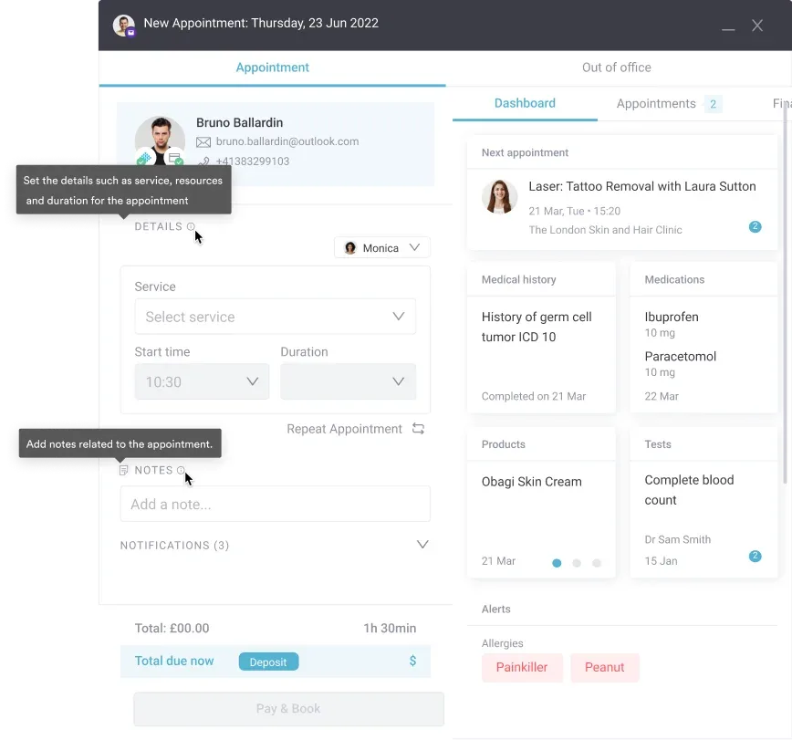
Practice management software like Pabau covers each stage of that workflow. Before the visit, digital intake forms collect history, symptoms, and current medications. The data lands in the patient record without a transcription step.
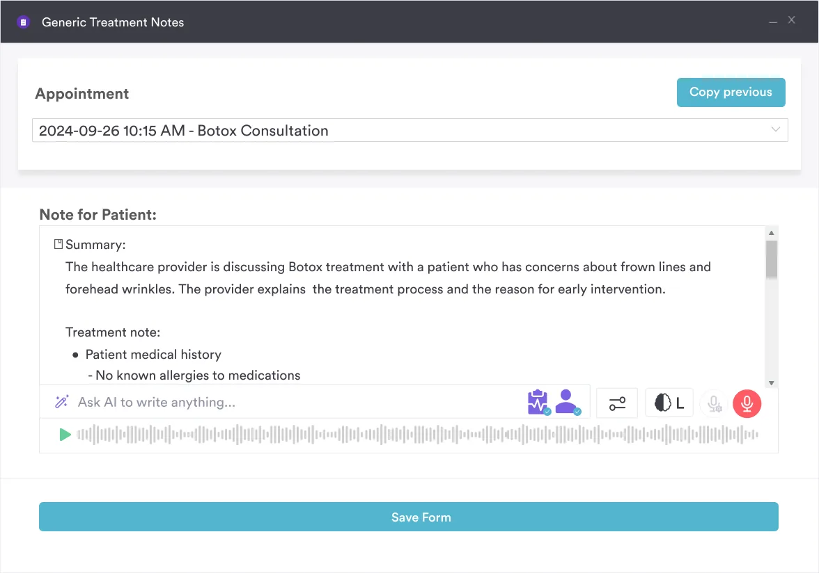
During the examination, Pabau Scribe, our AI scribe, captures notes in real time and structures findings across each component. The clinician never stops to type. For a practice seeing 20 to 30 patients a day, that time comes back as appointment capacity or an earlier finish.
After the appointment, recall is where eye care practices lose patients. A borderline IOP finding needs a 12-month review, and the patient relies on you to prompt it.
Pabau’s automated recall workflows send follow-up messages at set intervals, triggered by the appointment type or a clinical flag, with no manual chasing per patient.
The effect on patient care management shows up within a quarter. Patients return on schedule, findings are documented consistently, and clinicians spend examination time examining. Stronger patient engagement across the cycle, from reminders to post-visit education, means fewer patients drift out of the recall cycle altogether.

Pabau handles digital intake, AI-assisted documentation, and automated recall across every stage of the eye examination workflow. Book a demo to see it running on your own appointment types.

The clinical framework for an eye examination is settled. The variable is execution. Two practices can follow the same eight-point sequence and get very different outcomes. One books the review. The other hopes the patient remembers.
So choose a frequency policy, write it into your appointment types, and let the system chase the review. That one decision moves more of your at-risk patients from due for an exam to actually examined.
The trade-off worth remembering is time. A dilated exam costs 60 to 90 minutes of chair time, and nothing shortens that. What you can shorten is everything on either side of it. Book a demo to see how Pabau handles intake, documentation, and recall for eye care practices.

Screening for disease signs beyond the eye? Neurological eye exam covers the cranial nerve and visual pathway checks that can catch early disease.
Separating refractive error from disease fast? Pinhole test shows how to perform, interpret, and document the result in seconds.
Documenting a focus and convergence check? Accommodation eye test template is a free download for recording pupil, convergence, and lens findings.
Helping patients monitor vision changes at home? Amsler grid chart is a free printable tool for spotting early signs of macular damage.
Certifying a commercial driver’s vision? DOT eye test covers the visual acuity and peripheral vision checks federal standards require.
An eye physical examination is a structured clinical assessment of the visual system. It covers visual acuity, pupillary reflexes, eye movements, visual fields, intraocular pressure, the anterior segment, and the posterior segment. An optometrist or ophthalmologist performs it in 30 to 45 minutes, or 60 to 90 minutes with dilation.
The clinician tests vision with a Snellen chart, then checks pupil responses, eye movements, and peripheral vision. Intraocular pressure is measured with tonometry. The slit lamp examines the front of the eye, and ophthalmoscopy assesses the retina and optic nerve. If dilation is planned, mydriatic drops are given before that last step.
Eye exams detect glaucoma, cataracts, age-related macular degeneration, diabetic retinopathy, refractive errors, and hypertensive retinopathy. The retina also shows early signs of systemic conditions including hypertension and diabetes, which makes the examination useful beyond vision alone.
Low-risk adults aged 18 to 64 need an exam at least every two years, per the American Optometric Association. From 65 onward the recommendation is annual. Children need an exam before first grade and annually after that. Anyone with diabetes, a family history of glaucoma, or existing eye disease should be seen at least annually.
A routine eye physical examination takes 30 to 45 minutes. A comprehensive exam with dilation runs 60 to 90 minutes. Mydriatic drops need 20 to 30 minutes to take effect, and that wait sits inside the appointment.
Yes. Retinal examination can reveal signs of diabetic retinopathy and hypertensive retinopathy before a patient has a formal systemic diagnosis. The retinal vessels show hemorrhages, exudates, and caliber changes that point to metabolic or cardiovascular disease. Treat it as one indicator among many, not a standalone diagnostic test.