



GLP-1 diet plan
A GLP-1 diet plan is a structured eating framework for patients taking semagluti...

2
Last Updated: Oct 13, 2026

Pinhole test: a rapid clinical tool that distinguishes refractive error from organic/pathological causes of reduced visual acuity
Vision improving by 2+ Snellen lines through the pinhole indicates a correctable refractive cause; no improvement suggests deeper pathology requiring investigation
Dense cataracts can paradoxically worsen vision through the pinhole due to reduced luminance — a key limitation clinicians must know
Pabau’s digital consultation forms and structured client records let ophthalmic and allied health teams document VA and pinhole results in standard notation (e.g. 6/12 PH 6/6) with a full audit trail
Most practices see a patient with blurred vision and head straight for the Snellen chart. Instead, the pinhole test is the faster first step for telling whether the blur is refractive. If your eye care practice also runs aesthetic services, our comparison of software for med spas ranks eight platforms on charting and checkout.
The pinhole test is the 30-second step that tells you whether you’re looking at a lens problem or something deeper. Without it, a practice risks sending a patient away with a new prescription. Instead, what the patient may actually need is an OCT scan.
This guide covers the optics and the step-by-step technique. In addition, it explains how to read the results and how to document your findings in a structured patient record.
You might run the pinhole eye test as a quick screen. Or you might use it as a formal part of an eye exam. So, either way, it belongs in every visual acuity workflow. That holds whether you are an optometrist, ophthalmologist, or allied health professional running a clinical consultation. It typically sits within a broader eye physical examination that also checks pupils, motility, and ocular alignment.
First, the pinhole works on a simple principle known as the pinhole effect. Specifically, restricting the diameter of the entering light beam to roughly 1-2 mm eliminates the peripheral rays. Those rays are what cause a blurred retinal image in an uncorrected eye. What remains is a narrow central beam. That beam lands more precisely on the a blurred retinal image, reducing what optometrists call the “circle of confusion.”
Aperture size matters here. Around 1.2 mm is widely cited as the most effective diameter. Go much smaller, however, and diffraction starts to degrade the image rather than sharpen it.
In a patient with myopia, hyperopia, or astigmatism, that circle of confusion is the primary reason acuity is poor. That’s why the pinhole test and astigmatism go hand in hand. Removing most of the off-axis rays sharpens vision, often markedly, when the cause is refractive.
Some patients have blur from a structural problem, such as damaged photoreceptors, optic nerve disease, or a dense lens opacity. In these cases, the pinhole changes nothing useful. The damage sits downstream of where light enters the eye, so restricting the aperture can’t correct it.
This is the fundamental clinical value of the pinhole test. In practice, it partitions causes of reduced acuity into two categories before a refractionist has touched the patient.
A pinhole occluder test requires minimal kit, which is part of its value in triage and allied health settings.
In fact, no specialist equipment is needed beyond what is already in any ophthalmic or allied health consulting room. That accessibility is exactly why a 2019 PMC/NIH guide recommends the pinhole test as a frontline tool. Specifically, the guide points to ophthalmic nurses and allied health personnel performing myopia screening.
In general, the technique is consistent across professional bodies, with minor variations in chart distance. Follow these steps for each eye separately.
A practical clinical tip: patients sometimes struggle to align the pinhole with their visual axis on the first attempt. Giving them a few seconds to “hunt” for the clearest aperture position reduces false negatives. As a result, this approach is standard practice at most ophthalmic units.
In practice, interpretation comes down to comparing the pinhole acuity with the unaided (or current correction) acuity. A population-based validation study on pinhole accuracy set a clear clinical threshold. It found that an improvement of more than two logMAR lines predicts significant refractive error with high specificity. That is the threshold most clinicians apply when reading Snellen-based pinhole results.
Keep in mind that these interpretations are probabilistic, not diagnostic. However, the pinhole test indicates where to look next, not what the diagnosis is. Professional body guidance in your jurisdiction will define specific referral thresholds.
In practice, non-improvement is clinically significant and should prompt structured follow-up, not reassurance. Several conditions are commonly associated with failure to improve through the pinhole.
Structured documentation of these findings in a patient compliance and clinical audit context matters here. A clear record of which eye showed non-improvement, the baseline VA, and the date is essential. It provides the foundation for tracking progression and, in addition, for justifying an urgent referral.
The pinhole test is often the first step an eye clinician takes when macular disease is suspected. However, it plays a specific role: differentiation, not diagnosis. A result showing no improvement through the pinhole raises the index of suspicion for macular pathology without confirming it.
A patient may present with central vision loss, distortion, or reduced reading acuity. When the pinhole test shows no improvement in this situation, the recommended follow-up pathway includes:
Healthline’s overview of the pinhole test for macular degeneration makes a similar point. The test helps direct the investigation rather than conclude it. In most cases, that means a dilated eye exam to check the retina and optic nerve directly.
For practices running allied health screening programs, the pinhole test offers a practical triage decision point. Improvement suggests a spectacle referral; non-improvement, by contrast, suggests an ophthalmology referral. That simple binary holds up well in high-volume or resource-limited settings. It works everywhere, from wellness screening days screening days to dedicated eye practices.
Indeed, the same logic applies across specialties. Structured skin assessment tools guide triage decisions in dermatology. In the same way, a positive or negative pinhole result guides an eye referral, just as the Steinman test steers meniscal referral decisions in orthopedic practice.
Document the specific aperture position the patient used if you are using a multi-aperture occluder and they achieved their best result off-center. Noting ‘eccentric fixation used’ alongside the VA reading adds clinical precision that matters if the case is later reviewed by a specialist.
Best-corrected visual acuity (BCVA) is the gold standard: the sharpest acuity achievable with optimal refractive correction. In practice, the pinhole test provides a rapid estimate of what BCVA might be, without a refractionist or trial frame.
In straightforward refractive cases, pinhole acuity and BCVA agree closely. For example, both show near-normal acuity in a myopic patient whose only problem is an uncorrected prescription.
Where they diverge is clinically informative. If the pinhole shows, say, 6/9 but BCVA with a trial lens reaches 6/6, the small residual difference is telling. It often reflects the limits of the pinhole method, such as reduced contrast sensitivity or patient alignment difficulty. If pinhole acuity is markedly worse than an expected BCVA, that discrepancy warrants further investigation.
In emergency and triage settings — A&E eye units, remote health outposts, military medicine — pinhole acuity serves as a BCVA proxy. This proxy matters whenever formal refraction equipment isn’t available. The same logic applies in everyday private practice and GP settings. A full refraction suite often isn’t on hand there either.
Allied health roles, including nurses and paramedics, are trained to use the pinhole occluder. That’s why it’s part of a basic ophthalmic assessment.
See how time-saving practice tools translate this same principle of rapid, structured triage into practice management workflows.
The potential acuity pinhole (PAP) test predicts vision after cataract surgery. When a cataract is blurring vision, patients and surgeons want to know how much sight is likely to return. That typically happens once the lens is replaced — a procedure billed for anesthesia under 00144.
The test answers that directly. The patient reads the chart through a pinhole held against a bright light source. This pushes enough light past the lens opacity to estimate the retina’s underlying acuity.
A good pinhole result behind a cataract suggests the macula and optic nerve are healthy. It also suggests that surgery should restore vision. A poor result flags coexisting pathology that surgery alone won’t fix, so expectations can be set honestly beforehand. Once surgery is booked, a structured post-op eye drop schedule helps patients follow the four-week regimen correctly.
The dedicated potential acuity meter (PAM) is marginally more accurate. However, the PAP test is faster, cheaper, and possible in any consulting room.
Specifically, double vision is where the pinhole earns its place in triage. Ask the patient to cover one eye. If the doubling disappears, it is binocular diplopia, usually a muscle or neurological cause the pinhole cannot address. If the doubling persists in one eye alone, it is monocular diplopia. That’s why the pinhole test genuinely helps here. Cases with a neurological cause often need a neurological eye exam to check cranial nerve and pupil function.
Monocular double vision that resolves through the pinhole points to an optical cause. Specifically, it could be uncorrected astigmatism, an irregular cornea, an early cataract, or dry eye disrupting the tear film. Doubling that persists even through the pinhole suggests the problem sits deeper than the optics. That single step tells you whether to reach for a refraction or a referral.

Pabau’s structured consultation forms let ophthalmic and allied health teams record VA, pinhole results, and referral decisions in one place, with a full audit trail for every patient encounter.

In fact, no clinical screening test is without limits, and the pinhole test is no exception. Understanding these helps avoid misinterpreting results and over-relying on a single data point.
For more on the optics behind aperture sizing, see Review of Optometry. It explains why apertures between roughly 0.94mm and 1.75mm work best. It also explains how anything much smaller introduces diffraction that degrades the image rather than sharpening it. It is a useful reading reference for allied health training programs.
In practice, documentation is where clinical continuity often breaks down. Poor documentation of pinhole findings leaves the patient record incomplete. This can delay referrals, confuse follow-up clinicians, and create medico-legal exposure.
The accepted shorthand is straightforward. Take a patient whose unaided VA is 6/12 in the right eye. If that improves to 6/6 through the pinhole, you record: RE: 6/12 PH 6/6. The “PH” abbreviation is universally understood in UK and Commonwealth ophthalmic records. In US notation, substitute Snellen fractions with 20/x equivalents: OD: 20/40 PH 20/20.
Record the following for every pinhole test encounter:
Overall, this kind of structured capture reflects general best practice in ophthalmic record-keeping. The College of Optometrists’ guidance on what to record calls for full, accurate patient records at the time of examination. That record should include distance visual acuity findings. It’s exactly what any CQC or HIS inspection will look for in an ophthalmic practice.
This is where practice management software like Pabau helps. Pabau’s digital consultation forms let ophthalmic and allied health practices build structured pinhole visual acuity recording templates. These templates fit directly into the consultation workflow. Clinicians enter unaided VA, pinhole VA, and clinical interpretation into pre-built fields — no freehand notes that get misread or lost.

The structured client record stores each encounter chronologically with a full audit trail. So, a follow-up clinician or a specialist receiving a referral can see the entire VA progression at a glance.
For practices managing large patient volumes, the ability to search and report on pinhole findings across the patient cohort matters. This capability supports clinical governance and audit requirements. Well-maintained client records also reduce the time spent reconstructing history when patients present urgently.
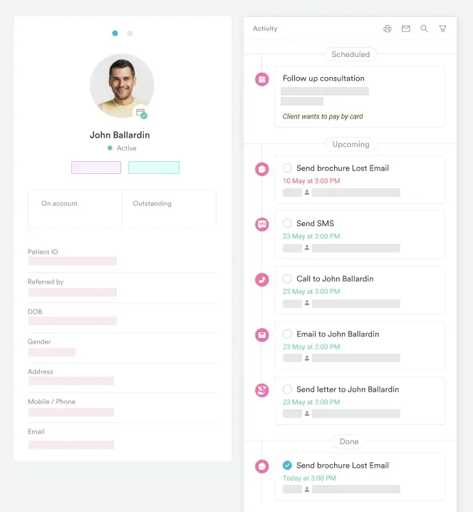
For teams building or refining their clinical documentation approach, writing safer clinical notes is a practical starting point. It helps structure ophthalmic findings in a way that meets regulatory and medico-legal standards. Practices wanting to move from paper-based VA recording to a fully paperless workflow have another option. They can explore how medical forms make the transition operationally straightforward.
In addition, capture forms are another option for practices that want to collect pinhole and VA data digitally. This works even before the patient enters the room. Pabau’s capture forms can be sent ahead of the appointment. This reduces in-room documentation time and gives clinicians a baseline before the consultation begins.

Overall, the pinhole test is one of the most efficient diagnostic partitions in ophthalmic practice. In under a minute, it separates refractive causes from pathological ones and guides the next investigation. It also gives every clinician a documented baseline for the patient’s visual function. Used correctly and recorded precisely, it protects patients and practices alike.
If your practice is still capturing VA and pinhole findings on paper or in unstructured notes, that’s a risk. Specifically, it’s a clinical governance risk worth addressing. Pabau’s consultation forms and client records make structured ophthalmic documentation straightforward. Book a demo to see how it works in an ophthalmic or allied health setting.

List Item #1
List Item #2
The pinhole test is a quick clinical procedure in which a patient views a Snellen eye chart through a small aperture (typically 1-2 mm) in a pinhole occluder. It works by restricting the entering light beam, reducing the circle of confusion on the retina and temporarily neutralizing uncorrected refractive error. If vision improves, refractive error is the likely cause of reduced acuity; if it does not, an organic or pathological cause is suspected and further investigation is needed.
In the context of macular degeneration, the pinhole test is used to determine whether a patient’s reduced vision has a refractive or structural cause. When macular disease has damaged the photoreceptors, no optical restriction of incoming light can improve the signal lost at the retinal level, so vision typically does not improve through the pinhole. A non-improving result raises the clinical index of suspicion for macular pathology and should trigger follow-up with Amsler grid testing, OCT, and ophthalmology referral where appropriate.
Non-improvement through the pinhole suggests that reduced acuity has an organic or pathological cause rather than a simple refractive one. Common causes include macular degeneration, optic neuropathy (including glaucoma), amblyopia, and corneal scarring. A dense cataract may cause paradoxical worsening through the pinhole. Any patient whose vision does not improve by at least two Snellen lines warrants further ophthalmic investigation — the specific referral pathway will depend on the clinical picture and local professional body guidance.
Seat the patient 6 meters from an illuminated Snellen chart and record their unaided or habitual VA. Occlude the fellow eye, then have the patient hold a pinhole occluder in front of the tested eye and read the chart through the aperture, adjusting position for the clearest view. Record the best VA achieved, note whether it improved, then repeat for the other eye. Document findings in standard notation (e.g. 6/12 PH 6/6 for the right eye).
A pinhole occluder is a clinical instrument, typically a card, paddle, or disc with one or more small apertures, used to restrict the diameter of light entering the eye during a visual acuity assessment. Its primary use is to determine whether reduced vision is caused by an uncorrected refractive error (which the pinhole can partially compensate for optically) or by an underlying pathological condition. It is distinct from consumer pinhole glasses, which are not clinical diagnostic instruments.
Refractive error causes a blurred retinal image because off-axis light rays create a circle of confusion at the retina. The pinhole eliminates most of these peripheral rays, so in a refractive case the image sharpens and acuity improves. In a pathological case, the blur does not arise from how light enters the eye but from damage to the retina, optic nerve, or visual pathway. Restricting the aperture has no meaningful effect on that type of blur, so acuity remains poor. Improvement of two or more Snellen lines is the clinical threshold that indicates a refractive rather than pathological cause.
Yes. Astigmatism is a refractive error, so the pinhole narrows the beam of light entering the eye and usually sharpens vision when astigmatism is the cause of the blur. If the pinhole test shows only partial improvement in a patient with significant astigmatism, the cause may be irregular astigmatism from a corneal problem such as keratoconus, which warrants corneal topography and a specialist opinion rather than a simple spectacle update.






