



GLP-1 diet plan
A GLP-1 diet plan is a structured eating framework for patients taking semagluti...

2
Last Updated: Oct 13, 2026
The cotton test is a manual orthopedic special test that detects instability of the distal tibiofibular syndesmosis.
The examiner grips the hindfoot and translates the talus from side to side inside the ankle mortise. Lateral translation beyond 3 to 4 mm, compared with the uninjured ankle, counts as a positive result.
It is one of the most specific bedside tests for high ankle sprain. Surgeons also use it during surgery, to confirm the syndesmosis has been reduced after fixation. Its weakness is sensitivity. A negative cotton test does not rule out a partial syndesmotic tear.

The cotton test assesses distal tibiofibular syndesmosis stability by translating the talus mediolaterally inside the ankle mortise.
A positive result is lateral talar translation exceeding 3 to 4 mm compared with the contralateral side.
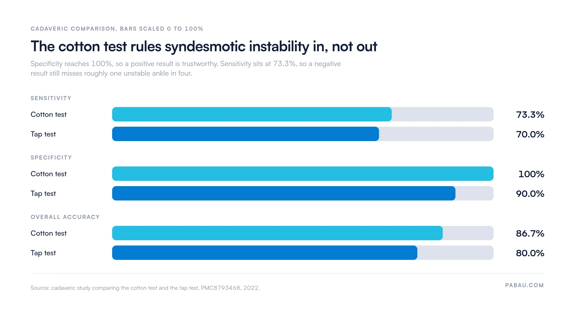
In a cadaveric comparison the cotton test reached 100% specificity but only 73.3% sensitivity.
Surgeons use the cotton test intraoperatively under fluoroscopy to verify syndesmosis reduction after fixation.
Pain-limited guarding in an acute ankle can produce a falsely negative test, so document the limitation.
The cotton test is a clinical special test that evaluates the integrity of the distal tibiofibular syndesmotic complex. The examiner stabilizes the tibia and fibula proximally, grips the hindfoot, and applies alternating medial and lateral force to the talus. Abnormal translation points to disruption of one or more parts of the syndesmotic ligament complex.
The test is also written as Cotton’s test, after the surgeon credited with describing it. Some texts use lateral translation test or lateral stress test for the same maneuver. The mechanism is identical either way. Stressing the mortise reveals instability that weight-bearing radiographs can miss in partial syndesmotic injuries.
The distal tibiofibular syndesmosis is a fibrous joint that binds the distal tibia and fibula together above the ankle mortise. Four main structures maintain its integrity:
Together, these structures keep the fibula seated against the tibial incisura and hold the mortise at the dimensions the talus occupies. When the complex is disrupted, the fibula displaces laterally and the mortise widens. Even 1 mm of lateral talar shift cuts tibiotalar contact area sharply, which is why subtle instability is worth detecting early.
Apply the cotton test whenever a syndesmosis injury is suspected and the Ottawa ankle rules have ruled out bony injury. Common clinical triggers include:
Fibular fracture level shapes how suspicious you should be. The Danis-Weber classification sorts ankle fractures by where the fibula breaks. Syndesmotic involvement becomes more likely the higher that break sits.
Avoid the cotton test when an acute fracture has not yet been excluded on plain films, since the translatory force risks displacing fragments. Be cautious when pain limits the examination, because guarding can produce a falsely negative result.
Correct hand placement and a controlled force decide whether the result means anything. Sloppy technique is the biggest single source of inter-examiner variability.
Position the patient supine or sitting, with the knee flexed to roughly 90 degrees and the ankle in neutral. Neutral position relaxes the Achilles tendon and removes the camming effect of the wider anterior talus. That lets you assess mortise laxity without the passive tensioning that plantar flexion creates.
Follow these steps in sequence:
The force should be firm but not violent. Stress the syndesmosis to its physiological limit and no further. Most authorities describe the force as roughly that of a firm handshake.
A positive cotton test is present when lateral talar translation exceeds 3 to 4 mm compared with the contralateral side. A soft, absent, or asymmetric endpoint counts as positive too. Record the finding alongside ankle circumference and neurovascular status, so the baseline is complete.
A negative cotton test does not exclude partial syndesmotic injury. The test is most sensitive for complete or near-complete disruption. A partial tear affecting only the AITFL may produce no detectable translation, because the remaining structures still stabilize the mortise under manual force.
Published evidence on the cotton test is limited, and most studies report it as part of a battery rather than in isolation. The clearest head-to-head figures come from cadaveric work.
A cadaveric study published in PubMed Central compared the cotton test with the tap test for coronal syndesmotic instability. The cotton test returned 73.3% sensitivity, 100% specificity, and 86.7% overall accuracy. The tap test returned 70%, 90%, and 80% on the same three measures.
Two caveats sit behind those numbers. They come from cadaveric ankles, which carry no pain and no muscle splinting. A bedside examination on a guarding patient will not perform as well.
No single special test settles the diagnosis. Each one loads a different part of the complex, which is why the examination runs several in sequence before imaging is ordered.
The cotton test has an important intraoperative application in ankle fracture surgery and isolated syndesmosis repair. After provisional or definitive fixation of the fibula, the surgeon applies the same mediolateral force to the talus under fluoroscopy. The aim is to confirm the syndesmosis is reduced and the mortise matches the contralateral ankle.
The intraoperative version differs from the clinical one. The patient is under general or regional anesthesia, which removes guarding as a confounder. The surgeon typically tests before and after syndesmotic screw or tightrope fixation, comparing mortise width on the fluoroscopic image.
A positive result after fixation should send the surgeon back to screw positioning, fibular length, or rotational alignment before closure.
When performing the intraoperative cotton test, image the contralateral ankle under the same fluoroscopic projection before surgery begins. Establish a baseline mortise width, so you have a reliable reference during fixation. Published normative values may not apply to this particular patient.
No single test is sufficient for diagnosing syndesmotic injury. The cotton test belongs to a battery, and each member interrogates a different aspect of syndesmosis integrity. Most examiners combine two or three before ordering advanced imaging.
The squeeze test compresses the fibula against the tibial shaft at roughly mid-calf level. The transmitted force stresses the interosseous membrane and, through ligamentous transmission, the AITFL at the syndesmosis. Pain reproduced at the distal syndesmosis rather than at the compression site is a positive result.
It is generally more sensitive than the cotton test for IOM injuries, because it loads the membrane over a longer lever arm. It is less specific for mortise widening, since it does not translate the talus directly.
The hook test is primarily an intraoperative maneuver. The surgeon places a bone hook beneath the distal fibula and applies anteroposterior traction to quantify fibular translation relative to the tibial incisura. Because the force is controlled and measurable under direct vision, some surgeons find it more reproducible than the cotton test.
The cotton test remains the more practical bedside option, since it needs no instruments beyond the examiner’s hands. Both are accepted options for intraoperative syndesmotic assessment among foot and ankle surgeons, including members of the American Orthopaedic Foot and Ankle Society (AOFAS).
The cotton test is easy to perform badly, and the failure modes are consistent. Knowing where technique breaks down matters as much as knowing the steps.
Documentation carries most of that weight. Recording the qualifier in a physical therapy EMR stops the next clinician from reading a guarded examination as a clean negative.
A positive cotton test needs prompt, structured follow-up. The pathway depends on injury grade, associated fractures, and the patient’s sport or activity demands.
Step four carries most of the rehabilitation planning. A staged return-to-running protocol gives you criteria to progress against, rather than a calendar date.
Imaging results also need somewhere structured to land. An ankle radiograph results template captures mortise measurements and clear-space values in the same fields every time, which makes the follow-up film directly comparable.
Imaging correlation is mandatory before any definitive management decision following a positive syndesmosis special test, per American Academy of Orthopaedic Surgeons recommendations. No single examination finding, the cotton test included, determines on its own whether surgery is required.
Most practices record a cotton test result as a line of free text in the visit note. That works once. It stops working when a clinician needs to compare today’s translation against the measurement taken three weeks ago, or against the uninjured side.
Practice management software like Pabau stores those findings as structured fields instead. Translation in millimeters, endpoint quality, and the contralateral comparison each get their own field on a custom assessment form. Imaging reports and operative notes attach to the same patient record.
The payoff shows up at the follow-up appointment. The clinician opens the record and sees the progression at a glance, so grading decisions rest on recorded measurements rather than recall. Every subscription includes those forms, so there is no tier to upgrade to.

Pabau stores structured assessment findings, imaging results, and rehabilitation milestones against the patient record. Physical therapy and sports medicine teams can compare side-to-side measurements across visits without rebuilding the history.

Trust a positive cotton test, and be careful with a negative one. Specificity near 100% means a positive result is worth acting on immediately, with imaging and orthopedic referral. Sensitivity in the low 70s means a negative result cannot close the case on its own.
The practical consequence lands on documentation. Record the ankle position, the force you could apply, and whether pain limited the examination. A guarded negative written down as a plain negative is how a Grade II injury gets missed.
Book a demo to see how Pabau keeps ankle assessment findings, imaging, and rehabilitation milestones in one patient record.

Assessing lateral ankle stability as well? Talar tilt test covers the calcaneofibular and anterior talofibular ligaments using the same side-to-side comparison.
Need a second syndesmosis maneuver? Fibular translation test assesses anteroposterior movement of the fibula at the tibial incisura.
Ruling out an Achilles rupture? Thompson test is the calf-squeeze screen for tendon continuity in the same painful ankle.
Documenting the posterior drawer? Ankle posterior drawer test template gives you a structured form for recording the result.
The cotton test assesses stability of the distal tibiofibular syndesmosis. The examiner translates the talus within the ankle mortise to detect abnormal mediolateral laxity. It is used clinically when high ankle sprain is suspected, and intraoperatively to confirm mortise reduction after fracture fixation.
A positive cotton test indicates syndesmotic instability. It is typically defined as lateral talar translation exceeding 3 to 4 mm compared with the contralateral ankle, or a soft or absent endpoint. The finding warrants imaging confirmation and orthopedic referral.
The cotton test translates the talus mediolaterally to stress the whole syndesmotic complex and detect mortise widening, which gives it higher specificity. The squeeze test compresses the fibular shaft against the tibia to load the interosseous membrane, which makes it more sensitive for IOM injuries. The two are usually performed together.
The cotton test is also written as Cotton’s test, after the surgeon credited with describing it. Some orthopedic texts use lateral translation test or lateral stress test for the same maneuver. They all describe the same mediolateral talar translation.
Recovery depends on injury grade. Grade I syndesmotic sprains managed conservatively typically allow return to sport in 6 to 12 weeks. Grade III complete disruptions requiring surgical fixation commonly need 4 to 6 months. Individual timelines vary with fixation method, rehabilitation compliance, and sport demands.






