Pabau GO app

The new Pabau GO is heredownload on the App Store

Download on the App Store
Book a demo Book a demo
Clinical guides

Neuro Exam Cheat Sheet

Key takeaways

Key takeaways

A neuro exam cheat sheet is a structured reference covering mental status, cranial nerves, motor (0-5 grading), sensory, reflexes, and gait – essential for systematic bedside assessment.

The MRC muscle strength scale ranges from 0 (no movement) to 5 (normal strength against full resistance); DTRs are graded 0-4+, where 2+ is normal and 4+ indicates hyperreflexia.

Urgent red flags requiring escalation include sudden neurological deficits, papilledema, meningism, acute severe headache, and new Babinski sign in adults.

Pabau’s digital templates transform paper cheat sheets into auditable, patient-linked clinical documentation that eliminates transcription error and supports multi-clinician workflows.

Download your free neuro exam cheat sheet

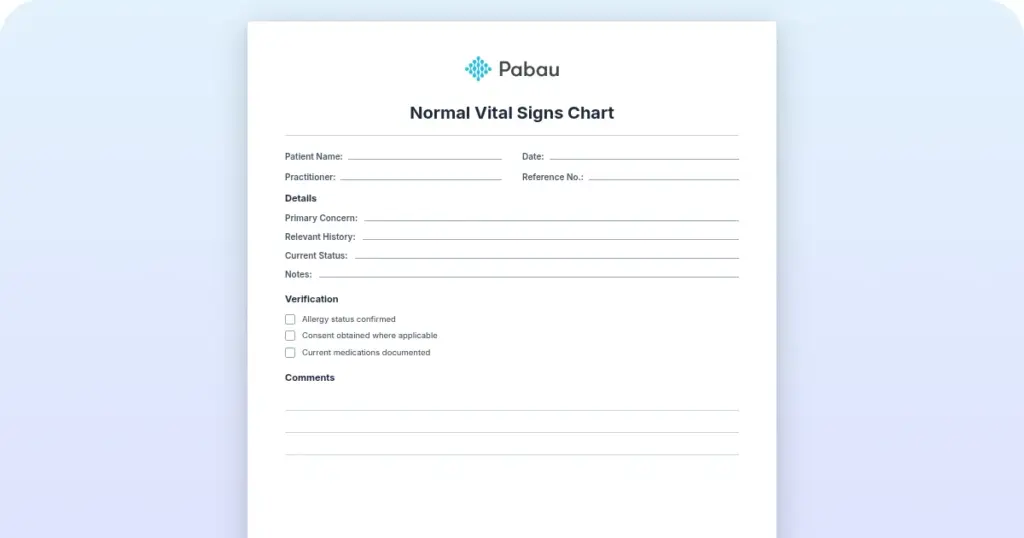
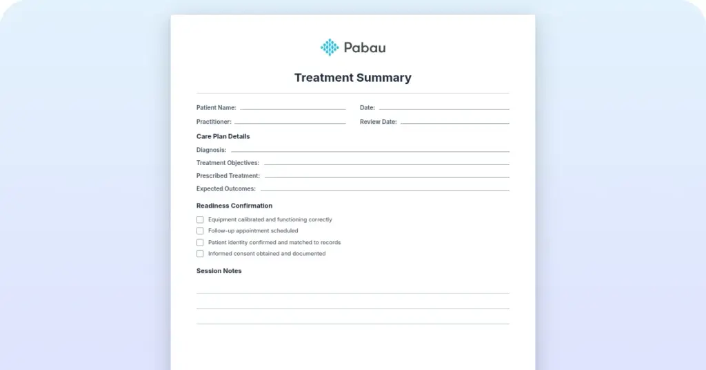
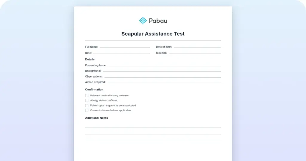
A 5-page fill-in exam form with a patient name and date-of-birth header, plus blank fields for recording findings across the mental status, cranial nerve, motor system, sensory, reflex, and coordination and gait examination — ready to print and complete at the bedside.

Download template

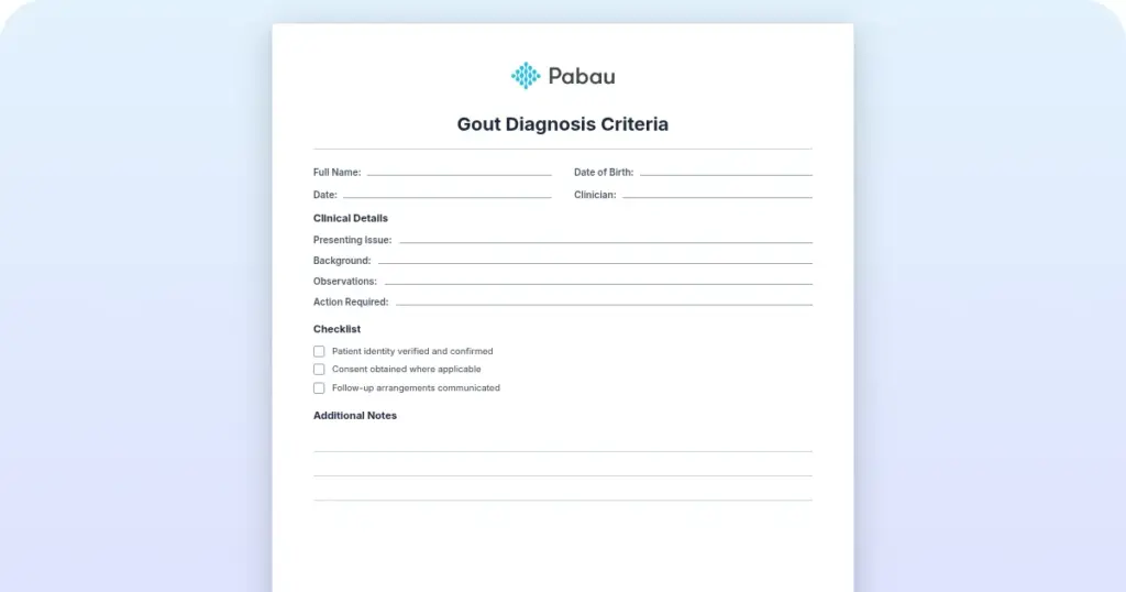
A structured intake form guides clinicians through every domain of the neurological examination – mental status, cranial nerves, motor strength, sensation, reflexes, coordination, and gait – ensuring no critical component is missed.
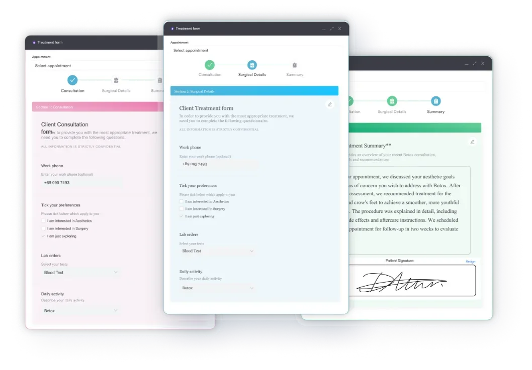
Customizable consent and intake forms
Pabau’s customizable intake forms turn each exam domain into its own structured section, so mental status, cranial nerve, and motor findings stay organized for the next visit.

Bedside time is limited. Furthermore, neurological findings are complex. As a result, a well-organized neuro exam cheat sheet transforms a potentially chaotic assessment into a systematic, repeatable process that clinicians can reference in seconds and document in real time. Whether you’re a junior resident, an emergency medicine physician, a neurologist, or a nurse conducting admission assessments, a portable neuro exam cheat sheet keeps you focused on the patient and confident in your findings.

This guide walks through every section of the complete neurological examination, including the grading scales clinicians use (muscle strength 0-5, reflex grades 0-4+), how to spot red-flag findings, and what to document for compliance and continuity of care. In addition, we cover nursing-specific variants and a quick NIHSS reference for acute stroke assessment.

What is a neuro exam cheat sheet?

A neuro exam cheat sheet is a clinical reference guide-often a single page or pocket card-that organizes all components of a complete neurological examination in a checklist or flowchart format. It serves clinicians who need to conduct a thorough neuro assessment at the bedside but lack time to consult bulky references.

Specifically, the primary purpose is systematic coverage: ensuring every domain (mental status, cranial nerves, motor, sensory, reflexes, coordination, gait) is assessed, documented, and communicated clearly. In addition, a neuro exam cheat sheet serves as a teaching tool for medical students and residents preparing for board exams or OSCE stations.

From a clinical safety perspective, standardized checklists reduce the risk of omission. A clinician reviewing a patient with acute headache who may have meningitis, for example, uses a neuro exam cheat sheet to ensure all cranial nerves, neck stiffness (Kernig and Brudzinski signs), and mental status changes are evaluated and documented-critical for diagnosis and liability protection under standards of care.

In addition, regulatory compliance matters. In particular, documentation of a systematic neurological examination is required by Joint Commission, CMS (for hospital billing), and state health boards. As a result, a cheat sheet ensures the documentation template captures all necessary elements to satisfy audit and accreditation requirements. For example, specific findings carry their own diagnostic codes: a traumatic cerebral hemorrhage is S06.344S, and a cervical spinal cord injury is S14.103D. In turn, accurate exam documentation supports correct coding downstream.

How to perform a systematic neuro exam

A structured neuro exam cheat sheet is designed to be used in real time during patient evaluation. Follow these five operational steps:

  1. Mental Status Assessment: Begin by assessing orientation (person, place, time, situation), attention (digit span, months backward), memory (recall 3 objects after 5 minutes), and language (naming, repetition, comprehension). Document whether the patient is alert, drowsy, lethargic, or unresponsive. Note any confusion, agitation, or memory gaps.
  2. Next, test the cranial nerves (CN I-XII): Systematically check each cranial nerve using the cheat sheet’s assessment methods: smell (CN I), vision fields and acuity (CN II), pupil reactivity and eye movements (CN III, IV, VI), facial sensation (CN V), facial motor symmetry (CN VII), hearing (CN VIII), palate elevation and gag reflex (CN IX, X), shoulder shrug strength (CN XI), and tongue midline and strength (CN XII). Record findings as normal or specify the abnormality.
  3. Then, perform the motor examination with strength grading: Check muscle strength in key groups (deltoid, biceps, triceps, wrist extensors, hip flexors, knee extensors, ankle dorsiflexors) using the 0-5 MRC scale. Assess tone (normal, increased, decreased) and bulk (normal, atrophy, hypertrophy). Document pronator drift (subtle upper-limb weakness). Use the grading table in your cheat sheet: 0 = no movement, 1 = visible contraction only, 2 = movement eliminating gravity, 3 = movement against gravity, 4 = movement against resistance but not full strength, 5 = normal strength.

Sensory, reflex, and coordination steps

  1. Sensory and Reflex Testing: Check primary sensory modalities: light touch (using a cotton swab), pain sensation (pin-prick or monofilament), vibration (128 Hz tuning fork), and proprioception (joint position sense). Test deep tendon reflexes using the 0-4+ grading scale (0 = absent, 1+ = hypoactive, 2+ = normal, 3+ = brisk, 4+ = hyperreflexia with clonus). Elicit the Babinski sign (stroke the lateral sole of the foot; normal = downward toe flexion, abnormal in adults = extensor plantar response).
  2. Finally, assess coordination and gait: Perform finger-nose-finger test, heel-shin test, rapid alternating movements, Romberg test (feet together, eyes closed – note loss of balance), and observe gait for smoothness, arm swing, and balance. Document cerebellar signs (dysmetria, dysdiadochokinesia, nystagmus) or gait abnormalities (shuffling, ataxia, spasticity).

Use the cheat sheet to record findings in real time or immediately after the examination. The goal is not to complete every test in 2 minutes. Instead, apply clinical judgment-a patient with stable vital signs and no focal neurological signs may receive a screening exam, while a patient with acute neurological symptoms requires a complete examination. Either way, your cheat sheet should support both workflows.

Mental status examination

Mental status assessment includes orientation, attention, memory, language, and executive function. First, start by noting the patient’s level of consciousness (alert, drowsy, lethargic, stuporous, comatose) and general appearance and behavior (cooperative, agitated, depressed, anxious).

  • Orientation: Ask for person (full name), place (current location), time (date and day), and situation (reason for visit or current event). Score orientation as: oriented to person/place/time/situation, or specify deficits (e.g., oriented to person and place only).
  • Next, test attention: Digit span forward (7±2 digits is normal) or recite months backward from December. Inability to concentrate or sustain focus indicates attention deficit.
  • Then, assess memory: Ask the patient to repeat 3 objects immediately (registration) and recall them after 5 minutes of other testing (delayed recall). Normal is 3/3; memory loss suggests delirium or dementia.
  • Language: Assess naming (show objects or pictures), repetition (“no ifs, ands, or buts”), and comprehension (follow a 3-step command). Aphasia (expressive, receptive, or global) is documented by type.
  • Finally, assess executive function: Ask the patient to list animals in 1 minute (typically ≥12 is normal) or perform serial 7s (100 minus 7, minus 7, etc.). Abnormality suggests frontal lobe or dementia pathology.

Common bedside screening tools (MMSE, Montreal Cognitive Assessment) can supplement your neuro exam cheat sheet assessment, but direct examination of orientation, attention, memory, and language is always the foundation.

Cranial nerve assessment chart (CN I-XII)

The 12 cranial nerves transmit sensation and motor control for the head, face, and neck, plus several critical functions (taste, hearing, balance, speech, swallowing). In short, a systematic assessment ensures you catch subtle deficits.

CN # Name Type Bedside test
I Olfactory Sensory Smell: coffee, mint, or alcohol. Often omitted.
II Optic Sensory Vision acuity, visual fields, pupils, fundoscopy.
III Oculomotor Motor Pupil size, reactivity; eye movements (up, down, medial). Ptosis = CN III palsy.
IV Trochlear Motor Eye movement (down and medial). Deficit = head tilt away from lesion.
V Trigeminal Sensory/Motor Facial sensation (light touch, pin-prick V1/V2/V3). Jaw strength (clench teeth). Corneal reflex.
VI Abducens Motor Eye abduction (lateral gaze). Deficit = convergent eye (turned inward).
VII Facial Motor/Taste Facial symmetry at rest and with expression (smile, close eyes, raise brows). Taste anterior 2/3 of tongue.
VIII Vestibulocochlear Sensory Hearing (whisper test, Weber, Rinne). Balance (Dix-Hallpike, HIT if vertigo).
IX Glossopharyngeal Sensory/Motor Gag reflex (on sides of soft palate). Taste posterior 1/3 of tongue.
X Vagus Motor/Sensory Soft palate elevation (say “ahhh”); uvula midline. Gag reflex. Hoarseness = dysphonia.
XI Accessory Motor Shoulder shrug (trapezius) against resistance. Head turn (sternocleidomastoid) against resistance.
XII Hypoglossal Motor Tongue protrusion (midline or deviation). Strength (push against cheek).

Abnormal findings in any cranial nerve warrant further investigation. For instance, CN VII palsy (one-sided facial drooping) suggests stroke or Bell’s palsy; CN XII deviation suggests a lower motor neuron lesion (deviation toward the lesion) or an upper motor neuron lesion (deviation away from the lesion).

Motor examination and muscle strength grading (0-5 scale)

Motor assessment includes strength grading, tone, bulk assessment, and pronator drift. Use the standardized MRC 0-5 muscle strength scale, documented consistently in clinical notes, to ensure consistency across examiners and visits.

Grade Definition Clinical meaning
0 No movement Complete paralysis; no visible muscle contraction.
1 Trace movement Visible muscle contraction, no joint movement.
2 Movement, gravity eliminated Joint movement only when gravity is removed (arm slides across table).
3 Movement against gravity Limb moves against gravity but not against resistance (arm lifts from table).
4 Movement against resistance Weakened; movement against resistance but not full strength. Patient yields to examiner pressure.
5 Normal strength Full strength against examiner resistance. Normal.

Test strength in major muscle groups: shoulder abduction (deltoid, C5), elbow flexion (biceps, C5-6), elbow extension (triceps, C7-8), wrist extension (C6-7), hip flexion (iliopsoas, L1-2), knee extension (quadriceps, L2-4), ankle dorsiflexion (L4-5), and ankle plantarflexion (S1-2). Assess tone (normal, spastic, rigid, flaccid) and bulk (normal, atrophy, fasciculation). Document pronator drift: with arms extended and eyes closed, a downward drift or pronation of the forearm suggests subtle upper motor neuron weakness.

Sensory examination

Sensory testing evaluates five primary modalities and maps potential neurological lesions (peripheral nerve, spinal cord, brain).

  • Light touch: Use a cotton swab or fingertip. Touch the skin and ask “can you feel this?” Test bilateral arms, legs, face, and trunk. Diminished sensation suggests peripheral neuropathy or dorsal column involvement.
  • Next, check pain (pin-prick): Use a sterile pin or monofilament. Lightly touch the skin (not penetrate) and ask “sharp or dull?” Hyperalgesia (increased pain) may indicate central sensitization or neuropathic pain.
  • Temperature: Often deferred if light touch and pain sensation are normal. Use warm and cold objects to assess.
  • Then, test vibration: Apply a 128 Hz tuning fork to bony prominences (fingers, toes, ankles, shins). Ask “do you feel the vibration?” Loss of vibration sense suggests dorsal column pathology or large-fiber neuropathy.
  • Finally, assess proprioception (position sense): Passively move the patient’s finger or toe up or down and ask “which way am I moving your finger?” Loss suggests dorsal column disease or sensory ataxia.

Sensory levels (a horizontal band below which sensation is impaired) suggest spinal cord pathology. Similarly, stocking-glove distribution (loss in distal limbs) suggests peripheral neuropathy. By contrast, one-sided sensory loss suggests brainstem or cortical stroke.

Deep tendon reflex grading scale

Deep tendon reflexes (DTRs) test the integrity of the reflex arc: sensory input → spinal nerve → motor output. Grading follows a 0-4+ scale.

Grade Description Clinical significance
0 Absent No reflex response. Suggests lower motor neuron or peripheral nerve lesion (e.g., diabetic neuropathy).
1+ Hypoactive/Diminished Reduced response requiring stronger stimulus. Common in neuropathy or older adults.
2+ Normal Brisk, prompt response. Normal reflex.
3+ Brisk/Hyperactive Exaggerated response. Suggests upper motor neuron lesion (spasticity) or hyperthyroidism.
4+ Hyperreflexic with clonus Repetitive involuntary jerking after the reflex. Indicates significant upper motor neuron disease (e.g., stroke, spinal cord lesion).

Key reflexes to test: biceps (C5-6), triceps (C7-8), brachioradialis (C5-6), patellar (L2-4), and Achilles (S1-2). Asymmetry (one side brisker than the other) is abnormal and localizes the lesion. The Babinski sign (stroking the lateral sole of the foot) elicits downward toe flexion in normal adults; an extensor plantar response (upward toe) is abnormal in anyone over infancy and suggests upper motor neuron lesion.

Coordination and gait assessment

Cerebellar dysfunction manifests as incoordination (dysmetria), irregular rapid movements (dysdiadochokinesia), and gait ataxia. Similarly, proprioceptive sensory loss causes ataxia. Testing separates the two.

  • Finger-nose-finger test: Patient touches their nose, then the examiner’s finger (at arm’s length), alternating. Dysmetria (overshoot or undershoot) suggests cerebellar disease.
  • Next, perform the heel-shin test: Patient places one heel on the opposite knee and runs the heel down the shin. Dysmetria or tremor indicates cerebellar involvement.
  • Rapid alternating movements: Patient rapidly alternates palm up/down on their lap or alternates tapping. Slowing or irregularity (dysdiadochokinesia) is cerebellar.
  • Then, conduct the Romberg test: Patient stands feet together, eyes closed. Mild sway is normal. Loss of balance (falling or requiring support) suggests proprioceptive loss (dorsal column disease) or vestibular dysfunction. Do not perform if patient is at risk of falling.
  • Finally, observe gait: Watch for smooth, symmetrical, steady walking. Note stride length, arm swing, posture, balance, and turns. Specific patterns: shuffling gait (Parkinson’s), wide-based gait (ataxia), steppage gait (foot drop from peroneal nerve), spastic gait (scissor-like).

Combination findings (Romberg positive + proprioception loss + dysmetria worse with eyes closed) = dorsal column disease. Cerebellar signs (dysmetria, dysdiadochokinesia, nystagmus, ataxia) = cerebellar lesion.

NIHSS stroke scale quick reference

The National Institutes of Health Stroke Scale, or NIHSS, is a 15-item structured tool for rapid assessment of acute stroke severity. Specifically, scores range from 0 (no stroke symptoms) to 42 (severe stroke), guiding treatment urgency and prognosis. This is an abbreviated reference only; always consult the official scoring manual for details.

  • 1a. Level of consciousness (LOC): 0-3 points. Alert and responsive (0) to unresponsive (3).
  • 1b. LOC questions: 0-2 points. Ability to answer “What month is it?” and “How old are you?”
  • 1c. LOC commands: 0-2 points. Ability to obey “Close your eyes” and “Make a fist.”
  • 2. Best gaze: 0-2 points. Eye movement: normal (0) to fixed/deviated (2).
  • 3. Visual: 0-3 points. Visual field assessment by confrontation: normal (0) to complete hemianopia (3).
  • 4. Facial droop: 0-3 points. Symmetry: normal (0) to complete paralysis (3).
  • 5. Motor arm (left and right): 0-4 points each. Strength 0-5 arm drift test: normal (0) to no movement (4).
  • 6. Motor leg (left and right): 0-4 points each. Strength 0-5 leg drift test: normal (0) to no movement (4).
  • 7. Limb ataxia: 0-2 points. Finger-nose and heel-shin coordination: normal (0) to present bilaterally (2).
  • 8. Sensory: 0-2 points. Light touch sensation: normal (0) to severe/absent (2).
  • 9. Best language: 0-3 points. Naming and comprehension: normal (0) to global aphasia (3).
  • 10. Dysarthria: 0-2 points. Speech clarity: normal (0) to unintelligible (2).
  • 11. Extinction/inattention: 0-2 points. Neglect or extinction to double stimuli: normal (0) to profound (2).

Total NIHSS score: 0-4 = minor stroke, 5-15 = moderate stroke, 16-20 = moderately severe stroke, 21+ = severe stroke. In practice, a score of ≥4 typically qualifies for thrombolytic therapy (tPA) if administered within 4.5 hours of symptom onset.

Red flags in neurological examination

Certain neurological findings demand immediate escalation for imaging or specialist evaluation. These red-flag findings may indicate acute, life-threatening pathology.

  • Acute focal neurological deficit: Sudden onset of weakness, vision loss, speech difficulty, or loss of coordination in one limb or side of the body. Suggests acute stroke until proven otherwise. Time-critical: call emergency services or arrange urgent imaging.
  • Altered level of consciousness: Drowsiness, lethargy, or unresponsiveness not explained by current medications or metabolic causes. Suggests intracranial pathology (hemorrhage, edema, infection, mass).
  • Papilledema (optic disc swelling): Blurred disc margins, absent spontaneous venous pulsations, or elevation of the optic disc on fundoscopy. Indicates raised intracranial pressure requiring urgent imaging.
  • Meningism (signs of meningeal irritation): Neck stiffness (inability to touch chin to chest), positive Kernig sign (resistance to knee extension when hip flexed), or positive Brudzinski sign (spontaneous hip/knee flexion when neck is flexed). Suggests meningitis or subarachnoid hemorrhage. Urgent lumbar puncture or imaging required.
  • Severe headache with neurological signs: Sudden, severe “thunderclap” headache (worst of life) or progressive headache with fever, stiff neck, focal deficits, or seizures. Consider meningitis, encephalitis, or subarachnoid hemorrhage.

Additional red flags requiring urgent evaluation

  • New Babinski sign (extensor plantar response) in adults: Upward (extensor) movement of the big toe when the sole of the foot is stroked. Normal in infants; abnormal in older children and adults. Indicates upper motor neuron lesion (stroke, spinal cord injury, brain tumor).
  • Acute bilateral symptoms: Weakness, numbness, or pain affecting both sides of the body. Suggests spinal cord pathology, brainstem lesion, or severe metabolic derangement.
  • Seizure activity: Generalized or focal seizures, especially if new-onset or prolonged. May indicate epilepsy, infection, toxic exposure, or brain lesion.
  • Unexplained loss of consciousness or syncope: Fainting or blackout without clear cause (not simple vasovagal syncope). Consider cardiac arrhythmia, stroke, or severe metabolic derangement.

In any of these cases, document findings carefully in the patient record, notify a physician or supervisor immediately, and ensure rapid access to emergency imaging or specialist input. In short, do not delay for additional tests if red flags are present.

Nursing neurological assessment checklist

Nurses often conduct bedside neurological screening as part of admission and ongoing assessments, often alongside a broader nursing review of systems adapted for the clinical setting. Notably, nursing assessments focus on rapid detection of acute change and clear documentation for continuity of care.

  • Level of consciousness (AVPU scale): Alert, Verbal (responds to voice), Pain (responds to pain), or Unresponsive. Simplified alternative to full GCS; use GCS (Glasgow Coma Scale) for more detailed assessment.
  • Pupils and eye response (PERRL): Pupils Equal, Round, Reactive to Light. Check size (mm), shape (round vs. irregular), and reactivity to light. Asymmetrical pupils or fixed pupils are urgent red flags.
  • Motor response: Ability to move all four limbs against gravity. Test grip strength bilaterally (ask patient to squeeze your hands). Document any weakness, drift, or paralysis.
  • Sensory testing: Light touch to bilateral upper and lower extremities. Any asymmetry or loss is significant.
  • Speech and language: Clarity of speech (normal, slurred, garbled, aphasic). Ability to understand commands. Confusion or difficulty speaking warrants physician notification.
  • Pain response: Ask “Do you have pain? Where?” Assess pain scale (0-10) and location. Acute neurological pain or sudden change in pain pattern may indicate new pathology.
  • Orientation: Person, place, time, and situation (reason for visit). Document as “oriented x3” (all three) or “oriented x2” (person and place only) if deficits.
  • Behavior and mood: Agitation, aggression, depression, or withdrawal; note any changes from baseline.

Nursing assessments are repeated at regular intervals (every shift, every 1-4 hours if acute illness) and documented in the patient’s clinical record. In practice, any acute change (new weakness, vision loss, confusion, difficulty speaking) is reported to the physician immediately and escalated for imaging or specialist evaluation.
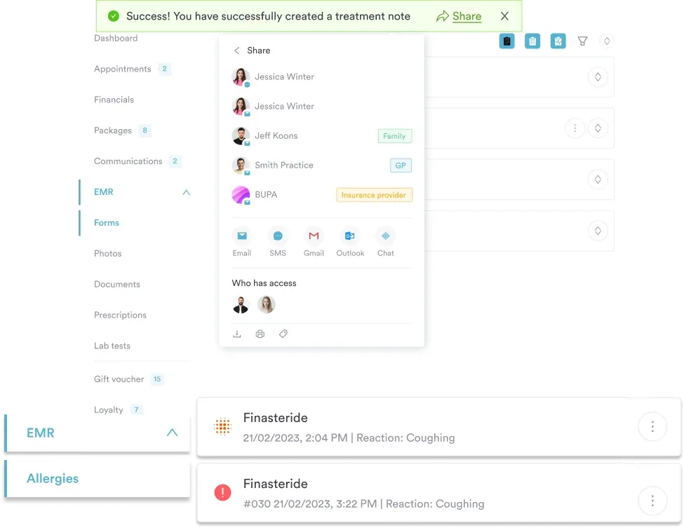
Comprehensive EMR & patient record management
Pabau’s EMR keeps every completed treatment note attached to the patient’s record and shareable with the wider care team, so a nurse’s neuro findings reach the covering physician without a second phone call.

How Pabau supports neurological examination documentation

Paper cheat sheets and static PDFs require a secondary transcription step: clinicians reference the checklist at the bedside, then later manually enter findings into the patient record. This workflow risks transcription errors, omission, and lost documentation.

In response, structured clinical workflows in Pabau replace that separate transcription step. Clinicians complete a customizable neurological examination template directly inside the patient record, capturing mental status, cranial nerve findings, motor grading, sensory results, reflexes, and red-flag observations in a single, auditable entry. As a result, every finding is timestamped, linked to the patient chart, and immediately available for future visits.
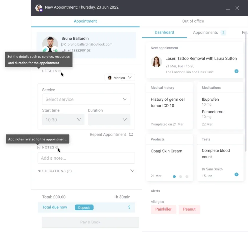
Appointment scheduling in Pabau
Pabau’s appointment screen surfaces medical history, medications, and allergy alerts alongside the visit, so clinicians can flag what changed before repeating a neuro exam at a follow-up.

In addition, Pabau Scribe, our AI scribe, can generate summary text from structured exam findings, allowing clinicians to spend less time typing and more time with patients. Meanwhile, multi-clinician teams see the same examination data instantly, ensuring continuity of care and reducing duplication of testing.
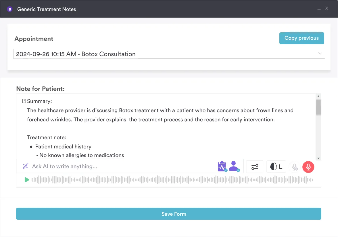
Creating treatment notes with Pabau Scribe
Pabau Scribe turns a recorded conversation into a structured treatment note, cutting the typing time clinicians spend writing up mental status and motor findings after a neuro exam.

For organizations serving neurology, primary care, physical therapy, or behavioral health practices, a digital neuro exam template embedded in the clinical workflow closes the documentation risk that paper checklists create. That combination makes documentation more complete, safer, and auditable for regulatory oversight (HIPAA, state board requirements).

If your team currently uses printed cheat sheets, a structured digital template in Pabau is a simple upgrade that pays dividends in accuracy, speed, and compliance.

Streamline neurological exam documentation and follow-up

Pabau's structured exam templates and Pabau Scribe, our AI scribe, turn a bedside neuro exam into a timestamped, patient-linked record so the next clinician sees the full picture at a glance.

Pabau clinic management dashboard

Conclusion

A printed neuro exam cheat sheet earns its place at the bedside because it is fast to consult and easy to carry. However, its limitation shows up afterward, when those handwritten notes still need retyping into the record, and that interval is exactly where errors and delays creep in.

Moving the same checklist into a structured, patient-linked template closes that interval. In turn, findings are captured once, at the bedside, and become visible immediately to the next clinician who opens the chart, whether that is a covering physician later the same shift or a specialist reviewing the case weeks later.

However, the trade-off is that a structured template asks for more upfront setup than a laminated card: the fields have to match how your practice actually documents each domain. Get that mapping right once, and every exam after it is faster to write up and easier to audit. Book a demo to see how Pabau turns a bedside neuro exam into documentation your whole team can rely on.

Continue your research

Continue your research

Need a nurse-focused cranial nerve script? Cranial nerve nursing assessment breaks CN I-XII testing into the exact bedside wording nursing staff use for admission screening.

Want a standalone cranial nerve reference? Cranial nerve examination checklist expands each of the 12 nerves into its own testing and documentation prompt.

Documenting a change in consciousness? Glasgow Coma Scale template scores eye, verbal, and motor response for tracking a patient’s level of alertness over time.

Screening for acute stroke? Cincinnati Stroke Scale template covers the three-item field test paramedics and ED staff use before NIHSS scoring.

Examining a pediatric patient? Pediatric neurological exam checklist adapts the adult exam domains for developmental stage and cooperation level.

Frequently asked questions

What does a full neurological exam include?

A full neurological exam includes mental status (orientation, attention, memory, language), cranial nerves (I-XII), motor examination (strength grading 0-5, tone, bulk, pronator drift), sensory testing (light touch, pain, vibration, proprioception), deep tendon reflexes (0-4+ grading), and coordination and gait assessment (finger-nose, heel-shin, Romberg, gait observation). Clinicians adapt the scope based on clinical context-an acute admission may require a complete exam, while a routine follow-up may use screening elements only.

How do you grade muscle strength in a neuro exam?

Muscle strength is graded using the MRC (Medical Research Council) 0-5 scale: 0 = no movement, 1 = trace movement only, 2 = movement with gravity eliminated, 3 = movement against gravity, 4 = movement against resistance but not full strength, 5 = normal strength against full examiner resistance. Test major muscle groups (deltoid, biceps, triceps, hip flexors, knee extensors, ankle dorsiflexors) bilaterally and document any asymmetry, which suggests focal neurological lesion.

What are red flags in a neurological examination?

Red-flag findings requiring urgent escalation include acute focal weakness or numbness (stroke), altered level of consciousness (intracranial pathology), papilledema (raised intracranial pressure), meningism with stiff neck (meningitis or subarachnoid hemorrhage), severe sudden headache, new Babinski sign in adults (upper motor neuron lesion), bilateral neurological signs (spinal cord or severe metabolic derangement), and seizure activity. Any of these warrant immediate physician notification and urgent imaging.

How long does a neurological examination take?

A screening neurological exam (level of consciousness, pupils, grip strength, gait) takes 2-5 minutes. A complete neurological examination (all seven domains with detailed testing) typically takes 15-30 minutes depending on the patient’s cooperation and complexity. In acute settings (stroke, suspected meningitis), clinicians prioritize the NIHSS or rapid focal assessment (5-10 minutes) to avoid delay to imaging or treatment.
×