Treatment summary
A treatment summary is a written record of what care was delivered during an epi...

2
Last Updated: Oct 16, 2026

Cranial nerve nursing assessment systematically evaluates all 12 CN for sensory, motor, and autonomic function to detect neurological deficits early.
The 12 cranial nerves are classified as sensory (CN I, II, VIII), motor (CN III, IV, VI, XI, XII), or mixed (CN V, VII, IX, X). This framework guides which tests to perform.
Abnormal CN findings in stroke (facial droop CN VII, tongue deviation CN XII) trigger urgent escalation and integration with FAST assessment protocols.
Pabau’s digital forms and patient record features enable nurses to document CN findings directly in the patient chart, supporting real-time clinical decision-making and team communication.
A ready-to-use assessment checklist covering all 12 cranial nerves, including normal findings, abnormal findings, equipment needed, assessment techniques, and documentation guidance for bedside use.
Download templateCranial nerve nursing assessment is a cornerstone of neurological examination that enables nurses to detect early signs of deterioration and communicate findings to the multidisciplinary team.
This guide walks you through a structured approach to assessing all 12 cranial nerves at the bedside, interpreting normal versus abnormal findings, and documenting your results clearly.

A cranial nerve nursing assessment is a systematic evaluation of the 12 pairs of cranial nerves (CN I-XII) to determine their sensory, motor, and autonomic function. Unlike a full neurological examination, cranial nerve assessment focuses specifically on these 12 nerves.
It’s performed to screen for focal neurological deficits, early signs of increased intracranial pressure, brainstem pathology.
Nurses perform cranial nerve assessments in acute care, stroke units, intensive care, and primary care settings. Physical therapy practices use them during stroke and traumatic brain injury rehabilitation.
The findings are time-critical: abnormal results can signal stroke, intracranial hemorrhage, or nerve damage that needs immediate escalation. Documenting baseline and serial assessments lets the team detect change over time, which is often the most clinically significant finding.
Understanding the 12 cranial nerves and their classifications (sensory vs motor vs mixed) guides which assessment techniques to use and what findings matter clinically.
The 12 cranial nerves are paired structures emerging from the brainstem and skull. Each has a Roman numeral designation (CN I-XII), a name, and a primary function. Here is the structured reference table all nurses learn:
Sensory nerves carry information from the body to the brain (smell, vision, hearing, taste). Motor nerves carry signals from the brain to muscles (eye movement, facial expression, tongue movement, shoulder strength). Mixed nerves do both, which is why they require more complex testing.
These are standard bedside items in most clinical settings. Having them ready lets nurses complete a cranial nerve assessment without leaving the patient’s room. Document the equipment used alongside baseline findings to support serial assessments.
A good patient management system files each completed scale under the right patient automatically.
Assessment proceeds from CN I through CN XII in sequential order. Each test is straightforward and takes seconds to perform.
CN I – Olfactory: Ask the patient to close one nostril and smell a familiar scent (coffee, mint). Normal: patient identifies the odor. Abnormal: unable to smell (anosmia).
CN II – Optic: Test visual acuity with a Snellen chart; assess visual fields by asking the patient to follow a penlight in all directions. Normal: 20/20 vision (or corrected), full visual fields. Abnormal: blurred vision, field cuts.
CN III, IV, VI – Oculomotor, Trochlear, Abducens: Perform PERRLA (Pupils Equal, Round, Reactive to Light and Accommodation). Test extraocular movements (up, down, left, right, diagonal). Normal: pupils 2-4 mm, constrict briskly to light, eyes track smoothly through all directions. Abnormal: unequal pupils, sluggish response, inability to move eyes fully.
CN V – Trigeminal: Test facial sensation by touching the forehead, cheek, and chin with a cotton ball. Ask the patient to report what they feel. Test jaw strength by asking the patient to clench teeth. Normal: intact sensation on all three divisions, strong jaw closure. Abnormal: numbness, weakness, difficulty chewing.
CN VII – Facial: Ask the patient to smile, raise eyebrows, and close eyes tightly. Normal: symmetric facial expression, equal eye closure, normal forehead wrinkles. Abnormal: unilateral facial droop, inability to smile on one side, eye stays open on affected side.
CN VIII – Vestibulocochlear: Perform whisper test: whisper a word at one ear while masking the other with your hand, then have the patient repeat it. Use Weber test (tuning fork at midline) and Rinne test (bone vs air conduction). Normal: hears whispered speech bilaterally, tuning fork heard equally at both ears, air conduction better than bone conduction. Abnormal: hearing loss, asymmetric tuning fork response.
In pediatric patients.
CN IX, X – Glossopharyngeal and Vagus: Ask the patient to say “ah” and observe uvula movement. Test the gag reflex by touching the soft palate with a tongue depressor. Normal: uvula midline, brisk gag reflex, clear voice. Abnormal: uvula deviates away from lesion, weak or absent gag reflex, hoarse voice.
CN XI – Accessory: Ask patient to shrug shoulders against resistance and turn head left and right against hand resistance. Normal: strong, symmetric strength. Abnormal: weakness on one side, shoulder drop.
CN XII – Hypoglossal: Ask the patient to stick out their tongue and move it side to side. Normal: midline protrusion, equal strength. Abnormal: tongue atrophy, weakness, and deviation toward the side of the lesion in peripheral or nuclear (lower motor neuron) palsies. In central lesions such as stroke (upper motor neuron), the tongue deviates away from the lesion, toward the side of hemiparesis instead.
This cheat sheet focuses on the most clinically critical findings.
In stroke patients, cranial nerve deficits are time-critical findings that guide triage and treatment decisions. The most common stroke-related CN deficits are:
Integrate cranial nerve findings with the FAST assessment and automated escalation workflows to ensure rapid notification of the stroke team. Serial assessments every 15-30 minutes detect change and inform thrombolytic or thrombectomy decisions. Before starting thrombolytic therapy, most stroke protocols require a signed consent for medical treatment, since the bleeding risk has to be discussed with the patient or their proxy first. Collected once and stored where clinicians read it, patient intake software removes the second round of questions.
Accurate documentation is essential for continuity of care and medicolegal protection. Use this format:
Cranial nerves are one of seven domains in a complete neuro note. The seven-domain neuro checklist adds consciousness, pupils, motor, sensory, reflexes, and gait, with example wording for each.
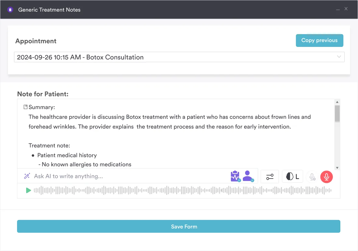
Use Pabau Scribe, our AI scribe, to quickly capture findings while maintaining detail. This reduces documentation burden and improves accuracy.
Chart notation example: “Neurological assessment: CN I-II intact, PERRLA, CN III-VI intact with smooth extraocular movements, CN VII symmetric, CN VIII intact to whisper test bilaterally, CN IX-X intact with brisk gag reflex, CN XI strong, CN XII tongue midline with normal strength.”
A classic mnemonic aids memorization of CN order and type (sensory/motor/mixed). Many nurses learn:
“Oh, Oh, Oh To Touch And Feel Very Good Velvet, Ah, Heaven” – maps to CN I through XII in order. Knowing the order helps you structure your assessment systematically and cross-reference findings quickly.
An additional memory aid for sensory vs motor: Some Say Money Matters, But My Brother Says Big Brains Matter Most – the first letter of each word indicates the CN type in order (Sensory, Sensory, Motor, Motor, Mixed, Motor, Mixed, etc.). This framework supports rapid classification during assessment and clinical rounds.
Systematic cranial nerve assessment turns a two-minute bedside routine into an early warning system for stroke, brainstem injury, and nerve damage. Getting the CN I-XII sequence, the mnemonics, and the documentation habits right means you catch subtle changes before they become emergencies, and you hand off findings the rest of the care team can act on right away.
A structured cranial nerve assessment is one of the most revealing bedside exams in nursing. Working through all 12 nerves in order, telling normal from abnormal, and recognizing the findings that signal an acute stroke lets you catch neurological changes early and escalate with confidence.
Download the free checklist above and keep it on hand during neuro rounds, so the assessment sequence stays consistent from shift to shift and nothing gets skipped under pressure.
Because nurses often record these findings in several places, Pabau captures all 12 cranial nerve results in one structured form, attaches photos or diagrams, and syncs them across the care team to cut errors and delays. Book a demo to see it in your workflow.
The 12 cranial nerves are paired structures emerging from the brainstem. They are the Olfactory (smell), Optic (vision), Oculomotor (eye movement), Trochlear (downward eye movement), Trigeminal (facial sensation), Abducens (lateral eye movement), Facial (expression and taste), Vestibulocochlear (hearing and balance), Glossopharyngeal (swallowing and taste), Vagus (voice and swallowing), Accessory (shoulder strength), and Hypoglossal (tongue movement). Each supports vital sensory, motor, or autonomic functions.
Nurses assess each CN in sequential order (I-XII) using simple bedside techniques. Test CN I by asking the patient to smell a familiar scent; CN II by checking visual acuity and fields; CN III-VI by performing PERRLA and tracking eye movements; CN V by testing facial sensation and jaw strength; CN VII by asking for facial expressions; CN VIII by whisper test and tuning fork tests; CN IX-X by observing swallowing and voice quality; CN XI by shoulder strength; and CN XII by assessing tongue movement. Each test takes seconds.
Standard bedside equipment includes a penlight (pupils and visual fields), cotton ball (facial sensation), tongue depressor (gag reflex), tuning fork (hearing tests), Snellen chart (visual acuity), and an ophthalmoscope (optional, for detailed eye exam). Most acute care units stock these items at every bedside.
CN VII (facial nerve, causing facial droop) and CN XII (hypoglossal nerve, causing tongue deviation) are the most frequently affected in acute stroke. CN IX and X deficits (weak gag or hoarse voice) indicate brainstem involvement and high aspiration risk. These deficits are part of standard stroke assessment.
Document the baseline assessment, specific deficits with CN number and finding, any changes from prior assessment, and the patient’s own report of symptoms. Use standardized format: “CN I-XII intact and symmetric” or list abnormalities by CN number with description. For serial assessments, compare to prior findings and note improvements or deterioration.
Unilateral facial droop (CN VII weakness on one side) is a classic sign of acute stroke affecting the opposite hemisphere. It is one of the three cardinal signs in the FAST (Face, Arms, Speech, Time) stroke screening tool. Facial droop warrants immediate stroke alert and neuroimaging.
In acute stroke, TIA, or traumatic brain injury, cranial nerves should be reassessed every 15-30 minutes during the acute phase, then every 1-4 hours depending on stability. Serial assessments detect change and guide clinical decision-making. Document all reassessments and report any deterioration immediately.
PERRLA stands for Pupils Equal, Round, Reactive to Light and Accommodation. It is the standard abbreviation for normal pupillary findings (CN III oculomotor function). Abnormal pupils (unequal, fixed, non-reactive) indicate serious pathology such as increased intracranial pressure, intracranial hemorrhage, or herniation and require urgent intervention.