CRM for chiropractors: how to manage patients, bookings and marketing
A CRM for chiropractors is a patient relationship management system built around...

Last Updated: Oct 13, 2026

The knee muscle diagram is an anatomical reference tool showing all muscles, ligaments, tendons, and cartilage structures of the knee joint for clinical education.
The diagram covers four main muscle groups: quadriceps (extension), hamstrings (flexion), gastrocnemius (calf), and popliteus (rotation), plus key ligaments for stability.
Physiotherapists and sports medicine clinicians use the diagram for patient education, treatment planning, rehabilitation protocols, and understanding injury mechanisms.
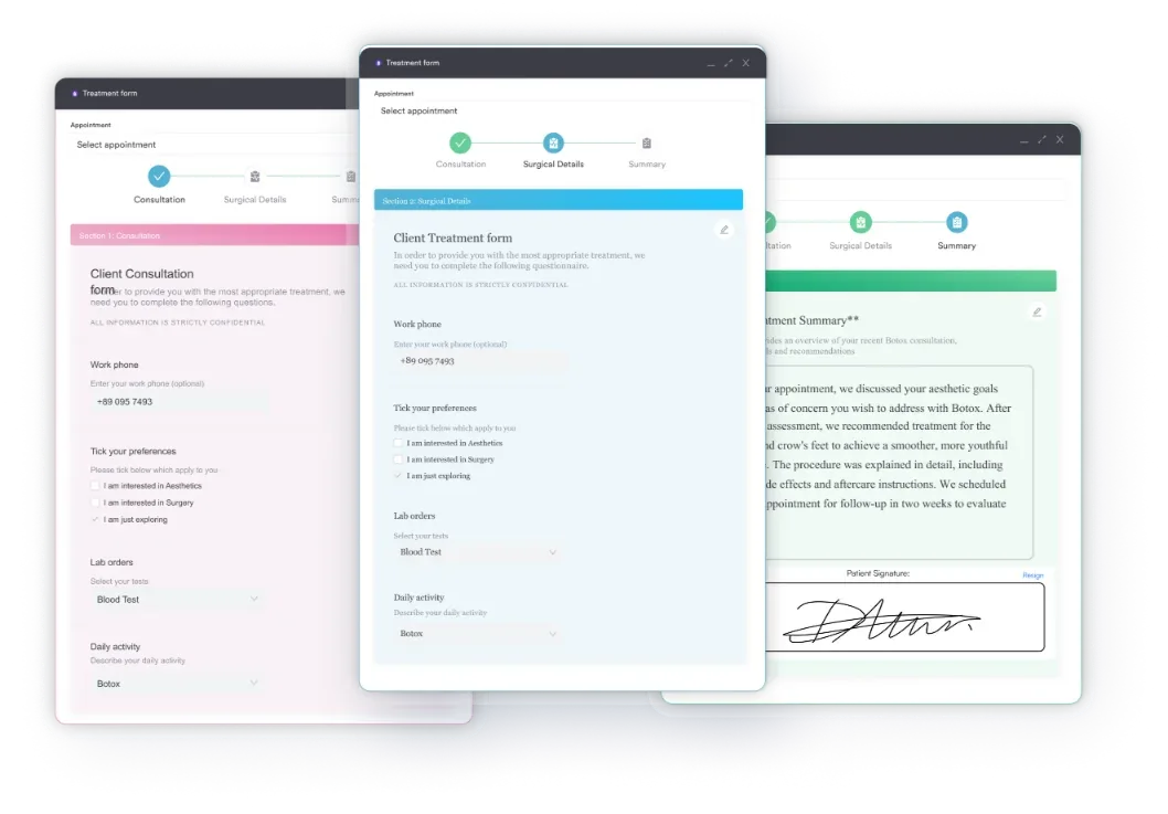
Pabau, a practice management software, integrates digital forms and a client portal directly into patient records and care workflows, improving documentation and patient engagement.
A ready-to-use anatomical reference covering all muscles acting on the knee joint, ligaments, tendons, cartilage structures, and their clinical functions. Perfect for patient handouts, treatment room reference, and rehabilitation planning.
Download templatePhysiotherapists and sports medicine practitioners spend valuable time explaining knee anatomy to patients during consultations and rehabilitation sessions. A comprehensive knee muscle diagram transforms that education — patients understand their condition faster, clinicians document treatment rationale more clearly, and treatment adherence improves because patients grasp why specific exercises matter.
This guide explains the anatomy covered in the knee muscle diagram, how to use it in clinical practice, and why anatomical clarity drives better patient outcomes.
A knee muscle diagram is a labeled anatomical reference illustration showing all muscles, ligaments, tendons, and cartilage structures surrounding the knee joint. It serves as both a teaching tool and a clinical reference — therapists use it to educate patients, document treatment reasoning, and plan rehabilitation protocols.
Unlike textbook anatomy, a clinical-grade diagram prioritizes clarity and functional grouping. Muscles are organized by action (extension, flexion, rotation); ligaments are color-coded by function (anterior/posterior stability, collateral support); tendons show attachment points relevant to common injuries. The result is a resource practitioners can reference in 30 seconds without requiring an anatomy textbook at the treatment table.
The knee joint is where three bones meet: the femur (thighbone), tibia (shinbone), and patella (kneecap). This hinge joint allows flexion (bending) and extension (straightening), plus minor rotation when the knee is bent. The complexity comes from the muscles, ligaments, tendons, and cartilage that work in concert to provide both stability and movement.
The quadriceps femoris is the dominant knee extensor — the group responsible for straightening the knee. It consists of four muscles working as a unit:
The quadriceps tendon connects the quadriceps to the patella (kneecap), which in turn connects to the tibia via the patellar tendon (also called the patellar ligament). This arrangement acts as a lever, multiplying the force the quadriceps can exert.
Weakness or imbalance in the quadriceps — especially the VMO .
Knowing how practice management software works helps you decide which forms to digitize first.
The hamstring group flexes the knee (bends it) and also extends the hip, making these muscles essential for running, climbing, and standing. Three muscles comprise the hamstrings:
The gastrocnemius (calf muscle) crosses both the knee and ankle. It flexes the knee when the foot is off the ground and assists with ankle plantarflexion. The popliteus muscle, a small but clinically important rotator at the back of the knee, initiates knee flexion from full extension — a movement therapists refer to as “unlocking” the knee.
While muscles create movement, ligaments provide passive stability. The four main ligaments are:
Key tendons include the patellar tendon (quadriceps to tibia), quadriceps tendon (quadriceps to patella), and hamstring tendons (attaching at the back of the knee).
The knee’s stability and movement depend on coordinated muscle action. During extension, the quadriceps contracts; during flexion, the hamstrings contract. But true knee function is far more nuanced: the VMO controls patellar alignment during extension; the gastrocnemius assists hamstrings during flexion; the popliteus initiates flexion from a locked, extended position.
Imbalances in these muscle groups create injury risk. A weak VMO combined with tight hip flexors often leads to lateral patellar tracking — the kneecap pulls outward — and anterior knee pain. Hamstring tightness restricts hip flexion, compensating through increased lumbar spine extension during bending activities. Understanding these functional interdependencies is essential for designing effective rehabilitation and preventing re-injury. Handing a patient an exercise program works better when the prescription and the review sit in one place.
During an initial consultation, a clear diagram helps patients visualize why they are experiencing pain — “Your vastus medialis is weak, so your kneecap is tracking laterally, irritating the cartilage underneath” is far more compelling than abstract jargon.
Patients who understand the mechanism are more likely to comply with strengthening exercises.
In treatment documentation, the diagram supports clinical reasoning. Rather than writing “knee pain,” clinicians reference specific muscles involved: “Quadriceps weakness (especially VMO) with lateral patellar tracking.” This clarity improves handoffs between providers and justifies treatment choices to payers.

During rehabilitation, the diagram informs exercise progression. Initial therapy targets VMO activation with isometric quads sets and, where indicated, E0745 neuromuscular stimulation; mid-stage therapy advances to step-ups and lateral band walks; late-stage therapy incorporates running mechanics and plyometrics — each progression tied to the muscle groups visible on the diagram.
Understanding the anatomy illuminates why conditions occur and how to treat them:
Each condition traces back to specific muscles visible on the diagram.
Audit your patient education materials. If you’re explaining knee anatomy verbally without a visual reference, you’re working harder than necessary. Print or digitally share the knee muscle diagram during consultations — it cuts explanation time and improves patient compliance because the ‘why’ becomes obvious.

Integrate anatomical diagrams, treatment plans, and rehabilitation protocols into your practice’s client portal — reducing consultation time and improving treatment outcomes.

A clear, anatomically accurate knee muscle diagram is more than a teaching tool — it is a clinical necessity. It connects textbook anatomy to real-world patient education, enabling physiotherapists and sports medicine clinicians to explain mechanisms of injury, justify treatment choices, and engage patients in their own recovery.
Download the knee muscle diagram above and integrate it into your clinical practice: print it for the treatment room, share it with patients in online appointment confirmations.
The clearer your patients understand their anatomy, the faster and more confidently they will recover, especially when backed by Pabau’s patient engagement tools, which reduce friction in your practice.

Wondering why a patient can’t tolerate exercise? cardiopulmonary exercise testing guide covers the five key variables, their normal values, and what each means.
Looking for a musculoskeletal intake form to pair with the diagram? Physical therapy intake form captures medical history, symptoms, and functional limitations before the first assessment.
Want a fitness-focused intake to pair with rehab handoffs? Personal training questionnaire captures activity level and contraindications before designing a strengthening program.
The primary knee muscles include the quadriceps (front thigh — extension), hamstrings (back thigh — flexion), gastrocnemius (calf — flexion and ankle plantarflexion), popliteus (back of knee — rotation and flexion initiation), and sartorius (crosses the thigh medially — assists flexion and rotation). Secondary stabilizers include gracilis and adductors.
The quadriceps and hamstrings are the primary dynamic stabilizers — they control extension and flexion, preventing excessive movement. Statically, ligaments (ACL, PCL, MCL, LCL) and the menisci provide stability. The VMO (vastus medialis oblique) is particularly important for patellar tracking and preventing lateral instability.
The quadriceps straightens (extends) the knee and also flexes the hip. It is the primary muscle used in standing, walking upstairs, jumping, and sprinting. Weakness in the quadriceps, especially the vastus medialis, is a common cause of anterior knee pain and patellar tracking problems.
The quadriceps femoris extends the knee. The hamstrings (biceps femoris, semimembranosus, semitendinosus) and gastrocnemius flex the knee. The popliteus initiates flexion when the knee is in full extension. Synergists like gracilis and sartorius provide secondary flexion assistance.
Print the diagram or share it digitally during consultations. Point to the specific muscle group involved in the patient’s condition and explain their pain mechanism — for example, show how a tight hamstring limits hip flexion, forcing compensation at the knee. Use the diagram to justify exercise selection and help patients understand why treatment works.