A chest pain nursing care plan turns assessment findings into NANDA diagnoses, timed interventions, and measurable outcomes.
Six diagnoses cover most presentations, from acute pain and decreased cardiac output to anxiety, activity intolerance, deficient knowledge, and risk for ineffective tissue perfusion.
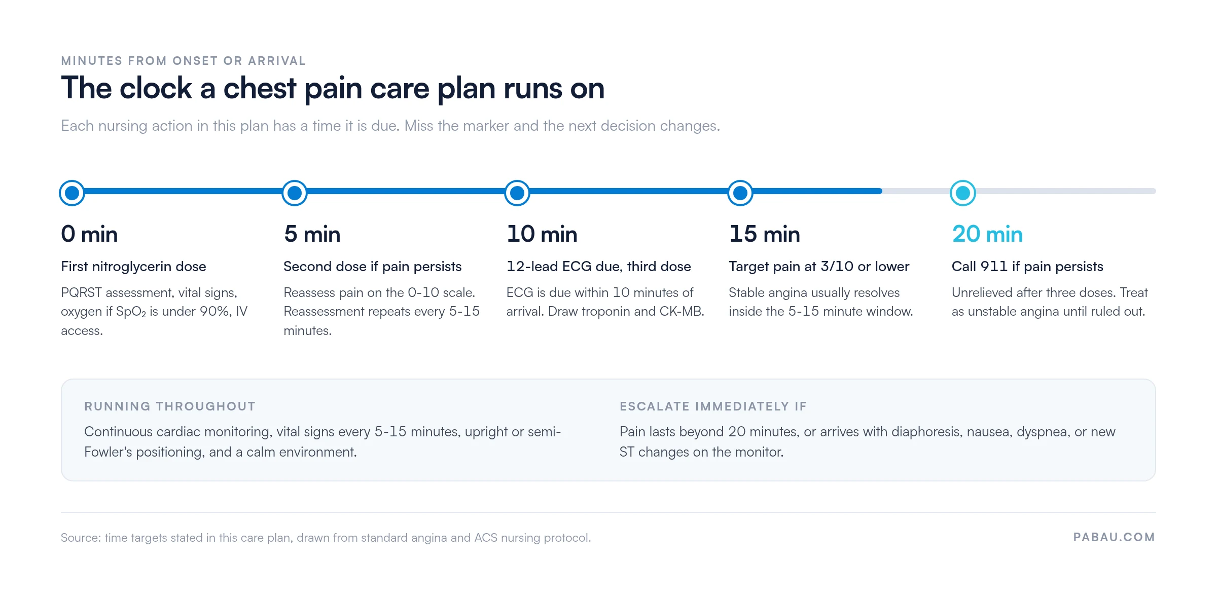
Timing drives the plan, with a 12-lead ECG due within 10 minutes and 911 if pain outlasts three nitroglycerin doses.
Core interventions span pain relief, hemodynamic monitoring, cardiac assessment, and patient education, each with a physiological rationale.
Pabau’s clinical documentation tools let nurses record and retrieve assessment data, intervention rationales, and outcomes in one place.
Download your free chest pain nursing care plan
A ready-to-use care plan covering the six NANDA diagnoses, PQRST assessment prompts, desired outcomes, and interventions with their rationales. It includes a care plan table you can adapt for emergency, inpatient, or outpatient settings.
A chest pain nursing care plan guides assessment, diagnosis, intervention, and evaluation for patients presenting with chest discomfort or angina. Angina is the discomfort that occurs when the heart muscle does not receive enough oxygen-rich blood, a state called myocardial ischemia.
The plan standardizes how nurses assess, which NANDA diagnoses take priority, which evidence-based interventions follow, and how outcomes get documented. It applies across hospital, emergency, and outpatient settings.
This plan covers stable angina, which is predictable and triggered by exertion, and unstable angina, which arrives unprovoked and signals acute coronary risk. Structured documentation records every assessment finding and intervention rationale in the same form each time.
That consistency is what carries a high-risk cardiac patient safely across clinician handoffs. Structured patient intake forms capture history and risk factors before assessment begins, so the nurse starts with the picture already assembled.
Found our content helpful?
Pathophysiology of angina pectoris
Angina pectoris develops when coronary artery disease reduces blood flow to the myocardium faster than oxygen demand rises. During exertion, stress, or emotional triggers, the coronary vessels cannot dilate enough, and the heart muscle is starved of oxygen.
That ischemic state produces chest discomfort, pressure, tightness, or pain radiating to the arm or jaw. It is the warning signal that myocardial oxygen supply no longer meets demand.
Stable angina: Triggered by predictable exertion or emotional stress. Symptoms resolve with rest or nitroglycerin within 5-15 minutes, and the underlying atherosclerosis is chronic rather than actively rupturing.
Unstable angina: Occurs at rest, on minimal exertion, or in a new or worsening pattern. It suggests active plaque rupture, thrombus formation, or vasospasm, and calls for immediate ECG and cardiac biomarker testing.
Vasospastic (Prinzmetal’s) angina: Caused by coronary artery spasm rather than atherosclerotic narrowing. It often occurs at rest or during sleep.
The mechanism sets the nursing priorities. Rapid pain relief reduces myocardial workload, continuous monitoring detects arrhythmia or ST changes, oxygen therapy restores tissue perfusion, and medication follows protocol.
Understanding it also helps nurses anticipate complications and teach patients which risk factors they can change, including smoking, hypertension, inactivity, and stress.
Nursing assessment for chest pain
A chest pain assessment gathers subjective and objective data to separate angina from non-cardiac causes and to stratify risk. Every diagnosis and intervention that follows depends on it.
Subjective data: Run the PQRST mnemonic at every reassessment. Record precipitating factors (exertion, stress, cold), quality (pressure, tightness, burning), radiation (jaw, arm, back), severity (0-10), and timing.
Objective cardiac assessment: A 12-lead ECG within 10 minutes of arrival, vital signs, oxygen saturation, and cardiac auscultation. Check peripheral perfusion for color, temperature, and capillary refill.
Cardiac biomarkers: Troponin, CK-MB, and myoglobin where acute coronary syndrome is suspected.
Risk factor history: Age, family history of coronary artery disease, smoking, hypertension, diabetes, hyperlipidemia, prior cardiac events, and current medications.
Documentation in comprehensive client records keeps pain characteristics, vital sign trends, and ECG findings retrievable for the next clinician on shift.
Pabau’s client records keep every ECG finding, vital sign trend, and pain score on one screen, so the next shift reads the same chart.
Priority NANDA nursing diagnoses
The NANDA International taxonomy provides standardized labels that turn assessment data into nursing priorities. A single patient with chest pain commonly requires six interrelated diagnoses, each with its own outcomes and interventions.
Diagnosis: Acute pain related to myocardial ischemia or coronary artery spasm, evidenced by reported chest discomfort, pressure, or tightness.
Desired outcome: Pain drops to 3/10 or lower within 15 minutes of intervention. The patient uses slow breathing and positioning, and can name their own angina triggers.
Assess pain with PQRST and the 0-10 scale every 5-15 minutes until stable. Rationale: Frequent reassessment catches escalation and shows whether the intervention is working.
Administer sublingual nitroglycerin per protocol, repeating every 5 minutes for up to three doses. Rationale: Nitroglycerin dilates the coronary vessels within 1-3 minutes and lowers myocardial workload. Call 911 if three doses bring no relief.
Provide supplemental oxygen if SpO₂ falls below 90%, or per protocol. Rationale: More oxygen reaches the ischemic myocardium and tissue oxygenation improves.
Position the patient upright or in semi-Fowler’s, with pillows for comfort. Rationale: Upright positioning reduces cardiac workload, eases breathing, and lowers anxiety.
Stay calm and keep the environment quiet, using therapeutic communication. Rationale: Anxiety raises sympathetic tone, heart rate, and blood pressure, which increases myocardial oxygen demand.
Teach slow, deep breathing and encourage relaxation techniques. Rationale: Controlled breathing lowers heart rate, prompts a parasympathetic response, and dulls pain perception.
Decreased cardiac output
Diagnosis: Decreased cardiac output related to reduced myocardial perfusion or impaired contractility, evidenced by hypotension, tachycardia, cool skin, or elevated jugular venous pressure.
Desired outcome: Blood pressure and heart rate return to baseline, and the skin stays warm and dry. Urine output holds at 0.5 mL/kg/hour or higher, with no new ST changes on the ECG.
Monitor blood pressure, heart rate, and respiratory rate every 5-15 minutes, and document the trend. Rationale: Hypotension and tachycardia signal falling cardiac output, and the trend guides escalation.
Run continuous cardiac monitoring and report arrhythmias, ST elevation, or T-wave changes immediately. Rationale: ECG changes mark ischemia progression, and early detection buys time for intervention.
Establish IV access and draw troponin, electrolytes, and a complete blood count. Rationale: IV access speeds medication delivery, and biomarkers confirm myocardial injury.
Give aspirin, beta-blockers, ACE inhibitors, and anticoagulants per protocol. Rationale: Each agent either reduces myocardial workload, prevents clot formation, or improves contractility.
Assess peripheral perfusion through color, temperature, pulses, and capillary refill. Rationale: Cool, clammy skin and delayed capillary refill point to reduced peripheral perfusion.
Monitor urine output and keep an accurate fluid balance. Rationale: Output below 0.5 mL/kg/hour signals reduced renal perfusion from falling cardiac output.
Falling cardiac output has its own set of expected outcomes and monitoring intervals. The decreased cardiac output template works through them in more depth than this section can.
Anxiety
Diagnosis: Anxiety related to fear of death or uncertainty about the diagnosis, evidenced by restlessness, trembling, or verbal expressions of worry.
Desired outcome: The patient reports less anxiety, engages in conversation, and uses a coping strategy without prompting.
Assess the anxiety level and ask open questions about fears and concerns. Rationale: Knowing the specific worry lets you target reassurance and interrupt catastrophic thinking.
Use therapeutic communication through active listening, validation, and presence. Rationale: Being heard lowers psychological distress, and physical presence communicates safety.
Explain procedures, findings, and the treatment plan in plain language, repeating as needed. Rationale: Information reduces fear of the unknown, and repetition works around anxiety-related memory loss.
Teach slow diaphragmatic breathing and progressive muscle relaxation. Rationale: These techniques lower heart rate, blunt the adrenaline surge, and break the anxiety cycle.
Encourage family presence and offer spiritual support where the patient asks for it. Rationale: Family connection and spiritual resources buffer anxiety in ways clinical reassurance cannot.
Administer anxiolytics such as lorazepam per protocol if other measures fall short. Rationale: Medication de-escalates quickly, which in turn lowers myocardial oxygen demand.
Activity intolerance
Diagnosis: Activity intolerance related to imbalance between oxygen supply and myocardial demand, evidenced by dyspnea, fatigue, or chest pain on exertion.
Desired outcome: The patient tolerates gradually increased activity, from sitting to walking, without chest pain or dyspnea, and vital signs stay stable on exertion.
Assess activity tolerance before and after each activity, recording vital signs and symptoms. Rationale: Baseline and response data guide safe progression and surface exertional triggers.
Plan graded activity from rest to sitting, walking, stairs, and self-care per protocol. Rationale: Gradual progression lets the myocardium adapt, while sudden demand triggers ischemia.
Teach energy conservation through pacing, scheduled rest, and avoiding exertion after meals. Rationale: These habits lower myocardial oxygen demand by reducing total workload.
Encourage range-of-motion exercises in bed and assist with daily living tasks at first. Rationale: Muscle tone is maintained without triggering ischemia, and confidence builds with it.
Instruct the patient to stop at chest pain, dyspnea, dizziness, or palpitations. Rationale: Stopping immediately prevents ischemia from progressing into a full anginal attack.
Deficient knowledge
Diagnosis: Deficient knowledge related to unfamiliarity with the disease process, medication use, and lifestyle modification, evidenced by misconceptions or repeated questions about triggers.
Desired outcome: The patient explains what angina is, demonstrates correct nitroglycerin use, and names three personal triggers before discharge.
Assess what the patient already understands about angina, the medications, and the warning signs. Rationale: Teaching pitched at the wrong level either overwhelms the patient or repeats what they know.
Teach nitroglycerin dosing, timing, and storage, then have the patient repeat it back. Rationale: Teach-back is the fastest way to catch a misunderstanding before discharge.
Name the triggers that apply to this patient rather than reading out a generic list. Rationale: Personalized triggers are the ones a patient recognizes and acts on at home.
Break teaching into short sessions and repeat the key points across the stay. Rationale: Pain and anxiety impair retention, so a single long session rarely holds.
Provide written instructions and confirm the patient can read and follow them. Rationale: Written material supports recall once the clinician is no longer in the room.
Risk for ineffective tissue perfusion
Diagnosis: Risk for ineffective tissue perfusion related to progressive coronary stenosis, thrombus formation, or vasospasm.
Desired outcome: The patient maintains stable blood pressure and heart rate, with no new ST changes and biomarkers that stay within range.
Track serial ECGs and troponin levels at the intervals your protocol sets. Rationale: One normal result early on does not rule out an evolving infarction.
Watch for chest pain that returns at rest or on less exertion than before. Rationale: A changing pattern is the marker that separates unstable angina from stable angina.
Keep blood pressure inside the ordered range and report readings outside it. Rationale: Uncontrolled hypertension raises wall stress and accelerates plaque instability.
Give antiplatelet and anticoagulant therapy on schedule, and check for bleeding. Rationale: These agents prevent thrombus extension, and bleeding is their main risk.
Reassess neurological status, pulses, and skin perfusion each shift. Rationale: Perfusion deficits elsewhere can be the first sign of an embolic event.
Key nursing interventions and their rationales
The table below consolidates the actions nurses take in the first minutes and hours of a chest pain presentation. Each one carries a physiological or psychological rationale grounded in cardiac pathophysiology.
What the table does not show is when each action is due. Several of these interventions are timed, and missing a marker changes the next decision.
The first 20 minutes decide whether this stays an angina episode or becomes an emergency. Timings are the ones stated in this care plan.
Patient education and discharge teaching
Patients who understand their condition, their medications, and their triggers have fewer ischemic episodes and recognize acute coronary syndrome sooner.
Nitroglycerin use: Keep it on hand at all times. Take one tablet sublingually at the first sign of chest discomfort. Repeat every 5 minutes for two more doses, then call 911 if the pain holds.
Angina triggers: Exertion, emotional stress, cold exposure, heavy meals, and smoking. Avoiding them or building tolerance gradually reduces how often attacks happen.
Activity guidelines: Start with light walking and increase duration before intensity. Stop at once if chest pain, dyspnea, or dizziness appears, and agree the progression with the physician.
Diet: Sodium under 2,300 mg a day, saturated fat under 7% of calories, plus adequate fiber and hydration.
Smoking cessation: Smoking sharply worsens coronary vasoconstriction. Point the patient at cessation programs, pharmacotherapy, and support groups.
Stress management: Teach deep breathing, progressive muscle relaxation, and meditation. Refer to counseling or cardiac rehabilitation where the practice offers it.
Sleep and rest: Adequate sleep supports cardiac recovery. Advise against lying flat immediately after meals.
When to call 911: Chest pain that is severe, lasts beyond 15-20 minutes, or does not respond to nitroglycerin. The same applies with dyspnea, diaphoresis, nausea, or palpitations.
How to adapt this care plan to your setting
This template works across emergency departments, intensive care, medical-surgical floors, primary care, and outpatient cardiology. Five steps make it fit your own clinical context.
Take a full chest pain history using PQRST. Document each element and separate stable from unstable features. Record baseline vital signs and the 12-lead ECG findings.
Select the NANDA diagnoses that match this presentation. Not every diagnosis applies to every patient, so prioritize by assessment data and acuity. The six above cover most chest pain care plans.
Match the desired outcomes to your setting’s acuity and length of stay. Emergency outcomes focus on pain relief and stabilization within hours. Inpatient outcomes run to activity tolerance, education, and discharge readiness over days.
Adjust the interventions for physician orders, facility protocol, and patient-specific factors. A patient in heart failure may need fluid restriction. A patient on several anticoagulants needs bleeding precautions.
Document each intervention, the patient’s response, and outcome progress every shift.AI-powered clinical documentation structures the notes consistently, so assessment history is quick to retrieve at handoff.
Most chest pain episodes resolve within 5-15 minutes of rest and nitroglycerin. Pain that lasts beyond 20 minutes is a medical emergency, and so is pain that survives three doses. Call 911. Not every chest pain is cardiac, but treat it as unstable angina until the workup says otherwise.
How Pabau keeps chest pain documentation consistent across shifts
In most practices the chest pain care plan lives in three places at once. Assessment findings go in one note, the intervention record in another, and the education checklist on paper that never gets scanned.
Practice management software like Pabau holds the whole plan in one patient record. Custom clinical forms carry the PQRST prompts, the NANDA diagnosis list, and the intervention fields your protocol requires. Nurses fill them in at the bedside rather than reconstructing the shift afterward.
Pabau Scribe, our AI scribe, drafts the narrative note from the encounter, so the nurse edits rather than types. Timed reassessments and vital sign trends sit on the same timeline, which is what a handoff or an audit actually asks for.
The outcome is a care plan the next clinician can read in seconds. The record also stands up when someone asks what was done and when.
Document care plans without chasing paper
Pabau’s clinical forms and documentation tools capture chest pain assessments, interventions, and outcomes in the patient record. Nurses hand off a complete plan instead of a partial one.
What makes this care plan work is the discipline of running the same sequence every time. Under pressure, with a patient who may or may not be infarcting, a fixed order is what stops steps from being skipped.
Adapt the outcomes to your setting and the interventions to your protocol, then leave the structure alone. A plan that changes shape between shifts stops being useful as a handoff document.
The trade-off worth remembering is that documentation quality decides how much of this survives past the shift it was written in. Book a demo to see how Pabau keeps care plan entries, timings, and outcomes in one retrievable record.
Continue your research
Managing an arrhythmia alongside the chest pain?Atrial fibrillation nursing care plan covers rate control, anticoagulation monitoring, and the outcomes to document.
Handing this plan over at shift change?SBAR report template gives you a fixed structure for situation, background, assessment, and recommendation.
Tracking vital signs every 5-15 minutes?Vital signs record keeps the trend in one place instead of scattered across separate notes.
Ready to evaluate whether the plan worked?Evaluation nursing care plan shows how to score outcomes and revise the plan for the next shift.
Reviewing how your team records clinical work?Clinical documentation software walks through what to look for before you change systems.
Frequently asked questions
What is a chest pain nursing care plan?
A chest pain nursing care plan is a clinical document that guides nurses through assessing, diagnosing, intervening, and evaluating care for chest discomfort. It uses NANDA diagnosis language, evidence-based interventions with rationales, and measurable outcomes. The discomfort is usually myocardial ischemia, either stable angina or a suspected acute coronary syndrome.
What are the priority NANDA nursing diagnoses for chest pain?
The six core NANDA diagnoses are acute pain, decreased cardiac output, anxiety, activity intolerance, deficient knowledge, and risk for ineffective tissue perfusion. Not every patient needs all six, so prioritize by assessment findings.
Which nursing interventions relieve chest pain fastest?
Give sublingual nitroglycerin and repeat every 5 minutes if pain persists. Add supplemental oxygen if oxygen saturation falls under 90%, position the patient upright, obtain a 12-lead ECG within 10 minutes, and establish IV access. Start cardiac monitoring and teach slow diaphragmatic breathing to lower myocardial oxygen demand. Follow facility protocol and physician orders throughout.
How does stable angina differ from unstable angina at the bedside?
Stable angina is predictable, triggered by exertion, and resolves with rest or nitroglycerin in 5-15 minutes. Care focuses on activity monitoring and education. Unstable angina is unprovoked, persistent, or worsening, and it calls for immediate ECG, biomarkers, and continuous monitoring.
What should a patient know before discharge?
Cover nitroglycerin use, including timing, dosing, and storage. Name the patient’s own triggers, set activity guidelines, and go through dietary changes, smoking cessation, and stress management. Finish with the emergency signs, which are pain beyond 20 minutes, dyspnea, diaphoresis, or nausea.
How often should a nurse reassess pain?
Reassess every 5-15 minutes during the acute episode, and immediately after any intervention such as nitroglycerin, positioning, or oxygen. Ongoing reassessment catches escalation and shows whether the intervention worked. Document intensity, quality, location, and associated symptoms each time.