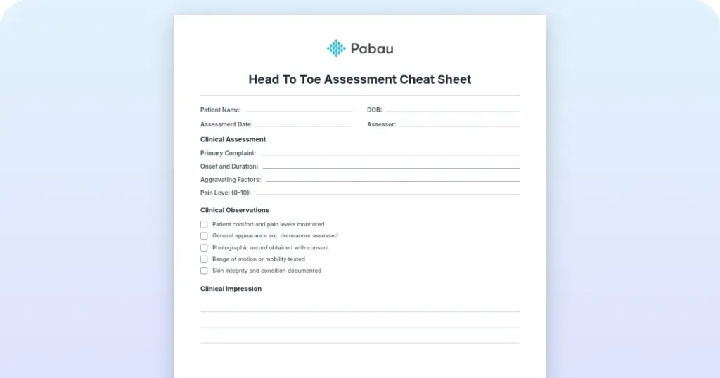
Head to toe assessment cheat sheet: Printable nursing guide
A head to toe assessment cheat sheet gives you one fixed order at the bedside, s...

2
Last Updated: Oct 23, 2026

An abdominal pain chart maps pain location to the organs beneath it, the likely diagnoses, and the ICD-10 code you record.
The four quadrants and the central zones each hold predictable organs, so location narrows the differential before any test is ordered.
Red flags such as guarding, rebound tenderness, and fever with focal pain override location and trigger immediate referral.
Code the pain location first with an R10 code, then add the underlying diagnosis once imaging or labs confirm it.
Practice management software like Pabau captures the same assessment fields every time, so escalation decisions stay documented and auditable.
A ready-to-use clinical reference mapping each quadrant to its organs and the differential diagnoses that present there. It also carries the red flag criteria for escalation and the matching ICD-10 codes. Print it for the consulting room or attach it to your assessment template.
Download templateAn abdominal pain chart maps where a patient hurts to the organs sitting under that spot. It then links each location to the conditions that commonly cause pain there, and to the ICD-10 code you document it with.
Most practices use the four-quadrant version: right upper, left upper, right lower, and left lower. The central epigastric and periumbilical zones sit alongside them.
Location narrows the differential quickly, but it never closes it. The chart earns its place as a prompt for systematic questioning and consistent documentation.
This guide walks through the quadrants, the red flags that force escalation, and the R10 codes for each region. The template below is free to download and use in consultation.
An abdominal pain chart is a structured clinical reference. It pairs each region of the abdomen with the organs that sit there and the conditions that commonly cause pain in that area. Some versions are quadrant diagrams, others are tables. Both do the same three jobs:
The formal anatomical model divides the abdomen into nine regions. Two vertical midclavicular lines cross two horizontal planes, the transpyloric and the intertubercular. Day-to-day practice mostly uses the simpler four-quadrant system instead. Either way, each region corresponds to specific organs, and many conditions present with predictable patterns. Gallbladder pain usually localizes to the upper right, appendicitis to the lower right.
A documented, systematic approach also demonstrates clinical competence and reduces liability. Capturing pain location, character, associated symptoms, and examination findings on a digital form keeps that documentation consistent from one clinician to the next.

The quadrant system splits the abdomen with two lines. One runs vertically down the midline, the other horizontally through the umbilicus. That leaves four quadrants, each holding predictable anatomy.
Central pain around the umbilicus or epigastrium usually comes from the small intestine, stomach, or aorta. One pattern is worth memorizing. Periumbilical pain that shifts to the right lower quadrant after a few hours points to appendicitis. Inflammation first irritates visceral pain fibers, felt at the umbilicus, before somatic fibers localize it over the appendix.
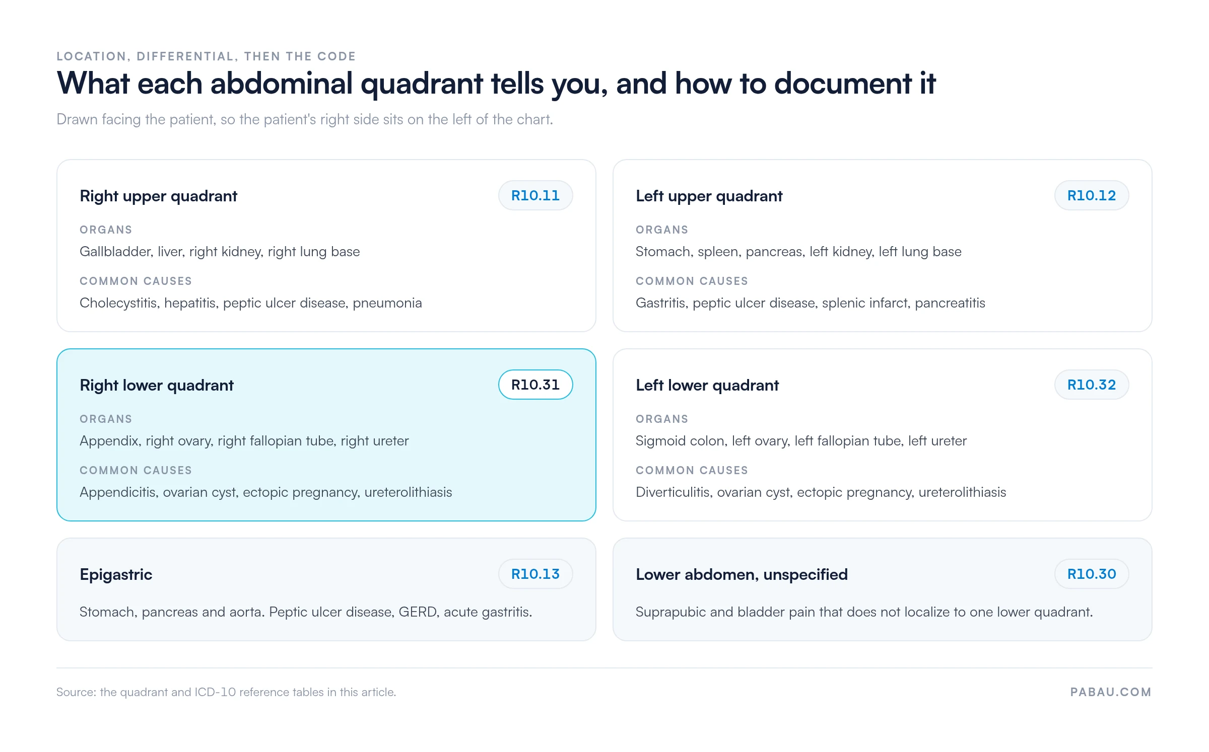
The chart below puts both halves of that reasoning in one view. It carries the organs in each region, the conditions that present there, and the matching R10 code.
Five steps take a patient from a vague complaint to a documented, coded assessment.
Writing those five fields the same way every time is what makes a later reader trust the note. Our guide to safer clinical notes covers wording that survives peer review. An abdominal exam documentation template gives you the fields already laid out.
The chart is a decision aid, not a diagnostic device. No single finding rules a diagnosis in or out. Appendicitis can present with left-sided pain, and renal colic can mimic biliary colic. What the chart does well is prompt complete documentation and flag which presentations need same-day imaging or referral.
Some findings demand urgent referral whatever the pain location. Recognizing them is the most safety-critical part of an abdominal assessment.
Structured patient records can carry the red flag fields and raise an alert when one is ticked. That reduces the chance of a missed urgent presentation. Documenting why a case was referred, or why it was not, protects both clinician and patient.

The female lower abdomen holds reproductive organs that can generate pain mimicking appendicitis, diverticulitis, or renal colic. A 25-year-old woman with right lower quadrant pain might have appendicitis.
She might equally have an ovarian cyst, an ectopic pregnancy, pelvic inflammatory disease, or mittelschmerz. Always ask about menstrual history, last menstrual period, the possibility of pregnancy, and gynecological symptoms.
A positive pregnancy test changes the whole differential, because ectopic pregnancy moves to the top of it. Your chart should flag female-specific diagnoses by quadrant so gynecological causes are not overlooked.
A pelvic examination is often warranted in females with lower abdominal pain, to check for cervical motion tenderness or adnexal masses.
Accurate documentation feeds directly into coding. The R10 series organizes codes by pain location and character, which mirrors the quadrant system you just used. The CDC/NCHS ICD-10-CM web tool confirms whether a code is still valid for the current fiscal year.
Code the location of the pain first with an R10 code. Then add a code for the underlying cause once it is identified, such as K80.20 for gallstones without cholecystitis. That two-code structure ties symptom documentation to diagnostic reasoning and supports billing accuracy.
The chart earns its keep anywhere abdominal presentations are triaged. Primary care physicians and nurse practitioners reach for it before referral.
Emergency clinicians use it to stream acute presentations, and gastroenterologists and colorectal surgeons use it when reviewing referrals. It doubles as a teaching tool for medical students and residents learning differential diagnosis.
The documentation side matters most in busy general practice, where the same complaint arrives several times a week. Software for primary care that stores the assessment template alongside the patient record keeps that reasoning in one place.
Standardized use of the chart pays off clinically and operationally. It reduces diagnostic error by prompting systematic questioning. That limits anchoring bias, the pull of a first impression that stops you looking further.
Operationally, it turns a vague presenting complaint into coded, searchable documentation. Hand-off notes to a specialist become easier to read, and the referral reason is already written down. Documented adherence to a systematic framework also supports peer review and accreditation.
Workflow automation closes the loop. When a red flag field is ticked, the record can raise a task instead of relying on someone remembering to chase it.
Most practices assess abdominal pain well and record it inconsistently. One clinician writes a paragraph, the next writes three words, and the referral letter has to be rebuilt from scratch.
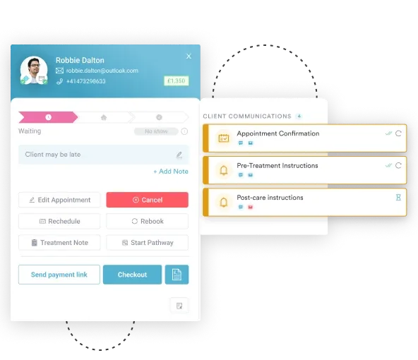
Practice management software like Pabau replaces that free text with a templated form. Pain location, character, severity, associated symptoms, and examination findings each get their own field, so the chart’s structure lives in the record. Location choices come from a dropdown, which cuts transcription error.
Red flags recorded in the form can trigger an alert and mark the case for same-day escalation. The result is an auditable assessment that another clinician can pick up, which matters most when the patient is referred onward.

Templated assessment forms capture pain location, red flags, and examination findings in set fields, so referrals leave your practice complete.

Use the chart as a prompt, not an answer. Its value is that it makes you ask the same questions in the same order. The note you write then ends up complete enough for the next person to act on.
One trade-off is worth remembering. Location is a filter, and a filter lets some cases through. Pair the chart with the red flag criteria and a low threshold for escalation, and the misses get rarer.
If your practice is tightening documentation more broadly, our HIPAA compliance checklist covers the storage and access side of the same records. Book a demo to see how Pabau turns the abdominal pain chart into a templated assessment your whole team fills in the same way.

Assessing chest pain by location too? Chest pain location chart applies the same mapping logic above the diaphragm.
Working with a patient who cannot describe the pain? Abbey pain scale scores pain from observed behavior when self-report is unreliable.
Need the numbers that back an escalation? Abnormal vital signs chart sets the thresholds that turn a concern into a referral.
Building a full assessment routine? Head-to-toe assessment template puts the abdominal exam in sequence with every other system.
An abdominal pain chart maps pain location to organs and likely diagnoses. It helps clinicians narrow the differential, document findings systematically, and identify red flags that warrant urgent referral.
There are four. Upper right holds the gallbladder, liver, and right kidney. Upper left holds the stomach, spleen, pancreas, and left kidney. Lower right holds the appendix and right ovary, lower left the sigmoid colon and left ovary. The central regions cover the epigastrium and the periumbilical area.
Guarding or rebound tenderness, fever with localized tenderness, and hypotension or shock all warrant emergency assessment. So does pain out of proportion to the examination findings. Pregnancy with pain and bleeding, and new pain in a patient over 65, both need urgent review.
Females have reproductive organs that generate pain mimicking appendicitis or diverticulitis. Ovarian cysts, ectopic pregnancy, and pelvic inflammatory disease must be part of the differential, especially in the lower quadrants. Menstrual history and pregnancy status are essential.
The R10 series codes pain by location: R10.11 (right upper), R10.12 (left upper), R10.31 (right lower), R10.32 (left lower), R10.13 (epigastric), R10.30 (lower abdomen). Add codes for underlying diagnoses such as cholecystitis or appendicitis once identified.
Yes. Early appendiceal inflammation stimulates visceral pain fibers, which the patient feels diffusely around the umbilicus. Once the parietal peritoneum becomes inflamed, somatic fibers localize the pain to the right iliac fossa where the appendix lies.