Sulcus Test: Shoulder Instability Assessment
The sulcus test is an orthopedic special test that checks for inferior glenohume...

3
Last Updated: Oct 22, 2026
The DRUJ instability test is a bedside exam that checks the distal radioulnar joint (DRUJ) for abnormal ulnar head movement against the distal radius. You stabilize the radius, shift the ulnar head dorsally and volarly, and repeat the maneuver in neutral, full pronation and full supination.
The test is positive when the ulnar head translates further than on the other wrist, usually with a soft or absent endpoint. According to PubMed-indexed hand surgery literature, DRUJ instability is among the most commonly missed wrist injuries, because standard radiographs underestimate ligament damage.
Physical therapists, hand therapists, and orthopedic clinicians use the test after wrist trauma or a distal radius fracture, before they order advanced imaging.

The DRUJ instability test measures how far the ulnar head translates against the radius, and tenderness alone doesn’t make it positive.
Perform the ballottement maneuver in neutral, full pronation, and full supination to identify which radioulnar ligament is disrupted.
Always compare both wrists, because constitutional laxity is the most common source of false-positive findings.
Diagnostic arthroscopy remains the gold standard, so treat clinical tests as screening tools rather than definitive confirmation.
Practice management software like Pabau records the same DRUJ exam fields at each visit, so grade changes show side by side at follow-up.
The DRUJ instability test assesses the mechanical stability of the distal radioulnar joint. You apply dorsal and volar translational stress to the ulnar head while the radius is held still.
The DRUJ is a pivot joint that allows forearm pronation and supination. Its primary stabilizers are the dorsal and palmar radioulnar ligaments, which form the deep limbs of the triangular fibrocartilage complex (TFCC), and the interosseous membrane.
When these structures are disrupted, the ulnar head translates excessively relative to the radius, producing the positive finding. Clinicians working in physical therapy practices see DRUJ instability most often after distal radius fractures, TFCC foveal tears, and Essex-Lopresti injuries. The test gives immediate bedside information on whether imaging or orthopedic referral is warranted.
The DRUJ is formed by the sigmoid notch of the distal radius and the ulnar head. Stability depends on three layers of restraint.
The cause of the injury tells you which position-specific maneuver to prioritize during the exam.
Several named maneuvers assess DRUJ stability, and each one isolates a specific restraint under a specific load. The table below summarizes the most commonly used tests alongside their positive finding.
The DRUJ ballottement test takes five steps, and precise hand placement matters most. Your grip has to isolate translational movement without compressing the joint.
Repeat the identical translational maneuver with the forearm in full pronation, then in full supination. This is the most clinically useful modification of the DRUJ instability test, because ligament tension changes dramatically across the rotation arc.
The piano key sign is a supplementary DRUJ exam that assesses dorsal ulnar head instability in pronation. It is positive when the prominently displaced ulnar head springs back after manual depression, like a piano key.
Unlike the ballottement maneuver, the piano key sign does not isolate palmar instability. Use both tests together for a complete DRUJ exam, and always compare the finding with the opposite wrist. If you want a form to record the result, download our piano key test template.
The same spring-back reading transfers to other joints. The rib spring test grades rib mobility from 0 to 4 on end-feel, recorded by rib level and side.
Always examine the contralateral wrist before interpreting any DRUJ instability test. Bilateral comparison is the most practical way to tell pathological laxity from constitutional hypermobility. It matters most in younger patients and women, where baseline joint laxity is more common.
Interpretation depends on three readings: the degree of translation compared with the other wrist, the endpoint quality, and which forearm position elicits the finding. Using validated clinical assessment tool grading frameworks helps standardize how results are recorded and shared across the treating team. The flow below shows how the three readings combine into a grade and a next step.
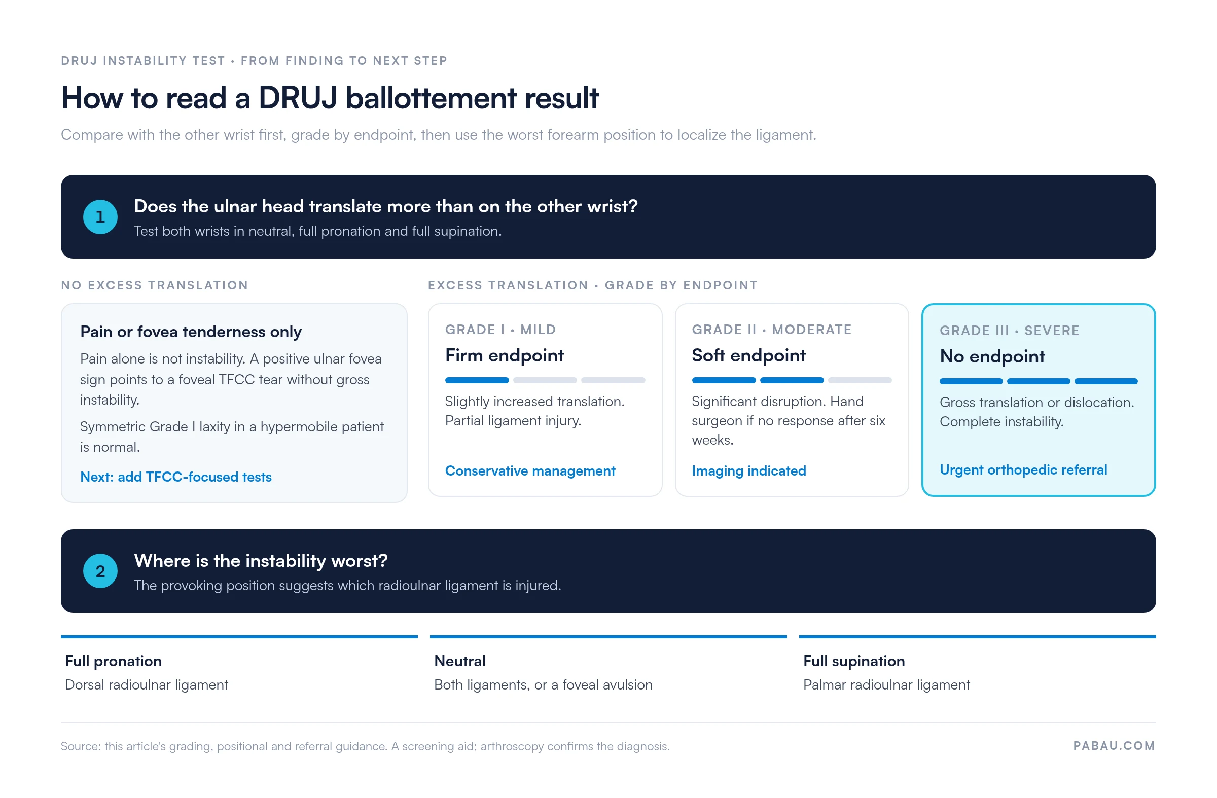
Published diagnostic accuracy data for the DRUJ instability test is limited by the small sample sizes typical of hand surgery studies. According to the Journal of Hand Surgery, no single clinical test reliably separates DRUJ instability from TFCC peripheral tears on physical exam alone. Accuracy improves when you combine several tests.
The ballottement test on its own has been described as moderately sensitive for clinically significant instability. Reported sensitivity and specificity values vary considerably across studies, though. Treat a positive DRUJ instability test as a reason for further investigation rather than a definitive diagnosis.
The American Academy of Orthopaedic Surgeons (AAOS) recommends combining clinical exam findings with advanced imaging to confirm DRUJ instability before surgical planning.
DRUJ instability and TFCC tears often coexist, but they are separate problems. Telling them apart at the bedside guides both imaging selection and management. Sports medicine clinicians see this overlap often in athletes with rotational wrist injuries.
DRUJ instability needs demonstrable translation, and pain on its own doesn’t meet that bar. A patient with ulnar-sided wrist pain, a positive ulnar fovea sign, and a negative ballottement test likely has a foveal tear without gross instability.
In that case, a TFCC compression test loads the disc directly. This distinction matters when you’re choosing between conservative management and surgical repair.
Clinical examination identifies instability, and imaging characterizes the anatomy before surgical planning. Record the grade and the provoking forearm position in the referral, so the imaging request is backed by your exam findings.
According to the British Society for Surgery of the Hand (BSSH), wrist arthroscopy is the reference standard for grading DRUJ instability and associated TFCC pathology. Clinical tests guide the decision to refer, and they don’t replace arthroscopic assessment. Referral pathways and imaging access vary by region, so treat these points as guidance rather than a rigid protocol.
Even experienced clinicians make predictable errors when performing the DRUJ instability test. Most errors fall into three categories: skipping the bilateral comparison, positioning the forearm incorrectly, and reading pain as instability.
When wrist exam findings go into free-text notes, one clinician writes “lax in pronation” and the next writes “Grade II.” Six weeks later, nobody can say for sure whether the joint has improved. That undermines the serial comparison this test depends on.
Practice management software like Pabau lets you build the DRUJ exam into a digital form. You record translation, endpoint quality, pain, and grade for each forearm position and both wrists. The completed form is saved to the patient’s record, alongside their treatment notes.

At the follow-up, you open the last exam in the patient’s record and repeat the same fields. Any change in grade shows up side by side, which turns the six-week conservative-care review into a quick decision.


Pabau’s digital forms and patient records let physical therapists and hand therapists capture structured exam findings at the point of care. Each entry links to the patient’s full history.

Run the ballottement test in three positions on both wrists before you trust any result. A single-position, one-wrist check is the quickest route to a false positive in a hypermobile patient, or a missed palmar ligament injury.
Treat the test as triage. It decides who needs imaging or a hand surgeon, while arthroscopy still decides the diagnosis. The full bilateral exam takes longer than a quick check, but it spares patients an unnecessary MRI or a delayed referral.
Recording the exam the same way at every visit makes the six-week review an easy call. Book a demo to see how Pabau’s digital forms keep DRUJ findings comparable from the first visit to the referral.

Seeing DRUJ disruption after a forearm fracture? Galeazzi fracture vs Monteggia fracture explains how to tell the two injury patterns apart.
Checking the radial side of the wrist too? Scapholunate ballottement test covers the step-by-step technique and its accuracy.
Assessing thumb-side wrist pain? Finkelstein test sets out the procedure, interpretation, and clinical workflow for De Quervain’s tenosynovitis.
Need a structured documentation workflow for your practice? Physical therapy practice management software covers the features that support organized patient records and referral tracking.
The DRUJ ballottement test detects anteroposterior instability of the distal radioulnar joint. It measures how far the ulnar head translates relative to the stabilized radius. It helps clinicians spot ligament disruption after wrist trauma or fracture, or in chronic ulnar-sided wrist pain.
A positive DRUJ instability test means the ulnar head translates dorsally or volarly further than on the other wrist, with a soft or absent endpoint. This suggests disruption of the radioulnar ligaments, often with a TFCC foveal tear. It is a screening finding that warrants MRI or arthroscopy, not a definitive diagnosis on its own.
Published data is limited and varies by study design and reference standard. The literature agrees on no single sensitivity or specificity figure. The ballottement test is considered moderately sensitive for clinically significant instability, so use it as part of a multi-test exam. Wrist arthroscopy remains the reference standard against which clinical tests are measured.
DRUJ instability requires demonstrable abnormal translation of the ulnar head. A TFCC peripheral tear may cause ulnar-sided pain and tenderness without gross mechanical instability. A patient can have a TFCC tear with a negative ballottement test, or both conditions together. The ulnar fovea sign and ulnocarpal stress test help identify TFCC pathology, while the ballottement test detects translational instability.
The piano key sign is positive when the dorsally prominent ulnar head springs back after being pressed down in forearm pronation, like a piano key. It indicates dorsal DRUJ instability. It is particularly associated with disruption of the dorsal radioulnar ligament and ECU subsheath.
Perform the DRUJ instability test in any patient with ulnar-sided wrist pain or a forearm rotation deficit. It also applies after a distal radius fracture, a Galeazzi fracture, or a suspected Essex-Lopresti injury. Athletes with rotational wrist injuries and post-operative patients with persistent symptoms after TFCC repair are candidates too.