Hyperbole and a Half pain scale: Every level + free template
The Hyperbole and a Half pain scale is Allie Brosh’s humorous pain chart, ...

1
Last Updated: Oct 5, 2026
The TFCC compression test is an orthopedic special test that loads the triangular fibrocartilage complex to reproduce ulnar-sided wrist pain.
The examiner takes the wrist into maximum ulnar deviation and pronation, then applies axial compression through the forearm. Pain that matches the patient’s familiar symptom counts as a positive result.
The test takes seconds, needs no equipment, and appears in most wrist assessment protocols. A 2020 BMJ Open systematic review of wrist injury diagnostics found no study reporting sensitivity or specificity for it.
So treat a positive finding as a reason to investigate further, never as evidence of a tear. This guide covers how to perform the test, how to read the result, what the evidence supports, and how to document it.

The TFCC compression test applies axial load to the wrist in ulnar deviation and pronation to reproduce ulnar-sided pain.
A positive result means the patient’s familiar ulnar-sided pain came back. It does not confirm a tear without imaging.
No published study reports sensitivity or specificity for the compression test, so treat it as a screening step rather than proof.
Record the position, the load direction, and the patient’s response in your SOAP notes, not just positive or negative.
Structured patient records help physiotherapy and sports medicine practices capture special test findings consistently.
The TFCC compression test is a clinical special test for triangular fibrocartilage complex tears and related pathology. It targets the TFCC, a fibrocartilaginous structure that stabilizes the distal radioulnar joint (DRUJ) and the ulnocarpal articulation. The trigger for using it is persistent ulnar-sided wrist pain after trauma, repetitive loading, or degenerative change.
Some clinical texts call it the TFCC load test. The principle is straightforward. Position the wrist where the TFCC is most compressed between the ulna and the carpal bones. If the structure is damaged, the patient’s familiar symptoms should reappear.
The triangular fibrocartilage complex is a multi-structure unit on the ulnar side of the wrist. Its components explain why the compression test provokes pain in specific positions. As described in the StatPearls review of TFCC anatomy, the complex contains the following structures:
The articular disc and the radioulnar ligaments bear the greatest compressive and rotational loads. Ulnar deviation of the wrist combined with pronation of the forearm tightens and compresses these structures. That is what the compression test exploits to reproduce symptoms.
Use this test when a patient reports ulnar-sided wrist pain alongside any of the presentations below. Overhead athletes, racket sport players, and manual workers account for most of them.
Practices carrying that caseload tend to run dedicated sports medicine software, so repeat assessments and progress measures stay in one record.
The test is not indicated in isolation. It forms part of a structured wrist assessment that includes palpation, range-of-movement testing, and a review of imaging where available.
Technique decides whether the result means anything. Inconsistent positioning or force direction is the most common source of false positives and false negatives in this test.
Seat the patient at a table with the elbow resting on the surface, flexed to approximately 90 degrees. The forearm starts in neutral rotation. The wrist and hand hang freely off the edge or rest lightly on the table.
The test is complete once you have held maximum ulnar deviation and pronation under axial load for 5 to 10 seconds. Some clinicians repeat it with the forearm in supination to compare symptom provocation across positions. That comparison helps narrow down which part of the complex is involved.
A positive TFCC compression test reproduces the patient’s familiar ulnar-sided wrist pain during axial loading in ulnar deviation and pronation. A click or clunk in the same position may also count as positive, particularly if it reproduces the reported symptom.
Interpreting the finding requires clinical context. A positive result raises suspicion for TFCC pathology but cannot confirm a tear. MRI or wrist arthroscopy remains the standard for definitive diagnosis. Arthroscopy allows direct visualization and is used both diagnostically and therapeutically for confirmed TFCC tears.
No published study reports a sensitivity or specificity for the TFCC compression test. The 2020 BMJ Open systematic review by Krastman and colleagues searched the wrist literature and found only two studies of physical examination for TFCC lesions. Neither one tested the compression test.
Reference sites that catalog special tests, including Physiotutors and OrthoFixar, note the same absence. Both rate the test as of questionable standalone value.
What the literature does offer are figures for other ulnar-sided provocation tests. They appear below for context only. None of them is the compression test, and none should be quoted as its accuracy. The chart sets that blank against what the neighboring tests report.
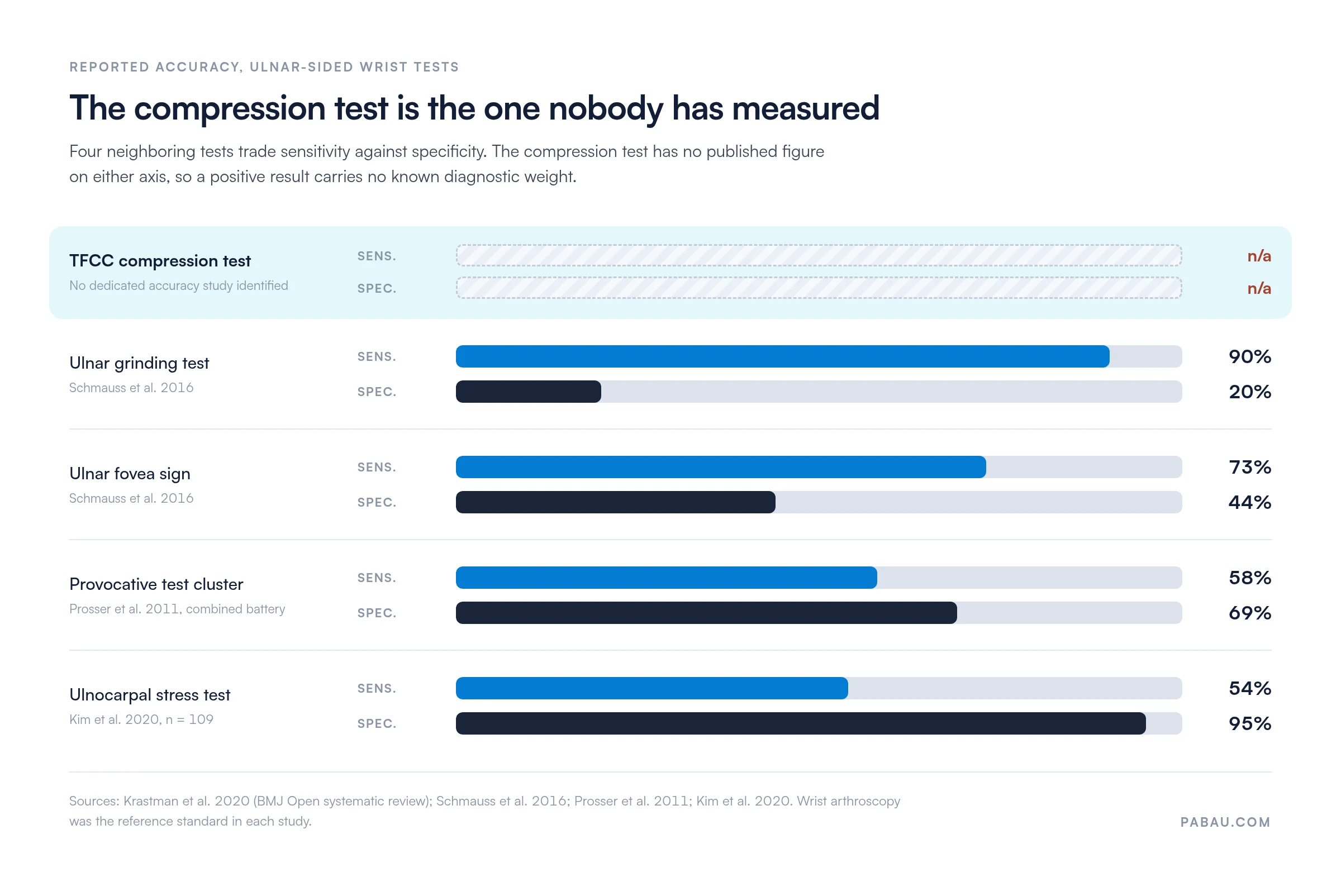
Wrist arthroscopy is the reference standard in every one of these studies, and no clinical test comes close to it. The practical consequence for the compression test is simple. Use it to establish whether ulnar-sided loading reproduces the patient’s familiar pain. Then let history, palpation, and imaging carry the diagnostic weight.
Quoting a sensitivity figure for this test in a report or a referral letter means quoting a number that does not exist. Where test-specific data is this thin, a validated decision rule does more work than any single maneuver. The Ottawa ankle rules are the standard example of what that looks like when the evidence is there.
A positive TFCC compression test does not exclusively point to TFCC pathology. Several conditions produce ulnar-sided wrist pain and can generate a false-positive result or a genuinely ambiguous presentation.
A detailed patient history, a thorough palpation map, and selective use of multiple special tests narrow the differential. When the compression test is positive but the clinical picture is unclear, MRI provides the most useful non-invasive information before considering arthroscopy.
Performing the test is half the job. What you write down afterwards decides whether the finding is useful to the next clinician, to a referring surgeon, or to you in six weeks. Practices that standardize this build it into their physiotherapy practice management system rather than leaving it to free text.
The principle is to record what you did, what you found, and what it means. A bare entry reading “TFCC test positive” gives a colleague nothing to work with six weeks later.
Below is a worked example for the Objective section of a SOAP note. The same structure adapts to any MSK special test result.
Structured digital forms let you turn that table into an assessment template the whole team fills in the same way. The fields prompt for position, load, response, and classification, so a rushed entry still carries the information a colleague needs.

Retaining a record of the clinical reasoning behind test selection matters for compliance too. A patient may later challenge a clinical decision, and a SOAP note showing the test performed, the finding, and the resulting plan is your protection. Consistent medical records management is what makes that defensible across a whole team.
Flag the TFCC compression test result in your assessment summary with a severity rating rather than just positive or negative. Recording the pain score at the moment of provocation gives you a measurable baseline for tracking improvement or deterioration across subsequent appointments.
The compression test is one of several TFCC special tests available to clinicians. Combining them improves diagnostic confidence, because no single test has enough sensitivity or specificity to stand alone. The table below summarizes the four most commonly used.
The press test was first described by Lester et al. (1995), with research available via PubMed PMID 7574285 as an office-based test for TFCC pathology.
The patient places both hands on the armrests of a chair and pushes their body weight upward, loading the wrist into extension. Pain on the ulnar side constitutes a positive result. Unlike the compression test, the press test is patient-driven and produces higher axial loads through the patient’s own body weight.
The supination lift test targets the dorsal radioulnar ligament, which makes it useful when you suspect a dorsal TFCC tear. The patient sits with elbows on the table and attempts to lift the table from below with their palms facing up.
Pain or weakness in the ulnar wrist region constitutes a positive result. Resisted supination stresses the dorsal structures that the compression test in pronation may underload.
The ulnar fovea sign is a palpation-based test rather than a provocation test. The examiner applies direct pressure into the fovea, the soft tissue depression between the ulnar styloid and the flexor carpi ulnaris tendon.
Focal tenderness here, particularly compared with the contralateral side, suggests a foveal TFCC tear or disruption of the ulnar DRUJ ligaments. In the one study that measured it, the fovea sign picked up 73% of confirmed lesions. It was also positive in more than half of the wrists without one. A positive fovea sign therefore narrows the likely location rather than confirming the diagnosis.
The TFCC grind test uses a similar starting position to the compression test but adds a circumductory rotation through the wrist while maintaining axial load. The grinding motion is thought to increase mechanical stress across damaged fibrocartilage.
Some examiners use it as a confirmatory step following a positive compression test. The closest published figure comes from the ulnar grinding test in Schmauss et al. 2016, which caught 90% of lesions at 20% specificity. A test that is positive in four out of five uninjured wrists cannot confirm anything on its own.
In most practices a special test result lands in a free-text note. The wording changes between clinicians, and the pain score is recorded only sometimes. Comparing today’s finding with one from six weeks ago means reading two paragraphs side by side.
Practice management software like Pabau replaces that with structured clinical forms. You build the wrist assessment once, with fields for position, load direction, patient response, and result classification. Every clinician then completes the same fields in the same order, and the entry saves straight into the client record.
What you get is a documentation trail you can compare across appointments. Treatment notes, uploaded imaging reports, and outcome measure scores sit in the same searchable file, so a handover or a referral letter takes minutes. For a multi-clinician MSK caseload, a physical therapy EMR that enforces one format beats any individual clinician’s note-writing habit.

Pabau’s digital forms and client records let physiotherapy and sports medicine practices capture position, load, and patient response in structured fields. Assessment findings stay with the patient record between appointments.

The TFCC compression test is a practical, low-equipment screening tool for ulnar-sided wrist pathology. Its accuracy has never been measured on its own, so it belongs inside a structured assessment battery rather than at the front of a diagnosis.
Record what the test reproduced, not just whether it was positive, and let imaging settle the question when the picture stays unclear. A practice that documents special tests the same way every time ends up with a record worth referring back to. To see how Pabau supports MSK assessment workflows, book a demo.

Ruling out a radial-sided cause first? Scaphoid fracture test walks through the maneuvers that separate anatomical snuffbox pain from ulnar-sided wrist pain.
Suspect a neurological contribution to the symptoms? Hand nerve tests covers the screening battery for ulnar, median, and radial nerve involvement at the wrist.
Need the radial-sided tendon screen as well? Finkelstein test explains how to provoke and interpret de Quervain’s tenosynovitis in the same assessment.
Building a full wrist examination sequence? Phalen’s test adds the carpal tunnel screen that most wrist pain protocols run alongside the special tests above.
The TFCC compression test is a clinical special test used to screen for triangular fibrocartilage complex tears or pathology in patients with ulnar-sided wrist pain. It applies axial load to the wrist in ulnar deviation and pronation to reproduce symptoms. A positive result raises clinical suspicion for TFCC damage and typically prompts referral for imaging or specialist review.
A positive result means axial loading in ulnar deviation and pronation reproduces the patient’s familiar ulnar-sided wrist pain. That raises suspicion for TFCC pathology. It does not confirm a TFCC tear. MRI or wrist arthroscopy is required for a definitive diagnosis. A click or clunk alongside pain may indicate a more significant mechanical lesion.
No study has published sensitivity or specificity for the TFCC compression test itself. The 2020 BMJ Open systematic review of wrist injury diagnostics found only two studies of physical examination for TFCC lesions, and neither assessed this test. Figures you may see quoted for it usually belong to other maneuvers. The fovea sign, for example, is reported at 73% sensitivity and 44% specificity. Treat the compression test as a screening step, with wrist arthroscopy as the reference standard.
The compression test is examiner-applied, using manual axial load in ulnar deviation and pronation. The press test is patient-driven. The patient pushes their body weight upward from a chair using their wrists, which generates much higher axial loads. Both target TFCC and DRUJ pathology. The press test loads the wrist into extension rather than the ulnar deviated, pronated position of the compression test.
The TFCC grind test uses the same starting position as the compression test, with axial load in ulnar deviation and pronation. It then adds a circumductory rotation through the wrist during loading. This grinding motion increases mechanical stress across the fibrocartilage. The compression test uses static axial compression without rotation. The grind test is sometimes used as a confirmatory step following a positive compression test result.
No. The TFCC compression test cannot replace MRI or wrist arthroscopy for definitive diagnosis. Because its accuracy has never been measured in a dedicated study, neither a positive nor a negative result carries a known diagnostic weight. MRI provides soft tissue visualization of TFCC integrity, while arthroscopy remains the gold standard for both diagnosis and treatment.