







HCPCS code S0510: Non-prescription safety lens billing guide
HCPCS code S0510: definition, descriptor, and code classification HCPCS code S05...

Last Updated: Sep 25, 2026


CPT code 92134 covers scanning computerized ophthalmic diagnostic imaging of the posterior segment, retina, with interpretation and report, unilateral or bilateral.
CPT 92133 is the other posterior segment code and it images the optic nerve, which is why glaucoma diagnoses belong to 92133 rather than 92134.
CPT 92132 is the anterior segment code, and it is not payable on the same day as 92133 or 92134.
One unit per session pays the same for one eye or both, so modifier 50 and split RT and LT lines both deny.
LCD L33751 supports one 92134 exam every two months, or one a month while a retinal condition is under active treatment.
Pabau’s claims management software and Claim.MD clearinghouse integration help ophthalmology practices submit clean 92134 claims, verify eligibility, and resolve denials faster.
CPT code 92134 bills retina optical coherence tomography, or OCT. The descriptor reads scanning computerized ophthalmic diagnostic imaging of the posterior segment, retina, with interpretation and report. One unit covers the session, whether you scanned one eye or both.
The rest of this guide works through the structure split, the covered diagnoses, the frequency ceilings, and the denials that repeat.
The AMA’s CPT code set places 92134 in the ophthalmology subsection of the medicine codes.
The descriptor ends with a semicolon and the word retina, and that last word is the whole distinction from 92133. A physician has to perform or supervise the imaging, read it, and sign a report. There is no separate bilateral code, so one session buys one unit.
Four structures sit on the retina side of that split:
The optic nerve head and the circumpapillary retinal nerve fiber layer are not on that list. Scans of those structures go out as 92133, even though both codes read posterior segment.
Cornea, iris, lens, and anterior chamber angle imaging belongs to CPT 92132. Side by side, the three codes divide the eye like this.
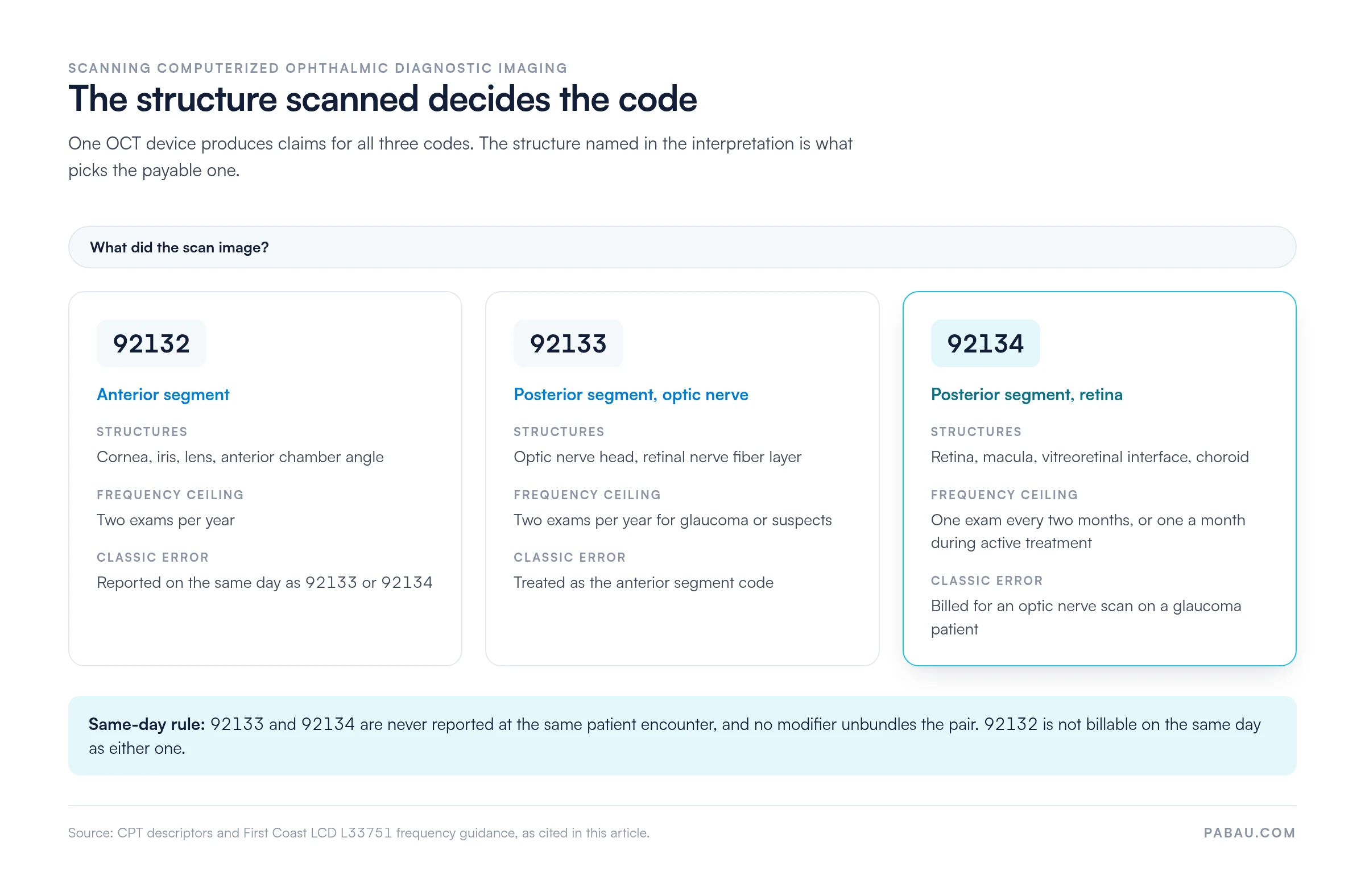
Coders call these the SCODI codes, after the opening words of the descriptor.
The structure scanned tells them apart, not the machine that scanned it. One OCT device can produce a claim for any of the three. So the descriptor you pick has to match the structure your interpretation describes.
92133 and 92134 are not reported at the same patient encounter. The pair carries a mutually exclusive edit, so no modifier unbundles it. Additionally, 92132 is not billable on the same day as either posterior code.
The AAPC CPT code lookup carries the official descriptor text for all three, which is worth putting in front of new billing staff.
Read the interpretation, find the structure, then pick the code. These five scenarios cover most of what an ophthalmology or retina practice sends out in a week.
Medicare covers 92134 when the record establishes medical necessity under the local coverage determination that applies to your practice. CMS delegates that authority to Medicare Administrative Contractors, so covered diagnoses and frequency limits vary by jurisdiction.
First Coast Service Options publishes LCD L33751 for scanning computerized ophthalmic diagnostic imaging. Its billing and coding article, A57804, carries the covered code and diagnosis lists. Other MACs publish their own SCODI policies, so confirm which one governs your claims before you bill.
Under L33751, posterior segment SCODI is reasonable and necessary for three groups of patients. Glaucoma and glaucoma suspects sit in the first group, and those diagnoses run through 92133. Retinal disease and macular abnormalities sit in the third group, and those are the 92134 claims.
Monitoring for chloroquine or hydroxychloroquine retinopathy sits in the second group, the one indication that looks like screening and is still covered.
The limitations matter as much as the indications. Screening a patient with no signs or symptoms is not covered. Nor is a scan run only to confirm a diagnosis that is already established. The exception is a record showing the result set a baseline or changed management.
So check coverage before the visit rather than after it. Eligibility checks flag patients whose plan or diagnosis will not support the test, and that is the moment to issue an Advance Beneficiary Notice.
Pairing 92134 with a diagnosis from the wrong list is the most common denial on the code. The billing article splits its diagnosis groups by CPT code. A glaucoma code on a 92134 line fails the medical necessity check, even though the same policy covers glaucoma under 92133.
Codes below sit at family level. Every claim needs the full character detail, so check the current ICD-10-CM codes for laterality and stage before it goes out.
Edge cases move between MACs. Ocular hypertension and glaucoma suspect codes are the usual examples, and they sit on the optic nerve side of the policy in any case.
Pull the current diagnosis group from your own MAC’s billing article rather than a cached list. The groups get revised most Octobers, alongside the ICD-10 update.
There is no single frequency limit for every 92134 claim. The utilization guidelines set a different ceiling for each clinical situation, and the record has to show why the interval was necessary.
A standing order for quarterly imaging will not survive a medical necessity review.
Those ceilings come from First Coast’s policy. Tracking scan dates per patient and per diagnosis is what keeps a high-volume imaging code inside them. The next billable date should be visible when the appointment is booked, not after the denial arrives.
CPT 92134 carries bilateral indicator 2 on the Medicare Physician Fee Schedule. Payment covers one eye or both, so the bilateral adjustment does not apply.
One unit per session is the whole claim. Most modifier denials on this code come from billing laterality as if it changed the money.
Commercial payers vary on whether they want -RT or -LT on a single-eye scan. A few reject the claim if it appears. That is a payer policy question rather than a coding one, so the answer belongs in your payer matrix.
Four same-day pairings account for almost every National Correct Coding Initiative (NCCI) edit a practice meets on 92134. Two of them cannot be modified away.
The encounter decides whether a 92134 claim is clean, long before it reaches the billing queue. Missing any element below gives a payer a defensible denial and an auditor a finding.
Capturing these fields during the encounter also saves the chart chase later.
Coding is one step in a longer chain, and most 92134 problems are created upstream of it. Here is the path a single claim takes:
Medicare pays 92134 under the Physician Fee Schedule, which CMS republishes every January.
The national unadjusted amount and the amount your practice receives differ, because the geographic practice cost index, or GPCI, adjusts each component by locality. Verify the current figure with the CMS Physician Fee Schedule lookup tool rather than a cached rate table.
Three inputs decide the number. Those are the code’s relative value units, or RVUs, the geographic adjustment for your MAC locality, and that year’s conversion factor. Look up the code, select the current year, then compare the facility and non-facility amounts for your locality.
Watching the paid rate matters as much as knowing it. Reconcile each 92134 payment against the expected locality rate. A steady shortfall usually means a payer is applying a different fee schedule or a global period restriction.
OCT denials repeat. The same handful cycles through accounts receivable month after month, which makes them straightforward to design out.
Reading the remittance closes the loop. Each claim adjustment reason code, or CARC, names the category behind the rejection. Staff can then route a 92134 denial to the right queue, instead of reading the remittance text for clues.
A denial and a rejection need different responses. A clearinghouse rejection never reached the payer, so fixing the data and resubmitting is enough. Once a payer has adjudicated the claim, though, the fix is a corrected claim or an appeal with the record attached. Sending a fresh original claim on a denied 92134 line usually earns a duplicate denial.
CPT 92134 sits inside a small family of ophthalmic diagnostic codes. Billing teams who know the family can sequence same-day services correctly and avoid unbundling errors.
Before you send 92134 and fundus photography together, check the current NCCI pair table for your MAC. Both tests can be necessary on one date, and both can be paid, but only where the note explains what each one answered. Our guide to 92250 covers the photography side of that decision.
A high-volume retina practice checks four details on every OCT claim. Those are the diagnosis group, the modifier, the signed interpretation, and the interval since the last scan. Done by hand at the end of the day, that is where revenue leaks. Practice management software like Pabau moves those checks into the encounter instead.
Our claims management software sits on the same record as the appointment and the chart. So the diagnosis, the last scan date, and the signed interpretation are already in front of whoever builds the claim.
In the US, claims run through our Claim.MD integration, which reaches thousands of payers. Eligibility checks, submission, and remittance sit in one place rather than a separate clearinghouse portal.
The connection handles 837 claim files and 835 remittance advice, so payment posting stays inside the workflow your front desk already uses.
That matters most on the denials you cannot design out. Electronic remittance advice gives staff the reason code on each rejected 92134 line, so the claim goes straight to the right fix.
That might be a corrected code, a records pull, or a frequency appeal. Reviewing the pattern monthly is what pulls the denial rate down.

Pabau integrates with Claim.MD to submit clean 92134 claims, verify eligibility in real time, and track denial patterns across your ophthalmology practice. See how it works.

Getting 92134 right is a point-of-care decision more than a billing one. Read the structure named in the interpretation, match it to the diagnosis group, then check the interval since the last scan. Retina goes to 92134, the optic nerve to 92133, and the anterior segment to 92132.
The practices that keep OCT revenue clean are the ones that surface those three checks while the patient is still in the chair. That takes a system rather than a note taped to a monitor. Book a demo to see how Pabau puts the diagnosis, the interval, and the interpretation in front of your billing team.

Need the workflow for resolving a denial? Denial management in healthcare walks through the path from reason code to appeal.
Want a reference for the codes on your remittance? Denial codes in medical billing maps common adjustment reason codes to the fix.
Wondering how a claim reaches the payer? Medical claims clearinghouse guide covers how 837 files move from practice to payer.
Checking coverage before the scan? Insurance eligibility verification sets out what to confirm before the patient sits down.
Chasing fewer reworks on ophthalmology claims? What makes a clean claim explains what a payer needs on the first submission.
Yes, where state scope of practice allows it. Medicare recognizes optometrists as physicians for services they are licensed to perform. The optometrist still has to supervise acquisition, interpret the scan, and sign the report.
No. A local coverage determination governs Medicare claims in that contractor’s jurisdiction. Commercial plans and Medicare Advantage plans publish their own imaging policies, with their own frequency limits and prior authorization rules.
Have the patient sign an Advance Beneficiary Notice before the scan, then append modifier GA to the 92134 line. That records the notice and shifts financial liability for the test to the patient.
The physician who supervises acquisition, interprets the images, and signs the report. Where the imaging and the interpretation are split between two entities, check the code’s professional and technical component indicator before either side bills.
No. Without interpretable images and a signed report, the record does not support the code. Document the attempt and the reason the scan failed, then bill once a usable study is captured.