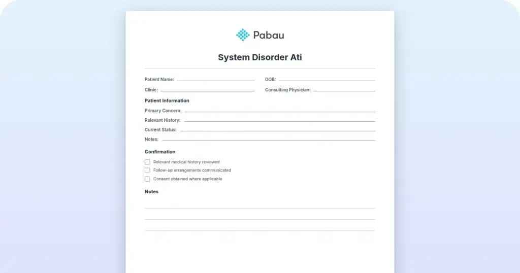
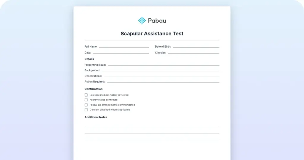
Nursing narrative note template
A nursing narrative note template is a structured PDF that guides you through re...

2
Last Updated: Oct 21, 2026

The hypoglossal nerve test (CN XII) is a purely motor cranial nerve examination assessing tongue movement, strength, and appearance for signs of neurological lesion.
In a lower motor neuron lesion, the tongue deviates toward the side of the lesion. In an upper motor neuron lesion, deviation is away from the lesion side.
Fasciculations and tongue atrophy indicate lower motor neuron pathology and are absent in pure upper motor neuron lesions.
Pabau’s digital clinical forms and AI-assisted documentation tools help neurology and speech therapy practices record CN XII findings accurately and efficiently.
Inconsistent examination technique causes most missed cranial nerve findings, more often than a lack of knowledge does. The hypoglossal nerve test takes under two minutes, yet inconsistent documentation and incorrect deviation interpretation persist across clinical settings.
Guidance on cranial nerve testing consistently lists the hypoglossal nerve examination among the assessments used for stroke workups and head and neck surgery follow-up. It also appears in suspected cranial nerve palsy and baseline neurological evaluation, as covered in general references such as StatPearls (NCBI Bookshelf).
This guide covers the anatomy, the step-by-step technique, and how to interpret what you find, including when an abnormal result warrants urgent investigation. It is written for clinicians, medical students, and healthcare practitioners performing neurological assessments.
CN XII, also called the 12th cranial nerve, is a purely motor nerve. Its name comes from the Greek for “under” and “tongue.” Hypoglossal nerve function covers motor innervation to nearly every tongue muscle, with one exception: the palatoglossus, which the vagus nerve (CN X) controls instead. A patient with a weak palatoglossus has a CN X problem, not a CN XII one.
The nerve originates from the hypoglossal nucleus in the medulla oblongata. This column of motor neurons runs the length of the dorsomedial medulla. From there, rootlets emerge anterolaterally between the pyramid and the olive. They converge and exit the skull through the hypoglossal canal, also called the hypoglossal foramen (anterior condylar canal), in the occipital bone.
Once extracranial, CN XII loops around the occipital artery and crosses both the internal and external carotid arteries near the angle of the mandible. A branch of the cervical plexus (C1 and C2 fibers) travels alongside the nerve here without merging into it. It eventually forms part of the ansa cervicalis. The nerve then passes lateral to the hyoglossus and fans out to complete its tongue innervation. The genioglossus is the primary muscle responsible for tongue protrusion, so impairment of CN XII affects speech articulation, swallowing, and chewing.
This hypoglossal nerve assessment follows a logical sequence: observe before you instruct, then layer in active tasks. Rushing to tongue protrusion without inspecting the tongue at rest misses fasciculations, which vanish once the muscle contracts. Knowing how to assess the hypoglossal nerve accurately starts with resisting that urge to skip ahead.
The digital clinical forms you use to document the exam need minimal equipment to perform it. For the bedside test itself:
Ask the patient to open their mouth and rest their tongue on the floor of the mouth. Do not ask for protrusion yet. Observe for 10-15 seconds. You are looking for:
Ask the patient to “stick out your tongue as far as you can and hold it still.” Normal finding is tongue protrusion in the midline. Any deviation is abnormal. Note the direction of deviation relative to the midline: this is the key interpretive step.
Ask the patient to move their tongue from side to side and then push it against the inside of each cheek. This assesses the full range of tongue lateralization and screens for subtle weakness that may not produce obvious deviation on straight protrusion alone.
Place a gloved finger or tongue depressor against the patient’s cheek (external surface). Ask them to push their tongue against that cheek from the inside. Compare side to side. Asymmetric weakness confirms unilateral hypoglossal nerve involvement and helps localize mild lesions missed on protrusion testing alone.
For subtle deficits missed on protrusion, ask the patient to repeat rapid syllables such as “la-la-la,” or words with hard “t” and “d” sounds. Slowed or slurred repetition points to the same side as the weakness.
The key interpretive decision is whether an abnormal finding reflects a lower motor neuron or upper motor neuron lesion affecting cranial nerve tongue movement. The pattern of deviation, atrophy, and fasciculations determines this. Tongue deviation direction is the most commonly misremembered rule in the whole cranial nerve battery.
Clinical rule for deviation: in an LMN lesion, the weak side cannot push the tongue across the midline. The tongue falls toward the weak (ipsilateral) side. In a UMN lesion, the lesion is in the brain or brainstem on the contralateral side to where the tongue deviates. The tongue deviates away from the brain lesion.
The discipline behind interpreting biomarkers without overpromising holds here too. Document exactly what you observe: deviation direction, presence of fasciculations, degree of atrophy. Then qualify your interpretation with the clinical context rather than jumping to a single diagnosis.
Before asking for tongue protrusion, always inspect at rest for 10-15 seconds with a penlight. Fasciculations are the most diagnostically significant finding in the hypoglossal nerve test and disappear once the patient contracts the muscle.
Hypoglossal nerve palsy can arise from central (intracranial) or peripheral (extracranial) pathology. Identifying the level of the lesion guides the investigation pathway. Central lesions typically produce UMN signs; peripheral lesions produce LMN signs.
Stroke survivors with this presentation often need a CVA nursing care plan that tracks swallowing and speech deficits alongside the neurological findings.
Clinicians working with patients who have dysphagia or dysarthria as sequelae of CN XII palsy frequently collaborate with speech-language pathology teams. Speech therapy practice management platforms that integrate clinical documentation across disciplines help coordinate this care pathway efficiently.
Reference sources rarely lay out a clear red-flag pathway for when an abnormal CN XII finding should trigger urgent investigation.
Use the following as a clinical decision framework, adapted from StatPearls neuroanatomy guidance (NCBI) and neurology clinical practice frameworks. The same principle used in crisis intervention strategies applies here: red flags drive urgency, context drives the modality.
Key imaging principle: MRI is preferred over CT for skull base and posterior fossa pathology. The hypoglossal canal and hypoglossal nucleus are poorly visualized on CT. CT remains the first-line choice when acute hemorrhage or stroke in the acute setting is the primary concern.
Cranial nerve XII is rarely examined in isolation. In clinical practice, CN XII is assessed as part of the lower cranial nerve battery. This battery also covers CN IX (glossopharyngeal), CN X (vagus), and CN XI (accessory). This combined cranial nerve test works because the four nerves share a brainstem origin, exit through adjacent foramina, and collectively govern bulbar function.
The standard sequencing in a full cranial nerve exam places CN XII last in the lower battery. It follows the palate elevation test (CN IX/X) and the shoulder shrug/head turn test (CN XI). This ordering matters.
Testing the gag reflex and palate before tongue protrusion puts any bulbar deficit in context. It also stops you from concluding that tongue weakness is isolated when it’s part of a broader bulbar syndrome.
Vagus nerve dysfunction and glossopharyngeal nerve function share the same gag-reflex pathway. Testing the glossopharyngeal nerve alongside CN X is faster than assessing each nerve on its own. The same logic applies to CN XI: a quick shoulder-shrug and head-turn check rules out an accessory nerve palsy before moving to the hypoglossal exam.
Clinicians using physical therapy clinical documentation platforms will recognize this structured battery approach: systematic, reproducible, with each step building on the last. The same principle applies to neurological examination: no finding stands alone.
For clinicians assessing bulbar function, also note that uvula deviation and tongue deviation can occur in opposite directions when lesion levels differ. A right CN X lesion pulls the uvula left, while a right CN XII LMN lesion pushes the tongue right. Distinguish these carefully before documenting combined findings.
This demand for precision in lateralization runs through the broader head and neck assessment that CN XII testing sits within.
Clear documentation of the hypoglossal nerve test is a regulatory and medico-legal requirement, yet most EHR systems provide no structured template for cranial nerve findings. Clinicians default to free-text entries that vary in completeness, making audit and follow-up comparison unreliable.
A standardized CN XII documentation entry should include:
Resources like SOAP note documentation frameworks offer adaptable structures for this kind of structured neurological clerking.
A neuro exam cheat sheet condenses the full cranial nerve battery onto one reference page for bedside use, while a nursing review of systems template extends the same structured approach across the rest of the physical exam. The full battery opens with CN I, and the olfactory nerve test is one of its most commonly skipped steps.
For practices managing multiple neurological assessments, a structured psychiatric evaluation template illustrates how to build repeatable structured assessment records across specialties.
The same field-by-field discipline transfers to very different assessments. It applies whether you are recording a bone marrow test, timing a Wingate test, or scoring a dark core personality test.
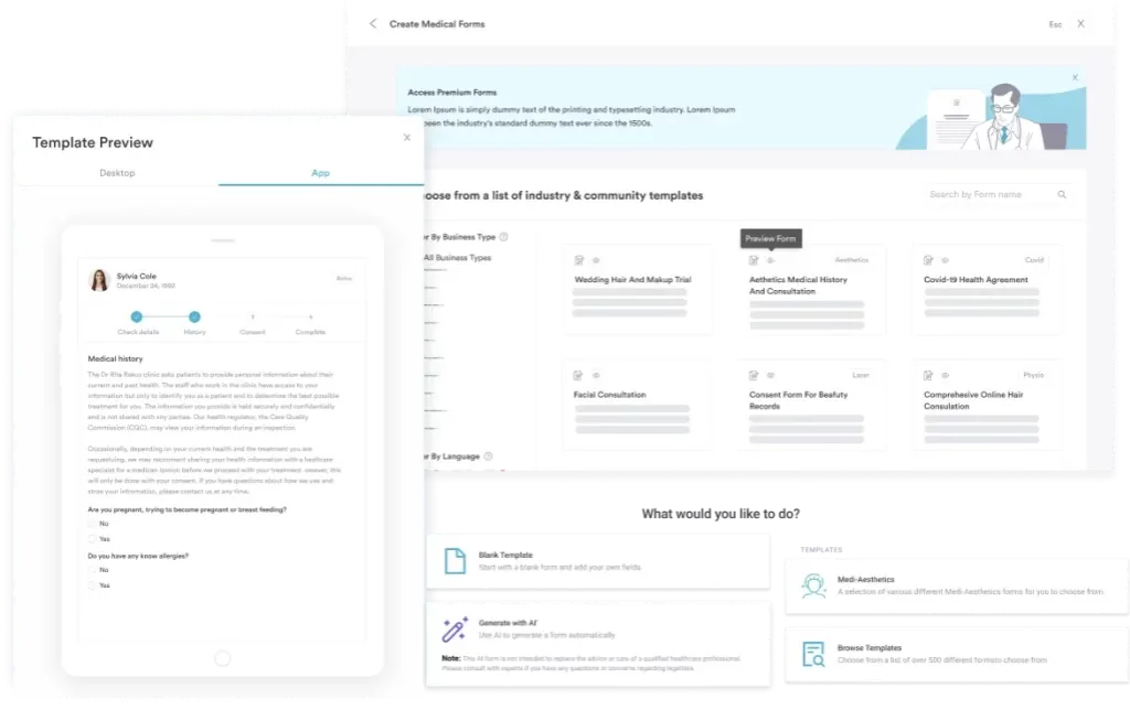
Pabau turns the six-field structure above into a reusable patient record template instead of a blank text box. Neurology, speech therapy, and private GP practices configure the fields once, so every clinician on the team documents CN XII findings the same way.
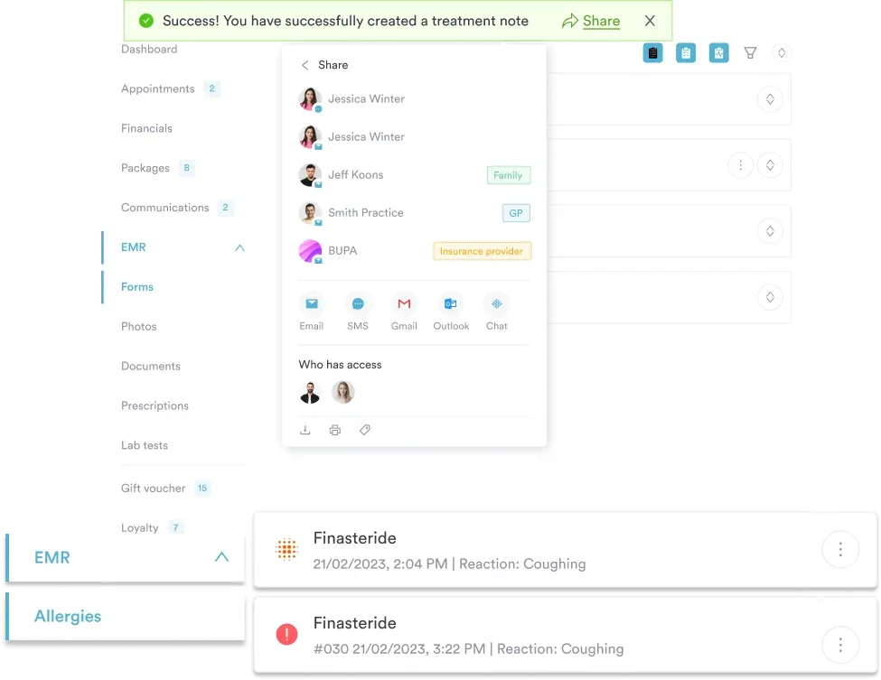
Pabau’s AI-assisted clinical documentation drafts the note directly from the encounter. The practitioner then only needs to confirm deviation direction, strength, and any red flags before signing off. Structured entries like this are what make an audit trail defensible and a follow-up comparison meaningful months later.

Pabau helps neurology, speech therapy, and private GP practices capture structured clinical notes, CN XII findings, and referral decisions in one place, with built-in digital forms and AI-assisted note generation.

Getting the hypoglossal nerve test right comes down to sequence and interpretation, not equipment. Inspect at rest before asking for any movement, then read the direction of deviation against the correct reference point. Treat an isolated abnormal finding as a reason to look further, rather than wait it out.
That discipline is what turns a two-minute bedside test into a reliable part of the neurological exam, instead of a step that gets rushed.
Structured documentation is what makes that discipline repeatable across a whole team, not just reliable in your own hands. Book a demo to see how Pabau’s digital forms and AI-assisted note-taking help neurology, speech therapy, and primary care teams document CN XII findings consistently.

Assessing balance in a younger patient? Pediatric balance scale gives a 14-item scoring template for tracking a child’s balance and motor function.
Need an intake form for a fitness client? Personal trainer assessment template covers client history, movement, and body composition across seven sections.
Screening a client before a training program? Personal training questionnaire covers medical history, PAR-Q screening, and fitness goals before a program starts.
The hypoglossal nerve test is a bedside cranial nerve examination of CN XII that assesses tongue motor function through four sequential steps. These are resting inspection for fasciculations and atrophy, midline protrusion, lateral movement, and strength testing against resistance. It takes under two minutes and detects upper or lower motor neuron lesions affecting tongue movement.
Inspect the tongue at rest on the floor of the mouth for fasciculations and atrophy. Then ask the patient to protrude their tongue in the midline and observe for deviation. Ask for lateral tongue movements and finally test tongue strength by asking the patient to push against resistance applied to the cheek. Record the direction of any deviation, which side is weak, and whether fasciculations or atrophy are present.
The hypoglossal nerve is purely motor. It carries somatic motor fibers to all intrinsic and extrinsic muscles of the tongue, except the palatoglossus. The vagus nerve (CN X) innervates that muscle instead. CN XII carries no sensory fibers for taste or tongue sensation. Those are carried by the chorda tympani (branch of CN VII) and the lingual nerve (branch of CN V3).
Tongue deviation indicates weakness on one side of the tongue. In a lower motor neuron lesion, the tongue deviates toward the side of the lesion. The weak genioglossus cannot push the tongue away from that side. In an upper motor neuron lesion, deviation is away from the side of the brain lesion. UMN fibers cross before reaching the hypoglossal nucleus.
Lower motor neuron lesions produce ipsilateral tongue deviation, fasciculations, and atrophy with reduced tone. Upper motor neuron lesions produce contralateral tongue deviation (relative to the brain lesion), no fasciculations, no atrophy, and increased tone (spasticity). Bilateral UMN lesions cause a small spastic tongue without wasting, as seen in pseudobulbar palsy.
Common causes include stroke (a UMN pattern from corticobulbar tract involvement) and ALS or motor neuron disease (bilateral LMN involvement with fasciculations). Other causes are skull base tumors compressing the hypoglossal canal, neck dissection, carotid artery dissection, penetrating neck trauma, and demyelinating disease. Isolated unilateral hypoglossal nerve palsy without a clear cause should prompt MRI of the brain and skull base.
A penlight and good ambient lighting are the only essential requirements. A tongue depressor improves visibility for resting inspection. Non-sterile gloves are needed if direct resistance testing involves contact with the patient’s tongue or cheek. No specialist equipment is required, making CN XII assessment suitable for any clinical setting including primary care and bedside ward assessments.
A normal hypoglossal nerve assessment shows the tongue at rest without fasciculations or atrophy. On protrusion, the tongue stays in the midline, moves fully side to side, and pushes with equal strength against resistance on both sides. Any deviation, wasting, or asymmetric strength is abnormal and needs further interpretation.