



GLP-1 diet plan
A GLP-1 diet plan is a structured eating framework for patients taking semagluti...

2
Last Updated: Oct 13, 2026

A neurological eye exam checks cranial nerves II, III, IV, and VI, plus pupils, visual fields, eye movements, and the optic disc.
Afferent pupillary defects, papilledema, and homonymous hemianopia each localize damage to a specific part of the visual pathway.
Third nerve palsy with a dilated, unreactive pupil needs same-day emergency referral to rule out a posterior communicating artery aneurysm.
Practice management software like Pabau captures structured neuro-ophthalmic findings and turns them into referral letters the same day.
Most routine eye exams focus on refraction and anterior segment health, but a neurological eye exam goes further.
It tests the cranial nerves, checks the visual pathway, and looks for signs of disease the patient doesn’t yet know about. For example, stroke, multiple sclerosis, brain tumors, and raised intracranial pressure all leave early traces behind the eye.
This guide is written for optometrists, ophthalmologists, and allied eye care professionals. It covers each part of the exam, maps findings to likely causes, and flags what needs same-day action. Finally, the closing sections cover the documentation and referral workflow that carries those findings out of the exam room.
A neurological eye exam is a structured assessment of the nervous system, carried out through the eyes. Unlike a standard refraction, it tests cranial nerve function, pupil reflexes, eye movement, visual fields, and the optic nerve head.
The exam does not diagnose neurological disease on its own. Instead, it surfaces the findings that justify imaging, bloods, or an urgent referral.
According to the American Academy of Neurology, the eye offers direct, non-invasive access to the central nervous system. The optic nerve is an extension of the brain, and retinal blood vessels form from the same tissue as the brain’s own vessels. In addition, several cranial nerves meet in and around the eye socket.
Eye care clinicians are often the first contact for a patient with unrecognized neurological disease. That’s why working through a cranial nerve examination checklist at intake puts the symptom timeline and baseline findings in the record before the exam begins.
Almost every major intracranial structure has a matching sign in the eye. The optic nerve connects directly to the brain, and the nerves that move the eye start in the brainstem. In fact, vision itself traces a longer path, running from retina to occipital cortex and passing near most critical brain regions on the way.
Sequence is what makes the exam reliable. Similarly, hand nerve tests in peripheral neurology follow a fixed order so no deficit goes unrecorded, and the neuro-ophthalmic components reward that same discipline.
The conditions this approach catches range from the life-threatening, such as aneurysm and stroke, to the chronic and progressive, such as multiple sclerosis. Many carry treatment windows measured in hours. That is why every eye care practitioner needs to recognize them on sight.
The workup follows the anatomy. History comes first, then the afferent pathway of acuity, pupils, fields, and disc. Finally, the efferent pathway of eye movement comes last. Working in that order lets you pinpoint an abnormality to the most specific site the findings allow.
A careful history is the most powerful tool you have for pinpointing the problem. First, ask about onset, progression, and whether symptoms are monocular or binocular. Monocular visual loss suggests disease in front of the optic chiasm, while a binocular field defect points behind it.
Best-corrected visual acuity (BCVA) establishes baseline afferent function. A drop unexplained by refractive error or anterior segment disease flags optic nerve or macular disease. Next, test each eye separately before you go on. A comprehensive exam billed under 92004 needs the history and acuity findings documented in full.
The cranial nerve eye exam targets four nerves tied to vision and eye movement. A fifth, CN VII, is checked when facial asymmetry or a lid abnormality shows up. Also, any diplopia you find belongs in the record, coded as H53.2.
The pupillary reflex test is among the highest-yield parts of the exam. Check pupil size, shape, and symmetry in ambient light. Then test the direct and consensual light reflexes with a bright penlight. A PERRLA eye exam form keeps those findings comparable between visits.
The swinging flashlight test detects a relative afferent pupillary defect (APD). Swing the light between the eyes at roughly two-second intervals. A positive APD is present when the illuminated eye dilates instead of constricting. As a result, that signals reduced input from that eye’s optic nerve compared with its fellow.
The American Academy of Ophthalmology lists optic neuritis and multiple sclerosis among the causes. Severe glaucoma, retinal detachment, and other unilateral optic nerve disease can also produce one.
Ask the patient to follow a target through the nine cardinal positions of gaze. Then watch for smoothness, symmetry, diplopia, and nystagmus in each direction.
Gaze-evoked nystagmus on lateral gaze suggests a cerebellar or brainstem lesion. Similarly, internuclear ophthalmoplegia, marked by slow adduction with contralateral nystagmus, is a classic sign of demyelinating disease.
Field testing pinpoints disease along the whole visual pathway. Confrontation at the chair is fast and sensitive for dense defects. By contrast, automated perimetry gives you precise numbers for documentation and monitoring. The pattern of loss is the clue that names the site, as the table below shows.
Fundoscopy assesses the optic nerve head directly. Specifically, look for disc swelling, disc pallor, cup-to-disc asymmetry, and spontaneous venous pulsation.
Optical coherence tomography (OCT) puts a number on what you see by measuring retinal nerve fiber layer thickness. Thinning points to earlier optic nerve injury, while thickening can reflect acute inflammation or raised intracranial pressure.
Bilateral disc swelling in a patient with headache and no other diagnosis is papilledema until proven otherwise. As a result, it calls for same-day neuroimaging and neurological assessment.
The table below maps the conditions most often identified during a neuro-ophthalmic workup to their key eye signs and the urgency each one carries. Instead, treat it as a working clinical reference rather than an exhaustive diagnostic list.
Visual symptoms that arrive alongside a psychiatric presentation deserve the same workup as any other. So, exclude neurological causes before anyone records the change as functional, especially for patients already under the care of mental health practices.
Some findings cannot wait for a routine appointment or an outpatient letter. Recognizing them, and having a referral pathway ready, is part of every eye care clinician’s duty of care. That’s why keeping a Cincinnati stroke scale to hand helps you describe the deficit clearly when you call for help.
Send the findings in writing rather than summarizing them over the phone. Stroke teams score deficits with the NIHSS score sheet, and a documented field defect or gaze palsy feeds straight into that score.
When a patient presents with new diplopia and headache, test the pupils before anything else. A dilated, non-reactive pupil in a CN III palsy changes the clinical management entirely. Document pupil size in millimeters and reactivity grade before the patient leaves the room.
Not every abnormal finding needs an emergency call. Many need a timely, structured outpatient assessment instead. Whether that referral goes out this week or slips into a backlog depends on your patient care management workflow.
Copy the letter to the patient’s physician too. Primary care teams also coordinate the imaging, bloods, and follow-up that sit outside eye care.
Refer routinely, within one to four weeks, for:
Refer urgently, within 24 to 48 hours, for:
Technology extends the reach of the clinical exam. In fact, three tools have moved from specialist-only to mainstream practice over the last decade.
AI-assisted retinal analysis is an emerging tool. Validated algorithms already screen fundus photographs for glaucoma and for the changes a diabetes eye exam looks for. Their neuro-ophthalmic role is still under investigation, so treat an AI-flagged finding as a prompt for clinical confirmation.
Documentation decides how much of your exam survives the trip to the specialist. So a thorough workup produces findings that need recording precisely, sending onward, and tracking over time.
Vague records slow specialist review and complicate coding for conditions such as optic neuritis, which bills as H46.8. They also create medico-legal exposure when a time-sensitive finding is never acted on.
Writing safer clinical notes for this exam means the same structured fields every time. Record acuity with correction, pupil sizes in millimeters, APD status, motility findings, and perimetry results. Finally, add the disc assessment with a photograph reference number where you have one.
A referral letter needs the exam date, the specific findings, your differential, the urgency level, and your contact details. Normal findings belong in it too, whenever they narrow that differential. Also, a diagnosis letter template keeps those fields in a fixed order.
In most practices this workflow runs by hand. First, the clinician writes findings into the notes, then dictates a letter later that day, while someone else tracks outstanding referrals on a spreadsheet. As a result, urgent findings depend on whoever remembers them.
Practice management software like Pabau closes that loop. First, patients complete a structured symptom checklist through digital intake forms before the consultation.
You walk in with a history to confirm and expand rather than build from scratch. As a result, that reduces history-taking time and keeps red-flag questions from being skipped.
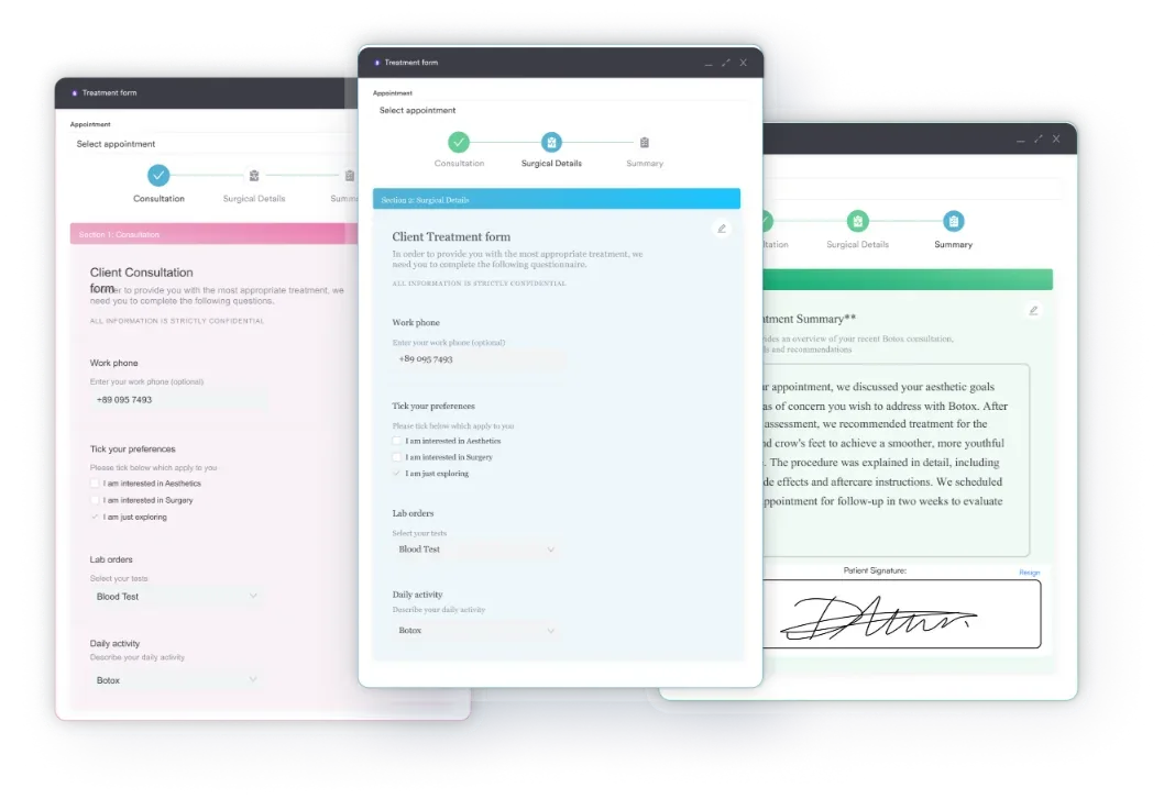
Automated workflows then build the referral letter from the clinical data you captured during the exam. The letter leaves the same day, and the outstanding referral stays visible until someone confirms the patient was seen.
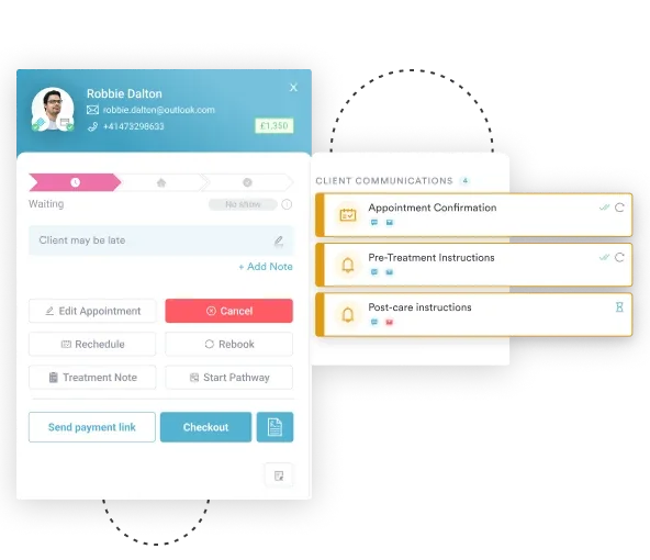
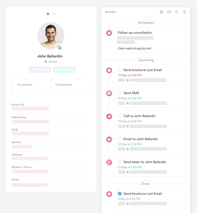
Follow-up is the part that quietly fails. For example, stable patients need interval perimetry and disc photography, while referred patients need a check that they were actually seen. Pabau’s patient records support follow-up flags tied to a specific finding, so complex cases are not lost in a busy optometry list.
Dictation is the other time sink. Pabau Scribe, our AI scribe, drafts clinical documentation from the consultation itself, so the findings are written while they are still fresh.

Pabau’s automated workflows and digital records help eye care practices build referral letters and track every outstanding referral. See how it fits your practice.

The hard part of neuro-ophthalmology is rarely the examination itself. Instead, it’s holding the same sequence on a busy day, when the patient in front of you booked in for new glasses.
Commit four findings to memory, because they change your next move. Those are a positive APD, bilateral papilledema, a dilated pupil with a third nerve palsy, and a fresh homonymous field defect. Know your emergency number and your neuro-ophthalmology route before the day you need them.
The workflow around the exam deserves the same discipline as the exam. So, book a demo to see how Pabau records neuro-ophthalmic findings and gets urgent referrals out the same day.

Need the full routine exam sequence? Eye physical examination walks through the eight components patients need and how often.
Assessing eye alignment and tracking? Eye movement test template is a free download for documenting saccadic and smooth pursuit findings.
Standardizing depth perception documentation? Stereopsis test template is a free download linking findings to strabismus or amblyopia diagnoses.
Screening a diabetic patient’s vision? Diabetes eye exam template is a free download covering retinopathy grading and referral fields.
A neurological eye exam is a structured clinical assessment of the visual pathway and the cranial nerves that serve it. It evaluates visual acuity, pupils, extraocular movements, visual fields, and the optic disc. However, it does not diagnose neurological conditions on its own. Instead, it identifies findings that require further investigation or urgent referral.
The examiner takes a structured history and measures best-corrected visual acuity. Next, they test the pupillary light reflex, including the swinging flashlight test for an APD. Extraocular movements are assessed through nine gaze positions, followed by confrontational or automated perimetry. The optic disc is then examined by fundoscopy or OCT.
Multiple sclerosis, ischemic stroke, brain tumors, posterior communicating artery aneurysm, optic neuritis, idiopathic intracranial hypertension, and pituitary adenoma all produce characteristic ocular signs. Each of those signs can show up during a neuro-ophthalmic workup. The exam does not diagnose the condition in isolation. Instead, it identifies signs that prompt neuroimaging and specialist referral.
An afferent pupillary defect (APD) is a difference in the pupillary light reflex between the two eyes, detected by the swinging flashlight test. The affected eye’s pupil dilates when the light swings to it, which signals reduced input from that optic nerve. It’s commonly associated with optic neuritis, often the first sign of multiple sclerosis. For example, severe glaucoma and other unilateral optic nerve disease can also cause it.
A standard eye exam mainly assesses refractive error and ocular health, including the anterior segment, intraocular pressure, and a basic fundus review. A neurological eye exam adds cranial nerve function, the pupillary reflex pathway, and motility testing for gaze palsy or nystagmus. It also maps the full visual field for localization and inspects the disc for raised intracranial pressure. Its purpose is neurological rather than optical.
Refer same-day emergencies immediately through emergency services, including third nerve palsy with a dilated pupil, bilateral papilledema, and acute stroke signs. By contrast, refer urgently within 24 to 48 hours for new unexplained bilateral disc swelling or symptomatic visual field loss. Finally, routine referral within one to four weeks suits stable cranial nerve palsies with normal imaging, unexplained disc pallor, and RNFL monitoring after optic neuritis.






