GLP-1 diet plan
A GLP-1 diet plan is a structured eating framework for patients taking semagluti...

2
Last Updated: Oct 13, 2026

The accommodation eye test evaluates the eye’s ability to focus on near objects by observing three reflex components: pupil constriction (miosis), eye convergence, and lens thickening via ciliary muscle contraction.
The near-reflex triad forms the acronym PERRLA (Pupils Equal, Round, Reactive to Light and Accommodation), a standard clinical documentation convention used in patient records.
Testing involves moving a target (typically a clinician’s finger) toward the patient’s nose while observing all three components and measuring the near point of accommodation in centimeters or diopters.
Abnormal accommodation findings can indicate presbyopia (age-related accommodation loss), accommodative insufficiency, convergence insufficiency, or neurological conditions including relative afferent pupillary defects (RAPD).
Practice management software like Pabau uses structured clinical note templates and digital forms to standardize accommodation test documentation across practices, reducing errors and keeping results recorded consistently.
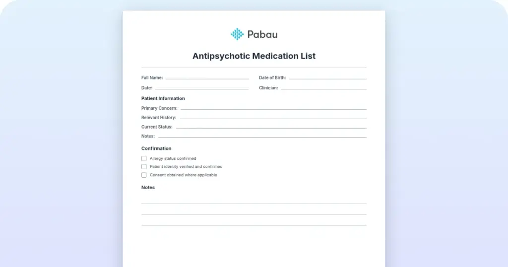
A ready-to-use clinical template for systematically documenting accommodation reflex findings, including pupil constriction observation, convergence assessment, lens change evaluation, near-point distance measurement, and PERRLA notation fields.
Download templateThe accommodation eye test is a clinical examination in optometry and eye care that assesses how well the eyes adjust focus from distant to near objects. For optometrists, nurses, and allied health professionals, performing this test accurately and documenting findings clearly helps identify vision disorders, detect age-related changes, and rule out neurological abnormalities.
This guide covers the test procedure, what normal and abnormal findings mean, and includes a downloadable template to standardize documentation in your practice.
Accommodation testing is part of every comprehensive eye examination. It sits alongside other reflex assessments, like the pupillary light reflex.
Whether you work in private optometry, occupational health.
The accommodation eye test measures the eye’s ability to change focus and adjust lens shape in response to a near target. The test works because accommodation is a reflex: the eye automatically responds when the brain signals a need to focus on something close.
This reflex involves three coordinated changes: the pupils constrict (miosis), the eyes turn inward (convergence), and the lens thickens (via ciliary muscle contraction). Together, these three components form the accommodation reflex triad, also known as the near reflex.
Clinically, the accommodation test is performed during routine eye examinations to detect whether the eyes respond normally to a near stimulus. When documented correctly, accommodation findings appear in patient records under the term PERRLA: Pupils Equal, Round, Reactive to Light and Accommodation.
The “A” in PERRLA refers to this accommodation response, making it a shorthand way to confirm all three components were assessed.
The accommodation reflex is a coordinated three-part response. Understanding each component is essential for accurate observation and clinical reasoning.
All three components occur simultaneously when accommodation is functioning normally. The pupils constrict, the eyes converge, and the lens changes shape, all in response to a single near target. If any component is absent or asymmetrical, an accommodation disorder or neurological problem may be present.
Performing the accommodation eye test is straightforward, but careful observation is critical. Follow this clinical procedure in your routine eye examination workflow:
The entire procedure takes 30-60 seconds per eye. The key is to move slowly, observe carefully, and record the near-point distance accurately. Many clinicians use a digital forms system to standardize these measurements and ensure consistent documentation.

With a tool that tracks clinical measurements, baseline and follow-up scores sit side by side in the patient record.
Clinical documentation of accommodation findings follows the same documentation standards used across other areas of practice, helping keep findings clear and consistent across practitioner teams.
Normal accommodation values vary significantly by age. The lens loses elasticity as we age, so accommodation amplitude decreases predictably throughout life.
Abnormal findings include: asymmetrical pupil constriction (one pupil constricts less than the other), impaired or absent convergence, reduced accommodation amplitude for age, or sluggish reflex response. These can signal presbyopia, accommodative insufficiency, convergence insufficiency, ocular nerve palsy, or systemic neurological conditions. Any abnormal finding warrants further investigation and possible referral to a specialist.
Book a demo to see how Pabau streamlines accommodation test documentation and clinical note standardization across your eye care team:
Abnormal accommodation findings often indicate specific clinical conditions that require monitoring, management, or referral.
Convergence and accommodative insufficiency are also routinely screened for in sports medicine practices, where near-point-of-convergence testing forms part of standard post-concussion vision assessments.
Many eye care practices document accommodation findings in handwritten notes or generic EHR fields, making it easy for details to be missed or inconsistently recorded across team members.
Pabau’s structured clinical records and customizable digital forms let you build a standardized accommodation test template with dedicated fields for pupil symmetry observation, convergence assessment, near-point distance (cm and diopters), PERRLA notation, and any abnormal findings.
This approach cuts down on missed details, ensures all practitioners record the same data points, and makes accommodation history easier to track across patient visits. A practice management platform is judged on the work it removes, not the modules it lists.
Use consistent lighting and a quiet environment during testing. Bright ambient light can artificially constrict pupils and mask pupil-response abnormalities. A dimmed, quiet exam room lets patients focus on your target without distraction.
Ensure your target (finger or near-point card) is clean, well-lit, and positioned at the exact midline. When measuring near point, move your target smoothly at a predictable speed. Sudden or jerky movements can startle the patient and disrupt focus.
Always perform the test bilaterally and compare sides. Asymmetrical findings are clinically significant and should never be dismissed as normal variation.
The downloadable accommodation eye test template above provides a structured workflow for documenting the test systematically. It includes step-by-step testing instructions, fields for recording pupil symmetry, convergence quality, near-point distance in centimeters and diopters, PERRLA notation, and a quick-reference chart of normal values by age.
Print the template and place it in your examination room, or scan it into your clinical note system as a checklist. Pair it with other eye care documentation tools.
Train all team members to use the same form so accommodation findings are recorded consistently across all practitioners and all patient visits. Over time, this standardization improves diagnostic accuracy and helps you identify trends in accommodation changes for individual patients.
The accommodation eye test is a quick, non-invasive clinical examination that provides valuable insight into lens elasticity, neurological function, and binocular vision alignment. Mastering the technique (smooth target movement, careful observation of all three reflex components, and accurate near-point measurement) helps you catch accommodation disorders early and identify potential neurological red flags.
The downloadable template above standardizes this examination in your practice, making sure every team member documents findings consistently. For optometrists, nurses, and eye care practitioners, this systematic approach transforms accommodation testing from a routine reflex check into a diagnostic tool that directly improves patient outcomes.

Need a visual to explain the anatomy? Labeled diagram of the eye gives a fully labeled PDF of eye structures for patient education and quick reference.
Need a structured way to log eye findings? PERRLA eye exam form replaces scattered handwritten notes with a structured template that flags abnormal findings faster.
Want the full exam this test fits into? Eye physical examination guide covers the eight components of a full eye exam and how often patients need one.
An accommodation eye test assesses the eye’s ability to focus on near objects by observing three coordinated reflex components: pupil constriction, eye convergence, and lens thickening. The test is performed by moving a target toward the patient’s nose and measuring the near point of accommodation in centimeters or diopters.
Position the patient at 30-40 cm distance with adequate lighting. Hold a target (your finger or a near-point card) at arm’s length in the midline. Ask the patient to focus on the target and slowly move it toward their nose. Observe both pupils constricting symmetrically, both eyes turning inward, and measure the distance (in cm) when the patient reports blur.
PERRLA = Pupils Equal, Round, Reactive to Light and Accommodation. The “A” confirms that the accommodation reflex (all three components: miosis, convergence, lens thickening) was tested and found normal. PERRLA is standard documentation shorthand used in patient records to confirm a normal eye examination.
Accommodation amplitude decreases with age. A 20-year-old typically has a near point of 8-10 cm (10-12.5 diopters), while a 50-year-old has a near point of 25-33 cm (3-4 diopters), and by age 60, functional accommodation is nearly absent. These values guide interpretation of accommodation findings.
Abnormal accommodation findings can indicate presbyopia (age-related lens stiffening), accommodative insufficiency (weak ciliary muscle), convergence insufficiency (impaired eye alignment), relative afferent pupillary defect (RAPD – optic nerve damage), or cranial nerve palsies including third nerve involvement.
Record “accommodates” if findings are normal, or specify near-point distance in centimeters (e.g., “near point OD 8 cm, OS 8 cm”). For abnormal findings, note which component is impaired (e.g., “poor convergence” or “reduced accommodation amplitude”). Include in PERRLA notation: “PERRLA – accommodates, near point 10 cm bilaterally.”