Head to toe assessment cheat sheet: Printable nursing guide
A head to toe assessment cheat sheet gives you one fixed order at the bedside, s...

2
Last Updated: Oct 23, 2026

A 12-lead ECG uses just 10 electrodes to produce 12 views of the heart’s electrical activity.
Placement accuracy decides the result, and misplaced precordial leads are the most common source of recording errors.
Normal values include a heart rate of 60-100 bpm, a PR interval of 120-200 ms, and a QRS duration under 120 ms.
Localizing a finding means reading the leads in groups, because the septal, anterior, lateral, and inferior walls each have their own set.
Practice management software like Pabau keeps ECG orders, interpretations, and follow-up actions on one patient record.
A patient with chest pain walks into your practice. Within minutes your team has to work out whether this is a musculoskeletal complaint or a STEMI. The ECG 12-lead test answers that question, and it does so in under ten minutes. According to the American Heart Association, it is still the first-line cardiac diagnostic test in use worldwide.
An ECG 12-lead test records the heart’s electrical activity from 12 angles at once. It uses 10 electrodes placed on the skin, four on the limbs and six on the chest, to produce those 12 views. Each view corresponds to a region of the heart muscle. Together they give a spatial map of conduction that no other resting test matches.
This guide covers electrode placement, including female and posterior leads. It also covers systematic interpretation, normal reference values, and how private practices build ECG workflows that cut administrative work.
ECG stands for electrocardiogram, the English abbreviation. EKG comes from the German Elektrokardiogramm, adopted early in cardiology’s history when German-speaking clinicians led the field. Both terms refer to identical technology and an identical procedure. In the UK and most of Europe, ECG is standard. In the US, you’ll see EKG used interchangeably, particularly in emergency medicine. This guide uses ECG throughout, and every point in it applies to an EKG.
A 12-lead ECG is performed to check the heart’s rate, rhythm, and conduction whenever symptoms or a planned procedure put cardiac risk in question. According to MedlinePlus, the most common indications include:
Where the trace points to ventricular dysfunction, the workup often moves beyond the heart. Heart failure drives fluid into the pleural space, and pleural fluid analysis separates a transudate from an exudate.
The test is painless and non-invasive. There is no radiation exposure, and the procedure carries no direct risk to the patient. Say that to the patient before you start. An anxious patient tenses up, and tense muscles spoil the trace.
Placement accuracy is where most recording errors originate. A misplaced V1 or V2 electrode can produce a false right bundle branch block pattern. Swapped limb leads produce mirror-image artifacts. Prepare the skin first, so that every site is clean, dry, and shaved where necessary. Cold or wet skin increases impedance and degrades signal quality.
Four electrodes produce the six limb leads. Place them on the fleshy parts of the limbs, avoiding bony prominences. Proximal placement (upper arms, thighs) is an accepted clinical variant when distal sites are unavailable, but it can affect waveform amplitude.
The six precordial electrodes are placed across the chest wall using intercostal spaces and sternal landmarks as anchors. V4 is the keystone: place it at the 5th intercostal space, midclavicular line, then work outward. All other chest placements reference off V4’s position.
Breast tissue displaces the electrode from the chest wall. That increases skin-to-electrode distance and attenuates signal amplitude. The clinical consensus is to place V3 through V6 beneath the breast rather than on top of it. That keeps skin contact at the correct anatomical level. Electrodes placed on top of the breast sit above the intended intercostal space and alter precordial waveform morphology.
Explain the placement approach to the patient before you begin. Use a same-gender clinician or a chaperone where patient preference or practice policy requires it. Document the approach in the patient record, so future recordings stay consistent and comparable. Practice management software like Pabau holds that note with the consent form, so the next clinician repeats the same placement.
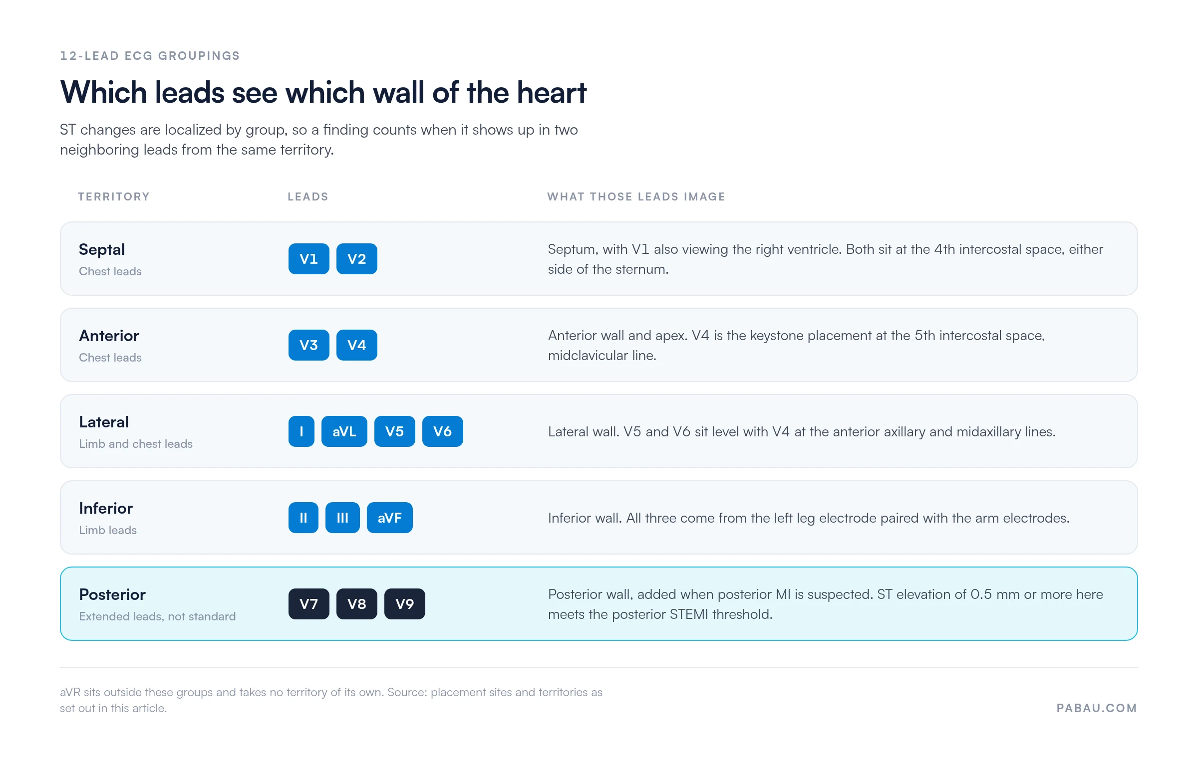
Posterior leads sit outside the standard 12-lead protocol, but they earn their place when posterior myocardial infarction is suspected. In a posterior MI the standard leads may show only ST depression in V1-V3, which reads like subendocardial ischemia. Adding V7, V8, and V9 images the posterior wall directly.
ST elevation of 0.5 mm or more in V7-V9 meets the threshold for posterior STEMI, according to cardiology literature. These are extended leads, and ordering them takes clinical judgment. Read them alongside troponin values and the clinical presentation, never in isolation.
Before applying any electrode, ask the patient to breathe normally and stay still. Instruct them not to speak during recording. Muscle artifact from shivering, talking, or Parkinsonian tremor is the most common cause of a non-diagnostic trace. A 10-second recording window requires genuine stillness.
Interpretation without a systematic framework invites missed findings. Work through the same sequence every time, whatever you notice first. The six-step system below follows the approach used in ACC/AHA clinical education, set out in the AHA/ACCF/HRS ECG standardization series.
Step six only works if the groupings are second nature. Leads II, III, and aVF look at the inferior wall, and leads I, aVL, V5, and V6 cover the lateral wall. V3 and V4 view the anterior wall and apex, while V1 and V2 view the septum. A finding in a single lead does not name a territory, which is why ST changes are judged in contiguous pairs.
Good notes capture all six steps in a structured format. Pabau’s electronic patient records let clinicians attach the interpretation straight to the patient’s file. Entries are timestamped and results can be flagged for physician review. That consistency is what makes serial ECGs safe to compare later.
Each waveform component reflects a phase of the cardiac cycle. The P wave represents atrial depolarization. The QRS complex reflects ventricular depolarization, usually the most prominent deflection on the trace. The T wave represents ventricular repolarization. A fourth component, the U wave, shows up less often. A prominent U wave can point to hypokalemia, which means checking the potassium value against a basic metabolic panel chart.
Normal duration thresholds matter for diagnosis. QRS duration above 120 ms suggests bundle branch block or ventricular conduction delay. A prolonged QT interval carries arrhythmia risk, particularly when it is drug-induced. Recording both values at every visit is what makes a slow drift visible on a patient taking cardioactive medication.
These values reflect consensus ranges from the Heart Rhythm Alliance STARS guidelines and mainstream cardiology references. Individual patient context always applies. A trained clinician’s judgment takes precedence over a single parameter falling outside these ranges.
An abnormal result does not automatically mean an emergency, though a few patterns need an immediate response. Context decides urgency. Give worried patients the honest version. An abnormal ECG needs clinical review, and most findings are not immediately life-threatening.
Document every finding clearly, including the clinical context and any immediate action taken. Structured notes protect both patient and clinician if a finding is reviewed or queried later.
Most patients arrive at the test with little preparation. A brief walkthrough before you begin reduces the movement artifact that anxiety and physical tension cause. The procedure takes 5-10 minutes from setup to result.
Practices running automated clinical workflows can send a preparation message before the appointment. It asks the patient to wear loose clothing and to skip body lotion on the chest and limbs that day. Both make a clean first trace more likely.
Collecting consent and preparation answers before arrival means the appointment itself can stay on the patient.
The ECG 12-lead test is fast. The administrative work around it does not have to be slow. Private practices and GP practices running ECGs hit the same friction. Request forms are printed, interpretations are handwritten and clipped to the trace, and results go out by phone or letter.
Digitizing that workflow changes the economics. Practice management software like Pabau ties test ordering, result documentation, and patient communication together, which removes three manual handoffs. The interpretation is typed straight into the patient record at the point of care.
Follow-up appointments are booked before the patient leaves. Abnormal results are flagged in the system and assigned to a reviewing clinician. The printed trace still needs a home in the record. Stored with Pabau’s organized clinical photo records, a scanned trace sits beside its interpretation instead of in a paper folder. An audit years later can still find both.
Practices running high ECG volumes, such as occupational health or sports medicine services, get more from clinical measurement tracking. It trends key parameters across serial recordings. Take a QTc interval that has crept from 430 ms to 455 ms over two years on the same medication. Paper files rarely surface that.

Pabau helps private practices and GP practices document diagnostic test orders, attach results to the patient file, and automate pre-appointment preparation. No lost paperwork, and no chasing results.

A 12-lead ECG is only as good as the ten minutes of preparation and documentation around it. A trace recorded from a misplaced V2 costs more time than it saved. So does one filed without its interpretation, because the next clinician has to read it cold.
Fix both ends of the process and the middle takes care of itself. Standardize placement, run the same six-step read every time, and store the result where the next clinician will look for it. Book a demo to see how Pabau keeps ECG orders, interpretations, and follow-ups on one patient record.

Need another point-of-care test workflow? Arterial blood gas test walks through sampling, interpretation, and the reference ranges to read a result against.
Chasing the electrolyte behind an abnormal trace? Basic metabolic panel chart collects the reference ranges an ECG finding sends you to check.
Monitoring patients on cardioactive medication? Daily medication chart records what a patient takes and when, so a QTc change can be read against the current regimen.
An ECG 12-lead test records the heart’s electrical activity from 12 distinct angles. It is non-invasive, and it uses 10 electrodes placed on the limbs and chest. It produces a trace of waveforms that clinicians use to assess heart rate, rhythm, conduction, and signs of ischemia or structural change.
EKG and ECG refer to the same test. ECG is the English abbreviation for electrocardiogram. EKG comes from the German Elektrokardiogramm. Both are used interchangeably in clinical settings, with ECG more common in the UK and Europe, and EKG frequently used in the US.
Four limb electrodes go on the wrists and ankles, or on the proximal limbs where distal sites are unavailable. Six precordial electrodes cross the chest. V1 and V2 sit at the 4th intercostal space, either side of the sternum, and V4 at the 5th intercostal space, midclavicular line. V3 goes between V2 and V4, with V5 at the anterior axillary line and V6 at the midaxillary line, both level with V4.
Not necessarily. Many abnormal ECG findings are incidental and clinically benign in context, such as an isolated right bundle branch block in an asymptomatic individual. Others, like ST elevation in multiple leads or a new left bundle branch block with chest pain, require urgent evaluation. Always have an abnormal result reviewed by a clinician who can interpret it alongside your symptoms and medical history.
The recording itself takes 10 seconds once electrodes are in place. The full clinical encounter, including patient preparation, skin preparation, electrode application, and initial result review, typically takes 5-10 minutes. More complex cases requiring repeat traces or extended monitoring may take longer.
There are three common reasons. The first is to investigate chest pain or shortness of breath for signs of ischemia or arrhythmia. The second is a pre-operative baseline cardiac assessment. The third is monitoring a known cardiac condition, or checking how a cardioactive medication affects rate, rhythm, and conduction.