CRM for chiropractors: how to manage patients, bookings and marketing
A CRM for chiropractors is a patient relationship management system built around...

Last Updated: Oct 13, 2026

An arterial blood gas test measures pH, PaO2, PaCO2, HCO3, and SaO2, plus base excess as a derived sixth value.
Normal pH runs 7.35 to 7.45, and anything outside that band needs urgent clinical assessment.
The radial artery is the first-choice site, and the modified Allen test comes before every radial puncture.
Read the result in five steps, starting with pH and ending with the clinical context.
Practice management software like Pabau keeps the order, the result, and the follow-up in one patient record.
A pulse oximeter tells you how saturated the blood is. An arterial blood gas test tells you why. It draws blood straight from an artery and reports six values at once, so you can see respiratory and metabolic function in the same snapshot.
That difference decides cases. A patient on supplemental oxygen can hold a reassuring SpO2 of 96% while carbon dioxide quietly climbs, and only an arterial sample will catch it. According to NCBI Bookshelf, blood gas analysis remains one of the most useful tests in emergency and critical care. It reads both systems from a single draw.
Getting value out of it depends on two things. You draw the sample cleanly, then read the numbers in a fixed order. Both are learnable, and both go wrong in predictable ways.
Every arterial blood gas report returns the same core dataset. The analyzer measures pH, carbon dioxide, and oxygen tension directly. It then derives the rest, including base excess, which most reports print as a sixth value underneath.
Those ranges come from the American Thoracic Society and NCBI Bookshelf. Treat them as benchmarks rather than absolutes. Altitude and analyzer calibration shift the figures slightly, the same caveat that applies to any abnormal vital signs chart.
Order an arterial blood gas test when you need a measured number, not an estimate, for ventilation, oxygenation, or acid-base balance. This is not a routine screen, and it hurts more than a venous draw. So the indication has to justify the needle.
Two of those account for most gases drawn on a medical ward. A patient in respiratory failure needs serial samples to show whether oxygen therapy is working. A COPD exacerbation needs one early gas before oxygen is titrated, which is also why a COPD care plan opens with a documented baseline.
Meanwhile, MedlinePlus notes the test earns its place whenever pulse oximetry is unreliable. Carbon monoxide poisoning is the classic example, because SpO2 reads falsely normal while the patient deteriorates.
Outpatient settings order fewer gases, but they do order them. Infusion suites and IV therapy practices escalating a deteriorating patient may need one before transfer. The result then belongs in the same record as the infusion notes, not on a loose printout.
Arterial sampling carries more risk than venous phlebotomy, so the sequence matters. Choose the site first, confirm the collateral circulation second, and only then puncture. Most clinicians work at the radial artery, with brachial and femoral sites held in reserve.
The modified Allen test confirms that the ulnar artery can supply the hand on its own. A negative result rules out radial sampling at that wrist, so pick another site instead of pressing on.
Once the test is positive, prep the site with antiseptic and load a pre-heparinized syringe. Enter the skin bevel-up at 30 to 45 degrees for a radial puncture. Brachial puncture is the site usually taught at a steeper 45 to 60 degrees.
After the draw, hold firm pressure for at least five minutes, and longer if the patient is anticoagulated. That single step prevents most hematomas. It is the same post-procedure discipline described in guidance on IV therapy complications, where recording what happened matters as much as doing it well.
Most avoidable problems start before the needle moves. Run through these eight points first, and the sample you send will be interpretable when it arrives.
Work through the report in the same order every time, and mixed disorders stop hiding. The sequence runs from the broadest question to the most specific, which is what stops you fixating on one abnormal number.
A 68-year-old with known COPD arrives breathless on 4 L/min of oxygen. The gas reads pH 7.28, PaCO2 68 mmHg, HCO3 31 mEq/L, and PaO2 58 mmHg. Running the five steps takes about 20 seconds.
Start with the pH, which says acidemia. A raised PaCO2 makes that respiratory acidosis. Because the HCO3 is raised too, you know the retention is not new. That PaO2 sits in the moderate hypoxemia band. Put together, this is an acute-on-chronic exacerbation, documented and coded J44.1, with an oxygen saturation target of 88 to 92%.
Numbers serve the clinical story, never the reverse. That principle sits at the center of interpreting clinical biomarkers honestly, and it applies just as much at 3am. Nursing frameworks such as an impaired gas exchange plan exist to turn a result like that into an intervention and a review time.
Each primary disorder leaves a distinctive fingerprint on the report. Match the pattern first, then work out the cause, because the pattern narrows the differential faster than the history does.
Compensation is always partial in an acute presentation. So a pH that has returned fully to normal points to one of two things. Either the state is chronic and compensated, or two disorders are canceling each other out.
When the pH looks normal but both PaCO2 and HCO3 are abnormal, suspect a mixed disorder. A patient with persistent vomiting and a COPD exacerbation can show a high PaCO2 from respiratory acidosis alongside a high HCO3 from metabolic alkalosis. The two abnormalities partly cancel in the pH reading. Treat both underlying problems, and do not let the reassuring pH slow you down.
Pick the test that answers the question in front of you. Pulse oximetry trends oxygenation continuously, a venous gas screens acid-base status, and only an arterial sample reports both properly.
If the choice still feels close at the bedside, three questions settle it.
The same triage logic shows up with other time-critical blood markers. A troponin levels chart earns its place in a chest-pain workup for the same reason, because it converts a raw number into a decision.
Blood work outside the respiratory system follows the same logic. Blood testing for depression turns vague symptom reports into an objective panel that guides treatment decisions.
Most complications of arterial puncture are minor and settle on their own. The ones worth worrying about are largely preventable with pressure, sensible site choice, and sterile technique.
Radial puncture is off the table with a negative Allen test. The same goes for infection or burns over the site, or an arteriovenous fistula in that arm. Anticoagulated patients need 10 minutes of pressure or more. Document each of those decisions, because they are exactly what gets questioned later.
A bedside analyzer returns a result in two to five minutes, while a central lab takes 15 to 60. In a deteriorating patient, that difference decides whether the gas guides treatment or simply confirms what already happened.
Pre-analytical errors produce more wrong results than analyzer faults do. Four of them recur constantly, and every one is preventable.
In most practices, the gas result lands in one place and the reasoning behind it lands in another. The printout gets scanned, the indication sits in a note, and the follow-up lives in somebody’s head. That works until a different clinician reviews the patient, or a complaint arrives 18 months later.
Practices that order blood work regularly feel it first. Good GP practice software treats a result as part of the record, rather than an attachment sitting beside it.
Practice management software like Pabau files the result against the consultation that prompted it. The indication, the ordering clinician, the collection site and time, and the immediate response all sit in the same patient record.
That trail supports continuity of care, and it is the backbone of safer clinical notes when a case gets reviewed.
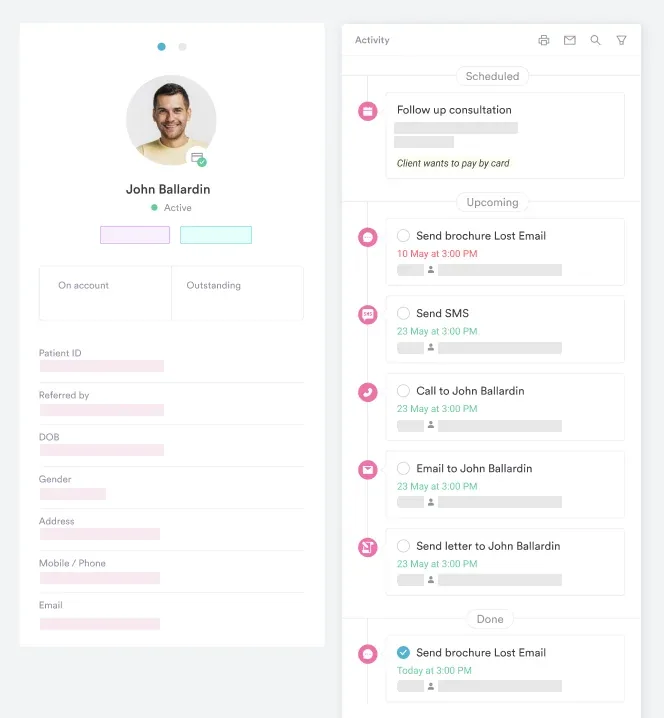
Documentation time is the other half of the problem. Pabau Scribe, our AI scribe, drafts the clinical note from the consultation itself. So the reasoning behind an urgent gas gets captured while it is fresh, rather than at the end of a long shift.
Teams comparing HIPAA compliant AI tools should check one thing above all. Does the tool write into the patient record, or into a document somebody then has to file?
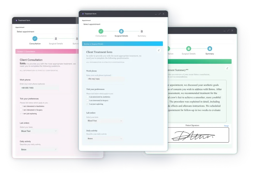
Consent and pre-procedure checks belong in the same file as the result. Digital intake forms capture the Allen test outcome, the site chosen, and the risks you discussed. Joining the order to the outcome is where software stops being a filing cabinet rather than a record.
Where the needle went matters beyond arterial sampling. Practices that give injectable treatments use Pabau’s injection plotting tool to mark each point on a patient photo.

Pabau files every result against the consultation that prompted it, with the indication, the clinician, and the follow-up in the same patient record. Nothing sits in a separate folder waiting to be found.

An arterial blood gas test is only as good as the sample and the sequence. Draw it cleanly, run the five steps in order, and the result tells you what the lungs and kidneys are doing right now. Skip either discipline and you get a number that misleads with confidence.
The clinical side of this is taught well. The documentation side rarely is, and that is where practices lose both time and defensibility. Decide now where a gas result lives, who reviews it, and how the follow-up gets recorded.
Book a demo to see how Pabau keeps test orders, results, and clinical notes together in one patient record.

Ready to cut prescribing errors and delays? Best e-prescribing software compares seven platforms on compliance, certification, and pricing.
Choosing an e-prescribing platform for your practice? Electronic prescribing software for US practices compares seven platforms on EPCS compliance and pricing.
Interpreting an unfamiliar blood panel? Rheumatoid arthritis test walks through what RF and anti-CCP results do and do not prove.
Want a fuller picture than one lab value gives? Body composition test explains the methods, results, and healthy ranges practices use to assess patients.
Explaining another invasive procedure to a patient? Bone marrow test explains what happens during the procedure and what the results mean.
Yes, more than a standard blood draw. The radial artery sits deeper and has more nerve endings around it, so patients often describe a sharp, cramping pain that fades within a minute. A small injection of local anesthetic before the puncture reduces it. Offer that when the patient is anxious or a repeat sample is likely.
No. Fasting makes no difference to blood gas values, so food and drink can carry on as normal. Timing matters instead. Wait 20 to 30 minutes after any change in oxygen flow or ventilator settings, so the sample reflects a steady state rather than the transition.
It splits metabolic acidosis into two groups, which changes the differential. Subtract chloride and bicarbonate from sodium on a paired electrolyte panel. Anything from roughly 8 to 12 mEq/L is normal. A raised gap points to lactate, ketones, renal failure, or a toxin. A normal gap points to bicarbonate loss, usually through the gut or the kidney.
Physicians, respiratory therapists, and nurses with documented competency all draw arterial samples in practice. The exact scope depends on state or country rules and on local sign-off, so check your own policy before delegating. Most services also require a number of supervised punctures before a clinician works unassisted.