Nursing narrative note template
A nursing narrative note template is a structured PDF that guides you through re...

2
Last Updated: Oct 21, 2026

Pediatric vital sign ranges shift with age, so 130 beats per minute is normal in a newborn and tachycardia in a teenager.
Heart rate and breathing rate fall as a child grows, while systolic blood pressure climbs toward adult values.
PALS puts the hypotension floor at 70 plus twice the age in years up to age 10, then a flat 90 mmHg.
Fever depends on route and age, from 38.0°C rectal under 3 months to 37.8°C oral in older children.
Record the child’s state next to the number, because a sleeping infant and a screaming toddler both read outside the resting band.
A one-page recording form with fields for heart rate awake and asleep, respiratory rate, blood pressure, temperature and oxygen saturation. Every entry has room for the reference range you applied, a low, normal or high reading, and notes.
Download templateA pediatric vital signs chart gives you the normal range for a child’s heart rate, breathing, blood pressure, temperature and oxygen saturation, by age. The most useful thing it tells you is that the numbers move.
A heart rate of 130 beats per minute is unremarkable in a newborn and a warning sign in a 12-year-old. Read a child against an adult range and you either escalate a healthy toddler or miss a school-age child sliding into shock.
Below are the ranges by age band, the cutoffs that trigger action, and the measurement details that make a number mean something.
The chart pairs two numbers for every measurement. One is the normal range for that age band. The other is the threshold that changes what you do next.
That pairing is what makes it a decision tool instead of a poster. A heart rate of 155 in a febrile toddler means little on its own. Set it against a normal band of 90 to 150, with the fever noted beside it. Now the next clinician sees exactly what you saw.
In practice it does three jobs:
Because a small heart moves less blood per beat. An infant delivers a fraction of an adult stroke volume. It has to beat faster to keep cardiac output up per kilogram.
Breathing follows the same logic. Small airways and a soft chest wall make each breath less efficient, so the rate compensates. Blood pressure runs the opposite way, rising as vessels stiffen and body mass increases through childhood. The physiology behind these shifting reference ranges is well documented.
Two groups sit outside the table. Premature infants keep neonatal numbers past their birth date, so correct for gestational age.
Children with a known congenital heart defect often run their own baseline. That baseline belongs in the record, not the reference chart. A pediatric review of systems is where that history usually surfaces.
Put all three trajectories on one age axis and the pattern is hard to miss.
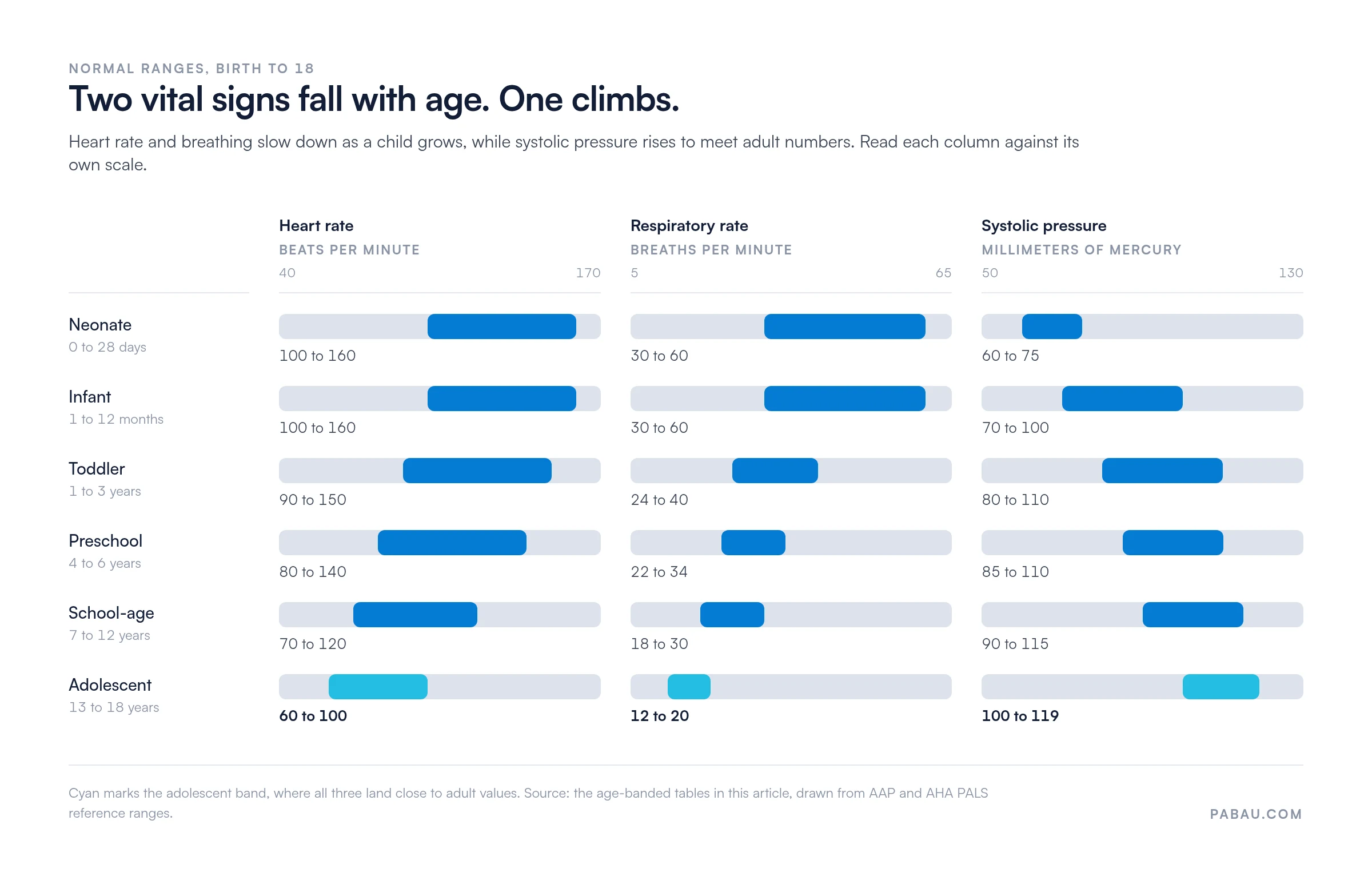
Neonates run 100 to 160 beats per minute. By 18, the same child sits at 60 to 100. Every band in between steps down, and the tachycardia cutoff steps down with it.
Bradycardia is the reading that should worry you more. A slow heart rate in a sick child is usually hypoxia or raised intracranial pressure, not fitness.
Because the band moves, the reading is only interpretable next to the date of birth. Practice management software like Pabau keeps both in the same client record, so anyone opening the file months later applies the right band.
Some practices lean on these bands weekly. Stimulant reviews in an ADHD clinic include a pulse and a blood pressure every time. The child ages out of one band into the next while still on the same prescription.
Respiratory rate is the most sensitive of the five and the easiest to get wrong. Count for a full 60 seconds, with the child settled and preferably not watching you do it.
A fast rate on its own is weak evidence. Fever, pain, anxiety and crying all push it up with no respiratory disease behind them. What turns a number into a finding is effort. Look for retractions, nasal flaring, head bobbing or grunting.
Sometimes the pattern matters more than the count. An ineffective breathing pattern care plan gives you somewhere structured to write that down.
Blood pressure is the one vital sign you cannot read from age alone. Normal also depends on height and sex, which is why the AAP publishes percentile tables rather than a single band. Treat the ranges below as orientation, not as a diagnosis of hypertension.
The hypotension floor is different. PALS gives you arithmetic instead of a table, and it is worth committing to memory:
A 3-year-old at 78 mmHg systolic is within range. A 9-year-old at the same number is hypotensive, because the floor has moved to 88. That is the argument for carrying the formula rather than one remembered number.
Cuff size decides whether any of it is worth recording. The bladder should cover about 40% of the arm circumference, and a cuff that is too small reads falsely high. A digital intake form can require the cuff size before it accepts the reading.

Well-child checks are where most pediatric blood pressures get taken. Primary care practices therefore hit the wrong-cuff problem more than anyone else.
Rectal is the reference standard under 3 months. At that age, 38.0°C rectal is a fever that needs same-day evaluation. From about age 4, oral becomes practical, with 37.8°C as the fever line.
Axillary and tympanic readings are convenient but less reliable, so neither should carry a fever decision in an infant.
The under-3-months row is the one that changes your afternoon. A rectal temperature of 38.0°C in a 6-week-old is a sepsis workup, not a follow-up call, however well the baby looks.
Record the route with the number, every single time. Without it, 37.6°C is either reassuring or a missed fever. Medical forms that make route a required field close that off before it reaches the chart.
Normal SpO2 is 95% or above from roughly 1 month onward, and it does not shift by age band. Below 90% is hypoxia at any age and needs immediate attention.
Newborns are the exception, and only for minutes. Targeted preductal saturation during transition is roughly 60% to 65% at 1 minute and 80% to 85% at 5 minutes. A baby reading 82% at four minutes of life is on track, so resist the urge to treat the number.
After that window, three things pull the reading down even with healthy lungs. Altitude, cold or poorly perfused fingers, and nail polish are the usual culprits. Check the waveform before you act on the digit.
The same child will give you three different heart rates in ten minutes. The chart assumes awake and at rest, and almost nothing in a pediatric clinic is awake and at rest.
None of that makes the chart wrong. It means a number without its context is not yet a finding. Writing “asleep” next to 118 bpm in an infant saves the next clinician a repeat measurement, and saves the parents a scare.
Pain drives the same rise, so a pain scale earns its place on the same form. And because the trend carries more signal than any single visit, tracking measurements across appointments beats a perfect one-off reading.
Five patterns change your next move rather than just your note.
The order matters. Adults drop their blood pressure early in shock. Children hold it until they cannot. Waiting for a low reading to confirm your suspicion is how a compensated child becomes a crash call.
Reference data only earns its place if it reaches the point of measurement. Here is the sequence that survives a busy clinic:
Pabau Scribe, our AI scribe, can drop spoken measurements straight into the assessment note. The numbers land in the record while you are still in the room.
Keep a printed copy at every clinical station, including the exam room and the bedside. In an emergency, staff read paper faster than they navigate a screen. A wallet-sized version in a coat pocket does the same job during a rapid assessment.
Most practices record vitals twice. Once on a paper form at the start of the visit, then again by hand into the record afterwards. The second copy is where the route, the cuff size and the child’s state quietly disappear.
Pabau, an all-in-one practice management system, keeps the whole thing in one place. Vital signs go into a digital form on a tablet, tied to the child’s file and date of birth. The reading and the age band travel together. Photos, consents and treatment notes sit in the same record, so nothing needs retyping at the end of a clinic.
The result is that the next clinician opens one file and sees the number, the route and who took it. Trends across visits are already plotted, so a slow drift in respiratory rate shows up as a pattern instead of a memory.

Pabau’s digital forms put heart rate, breathing, blood pressure, temperature and oxygen saturation straight into the child’s record. No paper form to transcribe, and no measurement route left behind.

The chart earns its keep the moment two people disagree about a reading. Age band, route, cuff size and the child’s state are what settle that argument. None of them survive in memory to the end of a long clinic.
So print it, download the form, and decide who fills in the reference range column. That last choice matters more than it looks. A blank reference field turns a careful measurement back into a number nobody can interpret.
If transcribing is where your numbers go missing, that is a software problem rather than a clinical one. Book a demo to see how Pabau captures pediatric vitals in the child’s record the first time.

Need the symptom history behind the numbers? Pediatric review of systems walks through each system in the order a pediatric examination follows.
Documenting abdominal pain at the same visit? Stomach pain chart template captures pain location, a 0-10 intensity scale, onset and associated symptoms.
Charting a fainting episode instead? Syncope nursing care plan covers assessment, NANDA-I diagnoses, interventions with rationales and evaluation criteria.
Ruling out appendicitis in the same exam? Tiptoe test for appendicitis documents the Markle sign with result fields and differential diagnosis notes.
Certifying a driver’s fitness instead? TLC medical certification form is the one-page NYC form recording the exam date and fit determination.
A fever in an infant under 3 months is a rectal temperature of 38.0°C (100.4°F) or higher. That is a medical emergency and needs immediate evaluation, even if the baby looks well.
Normal SpO2 in children over 1 month is 95% or above, at every age band. A reading below 90% is hypoxia and needs immediate escalation.
Use age for heart rate, respiratory rate and temperature. Blood pressure also depends on height and sex, so a percentile table beats an age band. Weight drives drug dosing, not vital sign interpretation.
Heart rate. Children compensate by beating faster and shunting blood away from the skin, so tachycardia and cool hands show up well before the pressure drops. Hypotension is a late sign.
The cuff bladder should cover about 40% of the arm circumference and 80% of its length. A cuff that is too small reads falsely high, and that is the most common cause of a wrong pediatric reading.