
 Tanja Lepcheska

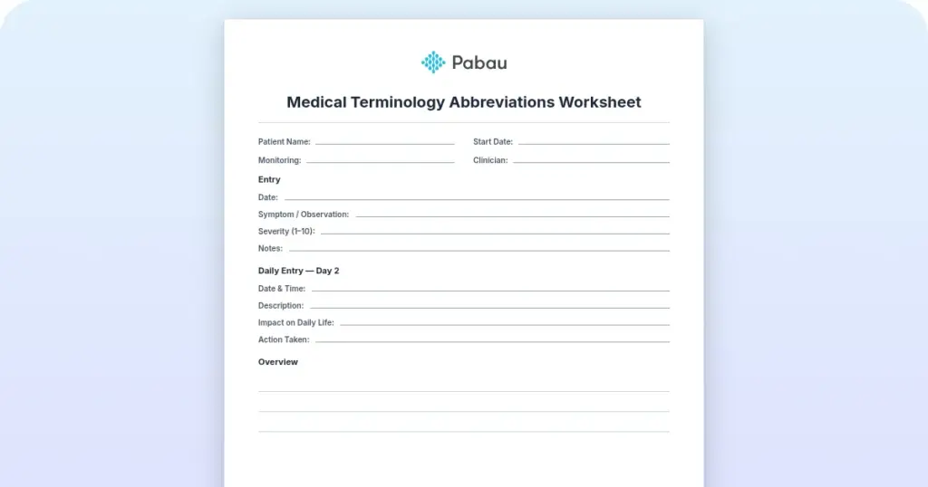
Medical terminology abbreviations worksheet
A medical terminology abbreviations worksheet is a printable practice sheet that...

Last Updated: Nov 11, 2026
Free PDF 1 page, print-ready Clinician-reviewed
A muscular anatomy chart is a reference diagram that labels the major muscles of the human body. Most charts show the body from the front (anterior view) and from the back (posterior view). That lets students and clinicians see which muscles sit on each side. Physical therapists and massage therapists use the chart to explain anatomy to patients and to agree on terms before treatment. This guide covers what the chart contains and how clinicians use it. It also shows how to link it to patient records in Pabau, the practice software we build.
A one-page chart with labeled front and back muscles, a muscle-group table, the three muscle tissue types and space for session notes. Print it to explain anatomy to patients.
Start from scratch, or upload this PDF or any form you already use, and Pabau's AI Form rebuilds it with tick boxes, text fields and a signature. Attach it to the treatment once, and every patient gets it on time.
AI Form rebuilds the consent, history and aftercare forms you already use. You review each one, then publish.
Patients complete forms on their phone before they arrive, so the front desk stops retyping paper.
Every signed form is versioned and attached to the treatment record, ready when you need it.
Set how long a consent stays valid. Returning patients skip forms they have already signed.
Supports HIPAA and GDPR compliance AES-256 encryption 4.8 on G2 4.7 on Capterra Trusted by 4,000+ practices worldwide

A muscular anatomy chart labels major muscles in front and back views.
Muscle tissue comes in three types, called skeletal, smooth, and cardiac.
Many physical and massage therapists use charts for patient education.
A chart supports patient education, but it never replaces a clinical assessment.
Pabau, the practice software we build, keeps chart-based explanations beside each patient’s treatment notes.
A muscular anatomy chart is a labeled visual reference for the body’s muscular system, usually presented as a full-body image.
Charts vary in detail. Some show only the superficial muscles, while fuller versions also label deeper muscles of the neck, pelvis, and limbs. Many teams that run a physical therapy practice keep a printed one in the treatment room.
A chart is a teaching and reference aid. It helps a practitioner name a muscle and see where it sits. It does not replace a clinical assessment such as a motor strength test, which grades muscle power on a scale from 0 to 5.
Muscle tissue comes in three types, called skeletal, smooth, and cardiac. Each type sits in a different part of the body and is controlled in a different way.
Skeletal muscles are the ones most charts label, because they attach to bones and produce voluntary movement at joints.
Skeletal muscles number more than 600 in most classifications, although the exact count ranges from about 600 to 650 depending on how muscles are grouped. For plain-language background, the MedlinePlus bones, joints and muscles topic from the U.S. National Library of Medicine is a good starting point.
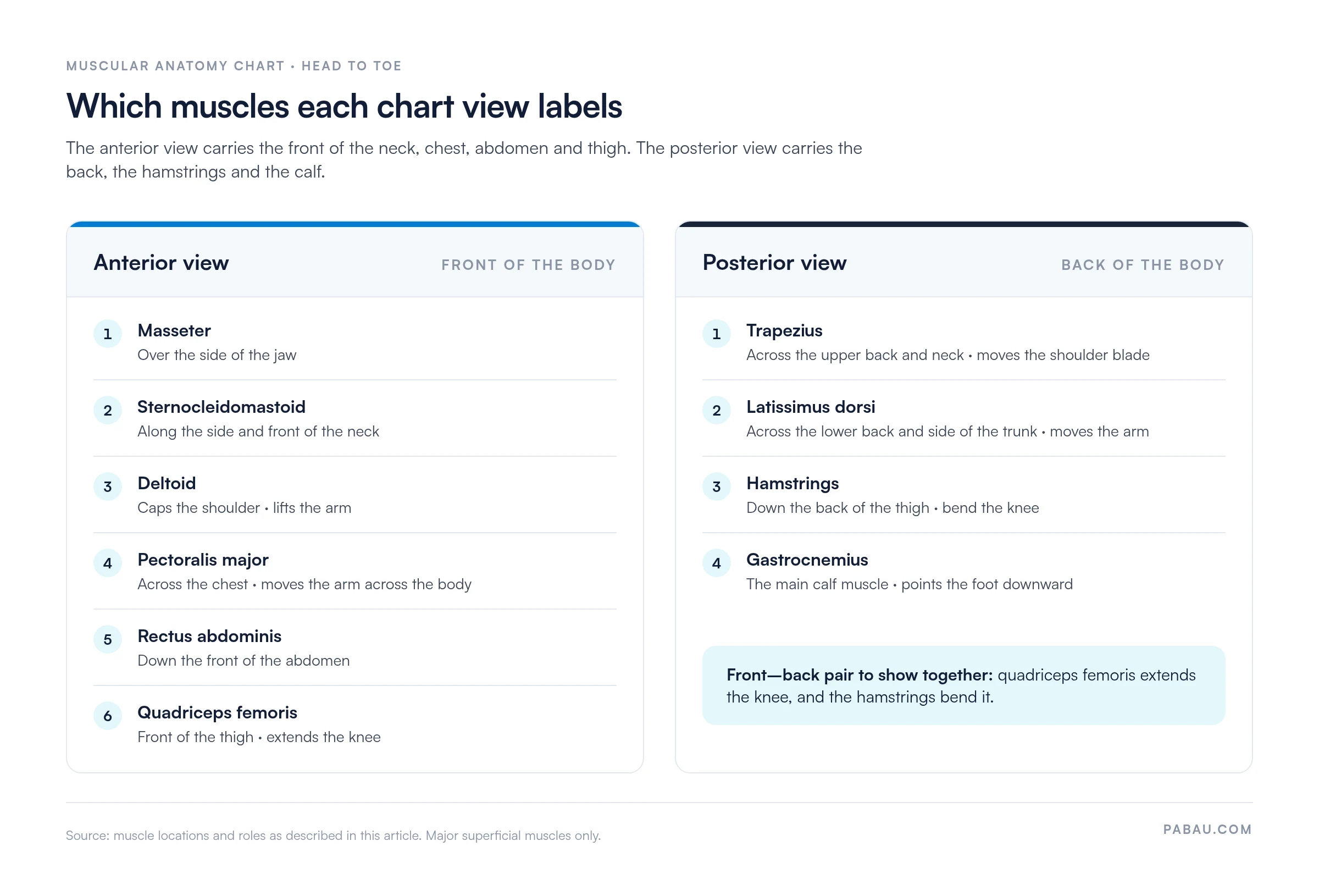
Most full-body muscular anatomy charts show two views, anterior and posterior, which together cover the whole body.
The anterior view labels muscles visible from the front. The pectoralis major crosses the chest, the deltoid caps the shoulder, and the rectus abdominis runs down the front of the abdomen.
The quadriceps femoris covers the front of the thigh. The sternocleidomastoid runs along the side and front of the neck, and the masseter sits over the side of the jaw.
The posterior view shows the trapezius across the upper back and neck. The latissimus dorsi spreads across the lower back and side of the trunk.
The hamstrings run down the back of the thigh, and the gastrocnemius forms the main calf muscle at the back of the lower leg. Side by side, the two views split the body into front and back muscle groups.
Want to see how a chart explanation can sit in the patient’s notes? Book a demo and our team will walk through the workflow with you.
The table below groups the muscles most often labeled on a chart, with the body region each group serves.
Use the table as a quick check, not a substitute for the detail on the chart itself.
Clinicians use a muscular anatomy chart mainly to explain anatomy, not to make a diagnosis. Patient education is a common use. A practitioner can point to the calf muscles while describing a stretching plan, which often makes the instructions easier to follow.
When you show a chart during a consultation, keep the focus on one body region at a time. Sports medicine teams use the same reference to explain where a strain sits. In a sports medicine practice, the chart helps an athlete see why a hamstring strain affects bending the knee.
Many physical therapists use a chart when they explain an exercise program, then note which muscles were targeted in the treatment record. A home exercise program gives the patient the same exercises in writing to follow between sessions. For professional context on the field, see the American Physical Therapy Association.
Massage therapists often use a chart to show patients which muscles lie beneath an area of discomfort, such as the trapezius or the hamstrings. Professional guidance for massage practice is available from the American Massage Therapy Association.
Without a link to the notes, the explanation you give at the chart stays in the treatment room. Pabau’s medical records software keeps each patient’s notes in one place, so the area you explained sits beside the treatment you documented.

Intake matters too. Pabau’s digital intake forms let patients complete their history before the first session, which gives you context before you open the chart.

Use the chart for education, the record for documentation, and the intake form for history. Keeping those three steps separate makes the workflow clear for the whole team.

Pabau keeps intake history, treatment notes and the areas you explained in one patient record. Your team sees what each patient was shown before the next session.

A muscular anatomy chart earns its place in the treatment room by doing one job. It helps a patient see the muscle you are talking about. Keep assessment and diagnosis with your clinical tests, and keep the chart for explanation.
The trade-off is detail. A chart of superficial muscles covers most consultations, but a fuller one that labels the deeper neck, pelvis, and limb muscles suits more specialized work. Whichever you choose, note the area you explained so the next session starts from the same point.
Book a demo to see how Pabau keeps chart-based patient education and treatment notes in one record.

Planning exercises after the explanation? Home exercise program gives patients a written plan for the muscles you showed them on the chart.
Need a record of each exercise session? Exercise worksheet covers what to record for each exercise a patient completes.
Testing muscle power as well as naming it? Motor strength test explains MRC grading and how to interpret the results.
Checking a hip muscle in detail? Gluteus medius test walks through how to perform the test and read the results.
Assessing the shoulder? Shoulder mobility test covers how to do the test and interpret what you find.
The three types are skeletal, smooth, and cardiac muscle. Skeletal muscle attaches to bones and moves voluntarily, while smooth and cardiac muscle work involuntarily in organ walls and the heart.
The anterior view shows muscles from the front, such as the pectoralis major and quadriceps femoris. The posterior view shows the back, including the trapezius, hamstrings, and gastrocnemius.
Physical therapists, massage therapists, and sports medicine teams use one most often. Students use it to learn muscle names, and practitioners use it to explain treatment to patients.
No. A muscular anatomy chart is a reference and teaching aid, and it does not replace an assessment by a qualified practitioner.
The human body has more than 600 skeletal muscles, and the exact count ranges from about 600 to 650 depending on classification.
Yes, free printable charts are widely available, but check the source before you use one with patients. Look for a named publisher, a clear date, and terms that allow educational use.