CRM for chiropractors: how to manage patients, bookings and marketing
A CRM for chiropractors is a patient relationship management system built around...

Last Updated: Oct 13, 2026

The gluteus medius runs from the outer ilium to the greater trochanter and is supplied by the superior gluteal nerve (L4, L5, S1).
The Trendelenburg test is the most widely used gluteus medius test, and a positive sign is contralateral pelvic drop in single-leg stance.
Manual muscle testing grades strength on the MRC 0-5 scale. A grade below 3 means the patient cannot resist gravity.
The Donatelli drop leg test isolates the posterior fibers, so it catches weakness that a mid-range abduction hold can hide.
Practice management software like Pabau keeps test findings, MMT grades and rehabilitation progress in one client record.
The gluteus medius test is a clinical assessment of hip abductor strength and pelvic control under weight-bearing or manual resistance.
Four tests do the work in practice. They are the Trendelenburg test, the single-leg stance test, manual muscle testing in side-lying, and the Donatelli drop leg test. Weakness here is linked to Trendelenburg gait, iliotibial band syndrome, patellofemoral pain syndrome, and low back pain. This guide covers how to perform each test, how to read the result, and where rehabilitation should start.
The gluteus medius test is any structured procedure used to detect weakness or inhibition in the muscle. Clinicians reach for it because the gluteus medius is the primary hip abductor during single-leg stance. When it fails, gait, knee alignment and lumbar loading all change to compensate.
The muscle sits on the outer surface of the pelvis, fanning from the ilium to the greater trochanter. Its upper fibers produce hip abduction, and its anterior fibers assist medial rotation. During walking it fires eccentrically on the stance leg, stopping the opposite side of the pelvis from dropping.
These tests are daily work in physical therapy, sports medicine and musculoskeletal rehabilitation. The four assessments below each return different information, and most protocols combine at least two. Recording them consistently matters as much as performing them, which is what physical therapy EMR software is built for.
The gluteus medius originates on the outer surface of the ilium, between the anterior and posterior gluteal lines. It inserts into the superolateral facet of the greater trochanter of the femur. Blood supply comes from the superior gluteal artery, a branch of the internal iliac.
Knowing the innervation level matters clinically. A patient with hip abductor weakness plus dermatomal changes at L4-S1 needs neurological screening first. Do not attribute the weakness to a musculotendinous cause before that.
The gluteus minimus lies deep to the medius and shares the same nerve supply. It originates between the anterior and inferior gluteal lines, and inserts into the anterior facet of the trochanter. Both muscles contribute to abduction, so telling them apart matters when you localize weakness.
Observational signs usually arrive before any formal test. They show up during gait analysis or basic functional tasks, and they tell you which test to prioritize.
These observations are prompts, not diagnoses. A patient with Trendelenburg gait still needs structured testing to establish how severe the weakness is. Testing also shows whether the cause is muscular, neurological, or pain-inhibited.
Gluteus medius weakness rarely presents on its own. It surfaces as a contributing factor in several common musculoskeletal presentations. That is why the test now appears in assessment protocols well beyond hip pain.
The low back pain link is the most studied of these. A systematic review of gluteus medius function compared people with and without low back pain, and reported differences in how the muscle works.
Screening for gluteus medius weakness belongs in the standard assessment battery for all of these presentations. Waiting for a hip-specific complaint delays the finding, and early identification changes the rehabilitation pathway.
The Trendelenburg test is the most widely validated gluteus medius test in practice. It assesses whether the muscle can hold the pelvis level during single-leg weight-bearing.
Positive result: the opposite side of the pelvis drops below the stance-side iliac crest. That means the stance-side gluteus medius cannot hold the pelvis level under load. Negative result: the pelvis stays level, or the non-stance side rises slightly.
Reported sensitivity and specificity are moderate when the test is checked against dynamometry. Treat it as a screen rather than a measure of strength grade. Pairing it with manual muscle testing gives the fuller picture.
The single-leg stance test evaluates balance, proprioception and hip abductor endurance. It differs from the Trendelenburg test by holding the position for a timed period. That exposes endurance deficits and compensatory strategies a brief Trendelenburg assessment can miss.
Interpreting findings: healthy adults usually hold single-leg stance for 30 seconds without marked compensation. Under 10 seconds on either limb, or clear asymmetry between sides, warrants further investigation. Trunk lateral flexion toward the stance leg suggests a compensated Trendelenburg pattern rather than a balance problem.
The timed score also works as a rehabilitation baseline. A patient who starts at 8 seconds with visible pelvic drop and reaches 28 seconds in six weeks has objective evidence of response.
Structured fields hold those timed scores better than free-text notes do. Storing them in medical records management software keeps each score attached to its visit. Outcome reporting for insurers then becomes straightforward, and continuity of care survives a change of clinician.

Manual muscle testing of hip abduction gives a graded, reproducible measure the observational tests cannot match. It uses the MRC 0-5 scale, and the patient lies in side-lying so gravity cannot assist the movement.
A grade of 3 or below is significant. The patient cannot resist gravity in functional positions, which explains both Trendelenburg gait and single-leg stance failure. Grades 4- to 4+ mark patients who function but fatigue under load, the common picture in runners.
MMT is a screening and grading tool, not a standalone diagnostic. Weakness that persists after eight weeks of targeted rehabilitation warrants imaging. Look for gluteus medius tendon pathology, a femoral neck stress fracture, or a neurological cause. Limited abduction range may come from tight tissue rather than weakness. The Phelps test shows whether a tight gracilis is the cause.
The American Physical Therapy Association recommends pairing MMT with functional outcome measures when you document rehabilitation goals. That combination supports both reimbursement and clinical governance.
When performing MMT for hip abduction, palpate the gluteus medius belly (just below and posterior to the iliac crest) during resistance testing. A Grade 4 that recruits tensor fasciae latae compensation produces a visible anterior hip crease during abduction. If the TFL fires prominently and the hip flexes forward, document it as Grade 4 with substitution. Adjust the rehabilitation plan accordingly.
The Donatelli drop leg test is a non-weight-bearing manual test performed in side-lying. It removes body weight from the equation and isolates the posterior fibers of the gluteus medius. The clinician passively places the hip at end-range abduction with slight extension, then asks the patient to hold it. Because the limb starts beyond the range most patients actively control, the test exposes weakness a mid-range hold can mask.
Positive result: the leg drops before the posterior fibers engage strongly enough to arrest it. Drop distances are commonly recorded between 2 and 12 inches, and a larger drop points to greater posterior fiber weakness. Negative result: the patient holds the limb at end-range abduction and extension with no appreciable drop.
The test isolates the posterior fibers rather than the whole muscle. That is why it often reveals a deficit in a patient who grades 4 or 5 on standard side-lying testing. Gowda and Donatelli published reliability data for the test in Physical Therapy in Sport in 2014.
Peer-reviewed sensitivity and specificity figures remain limited next to the Trendelenburg test. Treat the finding as supporting evidence within a multi-test assessment, rather than a decision point on its own.
No single gluteus medius test gives the complete picture. The value of a multi-test protocol is that each assessment reveals a different failure mode. The Trendelenburg test captures functional pelvic control, and MMT grades isolated strength. Single-leg stance reveals endurance and proprioception, while the drop leg test isolates the posterior fibers.
Refer for imaging and specialist review when significant weakness comes with neurological symptoms. Dermatomal pain, altered reflexes and sensory changes all qualify. Document the reasoning for that referral in the clinical record before starting high-load rehabilitation.
Exercise selection should match the failure mode the test battery identified. A patient at MMT Grade 3 starts with gravity-eliminated work. A patient with a marked leg drop starts with posterior fiber work, such as clamshells or side-lying abduction in slight hip extension.
The ladder below sets out where each grade band starts, and what the position is actually doing. The one hard gate sits between side-lying and standing work.
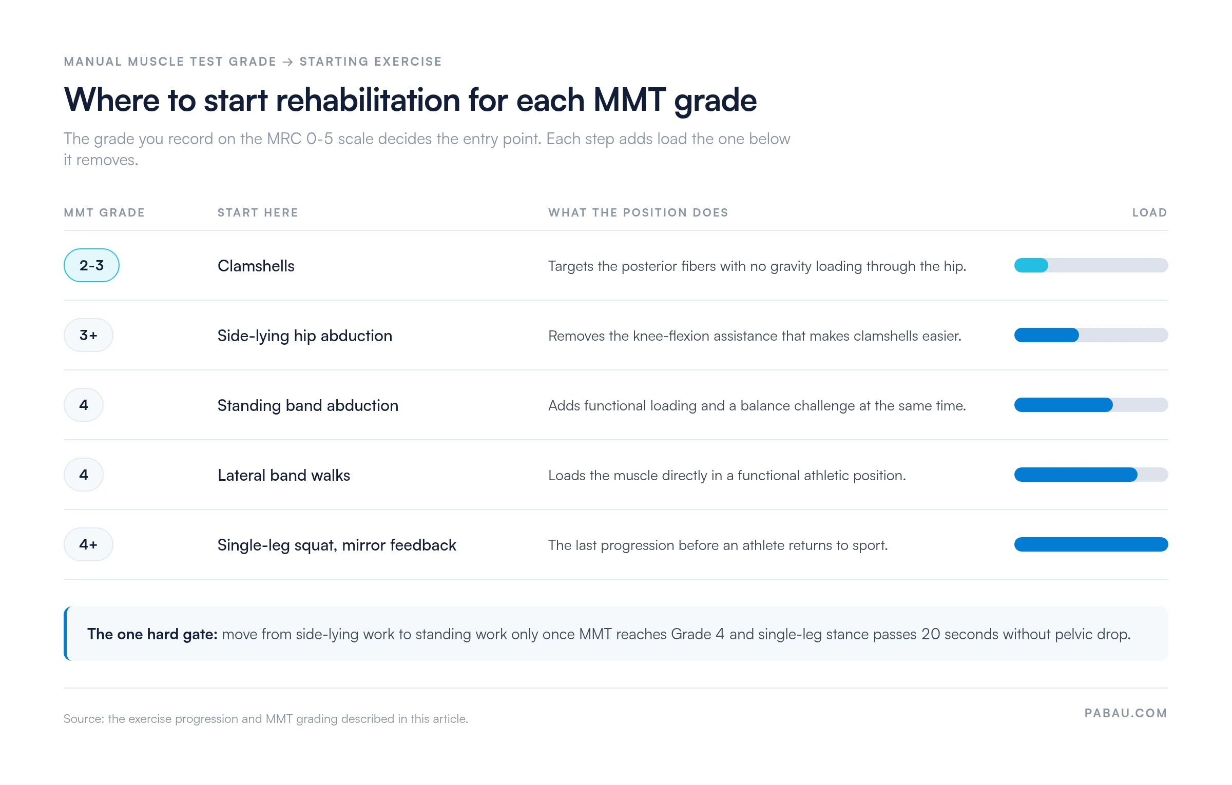
All exercise prescription should be individualized by a licensed clinician against the test findings. Progression criteria need to be explicit rather than assumed. Attaching the current program version to the client record keeps the patient on the right stage.
Occupational therapy and return-to-work settings use the same progression with different end points. Prolonged standing, stair climbing and lateral transfers replace athletic loading as the criterion. For athletes, the return-to-running protocol covers the loading criteria that follow this stage.
Documenting a multi-test assessment takes time. One session can produce a Trendelenburg finding, a timed stance score, an MMT grade and a page of gait notes. Free-text SOAP notes hold all of it, but they make comparison across visits slow.
Practice management software like Pabau replaces that free-text block with structured fields. Digital patient intake forms let you build an assessment template with dropdowns for MMT grades and numeric fields for timed scores. Tick-box sections cover the test findings themselves.
The same record feeds the treatment plan and the appointment notes, so nothing is re-entered between visits. Progress across MMT grades or stance time then reads as a series. Any clinician covering the session opens the full assessment history.

Dictation covers the sessions where typing gets in the way. Pabau Scribe, our AI scribe, transcribes a verbal summary after the appointment and drafts the note from it. Practices comparing platforms before they commit can start with our roundup of physiotherapy practice management software.
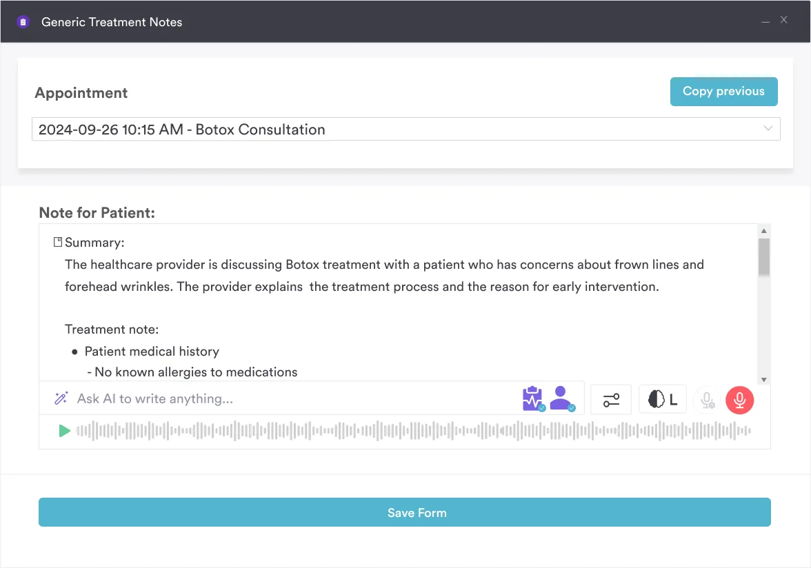
The result is one record that carries the finding, the grade and the plan together. Insurance reporting and outcome measure requirements then draw on data that is already structured.

Record gluteus medius test findings, MMT grades and rehabilitation milestones in one structured client record. Pabau’s clinical notes and treatment plan templates keep the whole team working from the same history.

Gluteus medius weakness sits behind more presentations than a hip-focused assessment would suggest. Knee valgus, ITBS, low back pain and trochanteric tendinopathy all trace back to it. One test will not find it reliably.
Run at least two, and choose them for what they reveal. The Trendelenburg sign reads functional pelvic control, while MMT grades isolated strength. Single-leg stance captures endurance, and the drop leg test catches posterior fiber weakness the other three can mask.
The trade-off is time, and structured documentation is what buys it back. Book a demo to see how Pabau keeps test findings, MMT grades and rehabilitation progress in one client record.

Assessing lateral knee pain in a runner? Noble compression test walks through the ITBS assessment that often sits alongside hip abductor weakness.
Need a balance measure to sit beside single-leg stance? 4-stage balance test gives you a graded static balance screen you can run in the treatment room.
Tracking lower-limb strength without a dynamometer? 30-second chair stand test offers a functional strength measure that repeats cleanly between visits.
Checking your documentation meets regulatory requirements? Mandatory compliance for physiotherapy clinics sets out the governance records a physical therapy practice has to keep.
The gluteus medius test is a clinical assessment of hip abductor strength, endurance and neuromuscular control. The common forms are the Trendelenburg test, manual muscle testing in side-lying, and the single-leg stance test. Each one reports on a different demand placed on the muscle.
Ask the patient to stand on one limb for 30 seconds and watch for the opposite side of the pelvis dropping. That is the Trendelenburg test. Confirm the severity with manual muscle testing in side-lying, graded on the MRC 0-5 scale. Two tests give a more reliable picture than one.
The Trendelenburg sign is a static test finding. The opposite side of the pelvis drops during a deliberate single-leg stance. Trendelenburg gait is the walking pattern that results from the same weakness. It shows as a lateral trunk lurch or pelvic drop with each step on the affected limb. The sign confirms the deficit, and the gait pattern is its functional consequence.
MMT Grade 3 means the patient can move the hip through its full abduction range against gravity but cannot resist any added manual resistance. The muscle is functional for basic movements but not for weight-bearing tasks. Holding the pelvis level against body weight is beyond it, which makes Grade 3 a significant finding for gait and sport.
Gluteus medius weakness is associated with iliotibial band syndrome, patellofemoral pain syndrome, greater trochanteric pain syndrome, low back pain, and medial tibial stress syndrome. The common mechanism is loss of pelvic and femoral control during single-leg loading. Stress then rises on structures from the lumbar spine down to the foot.
No. Activation describes the muscle’s recruitment pattern, which EMG measures directly. You can also infer it from the timing and quality of movement during functional tasks. Strength is force production capacity, measured by MMT or dynamometry. A muscle can activate normally and still produce too little force. It can also activate late and pass a strength test. A complete assessment covers both.