Nitrates medication list
A nitrates medication list is a point-of-care reference that sets out the main c...

Last Updated: Oct 6, 2026
A motor strength test grades how much force a muscle or muscle group can produce against examiner resistance. So clinicians use it to detect, localize, and monitor neuromuscular deficits in neurological, musculoskeletal, and rehabilitation settings. According to StatPearls (NIH/NCBI), grading follows the Medical Research Council (MRC) scale from 0 to 5.
This guide covers how to perform, grade, and interpret the test. It then walks through the MRC scale, upper and lower extremity technique, the pronator drift screen, objective options, and documentation that holds up on review.

A motor strength test grades muscle force on the MRC 0-5 scale, where 5/5 is full strength against resistance and 0/5 is no contraction.
Manual muscle testing (MMT) compares bilateral sides, and asymmetry is often more diagnostically useful than an absolute grade.
Upper motor neuron lesions produce spasticity and hyperreflexia, while lower motor neuron lesions produce flaccidity and hyporeflexia.
Read every strength grade alongside muscle tone and reflex findings before you decide where the lesion sits.
Pabau’s customizable clinical note templates let physical therapy and neurology practices document motor strength findings consistently across practitioners.
A motor strength test is the part of the neurological exam that measures how well a muscle or muscle group makes force. Then the examiner compares that output against gravity and against applied resistance. Clinicians perform it whenever a patient presents with weakness, altered gait, falls, sensory changes, or a recent neurological or musculoskeletal injury.
The motor exam has three distinct layers:
Together, these three components localize lesions, track disease progression, and inform rehabilitation targets. So a strength grade on its own says very little. Read it next to the tone and reflex findings from the same exam.
The MRC grading scale is the standard for documenting motor strength test findings. It runs from grade 0, meaning no muscle contraction, to grade 5, meaning full strength against full examiner resistance. So every grade carries its own descriptor and its own clinical implication.
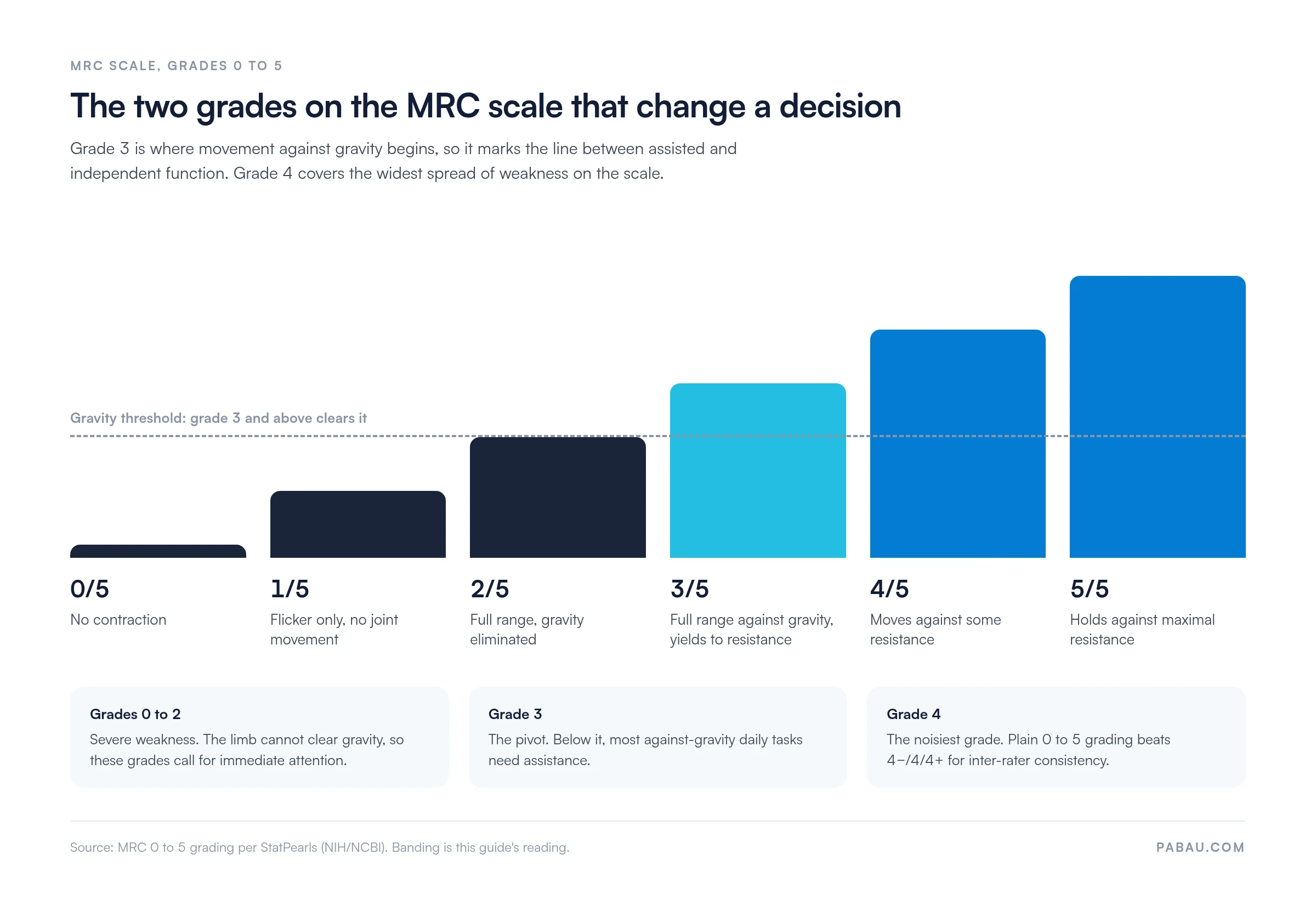
Grades 0 to 2 indicate severe weakness that needs immediate attention. Grade 3 is the pivotal threshold, because a patient who cannot move against gravity cannot perform most functional tasks. Grade 4 is the noisiest grade, covering a wide spectrum of mild-to-moderate weakness. Some practitioners subdivide it into 4-, 4, and 4+, but the plain 0 to 5 scale gives better inter-rater consistency. So the chart below marks where those two decision points sit.
Performing a motor strength test correctly requires patient positioning, consistent examiner technique, and bilateral comparison. The Merck Manuals Professional Edition describes the core procedure. First, inspect the limbs for drift or asymmetry, then apply resistance to specific muscle groups and compare sides.
Pain and patient effort both confound grading. If a patient stops resisting because of pain rather than weakness, note “effort-limited” or “pain-limited” instead of assigning a low grade. So that distinction changes what the next clinician does with the finding.
Upper extremity motor strength testing follows a proximal-to-distal sequence across the main nerve root levels. Standard muscle groups and their root levels are listed below.
Lower extremity motor strength testing follows the same proximal-to-distal principle, from hip flexion (L2-L3) down to plantarflexion (S1-S2). The standard sequence is:
The pronator drift test detects subtle upper motor neuron weakness that may still grade 4/5 or even 5/5 on standard testing. Ask the patient to extend both arms in front with palms facing upward and eyes closed. Then hold for 10-20 seconds. A positive test shows pronation, where the palm rotates inward, together with downward drift of the affected arm. That points to pyramidal tract involvement even when formal resistance testing looks near-normal.
The test is particularly valuable in early stroke, multiple sclerosis, and cervical myelopathy, where subtle upper extremity weakness may be the first presenting sign.
Assigning the grade takes seconds. Reading the pattern across muscle groups is where clinical judgment comes in. Isolated weakness means something different from a distributional pattern, and acute onset means something different from chronic. So read strength grades alongside tone and deep tendon reflexes to work out where the lesion sits.
Upper motor neuron (UMN) and lower motor neuron (LMN) lesions produce opposite examination findings. So the distinction drives the differential diagnosis and should be explicit in every motor exam note.
Three patterns in a motor strength test count as red flags:
So any of these warrants urgent further investigation.
Always test the unaffected side first. Patients calibrate their effort to your resistance style after the first limb, so the stronger side gives you a reliable baseline. That baseline matters most at grade 4, where weakness and pain-limited effort look alike.
Handheld dynamometry (HHD) and isokinetic testing are numeric options next to the MRC-based motor strength test. So both return a numerical force value in Newtons or kilograms rather than an ordinal grade. Physio-pedia describes both as providing objective data that MMT cannot capture, which matters most in sports medicine and research.
HHD is the more practical upgrade from MMT. So it is portable, affordable, and it produces values you can compare across serial visits. Isokinetic rigs are more reproducible again, but their cost keeps them in research and elite sports settings.
Manual muscle testing is the most widely used motor strength test in clinical practice. So it carries known limits worth understanding before you interpret a grade.
When serial measurements drive clinical decisions, such as rehabilitation milestones, return-to-work, or legal proceedings, supplement MMT with HHD or isokinetic testing. For those decisions an ordinal scale is a starting point rather than an endpoint.
Good motor strength test documentation does three jobs. So it tells the next clinician exactly what was tested, gives a baseline to compare against, and holds up under medico-legal review. Thin exam notes are a common audit finding when a practice is reviewed against physiotherapy compliance requirements.
Standard notation follows this pattern:
Standardize the sequence of muscle groups your practitioners test, so any clinician picking up the chart knows what was assessed and in what order. Our MRC scale template sets out that sequence as a printable grading sheet you can take into the exam room.
Standardizing the motor exam across a multi-practitioner practice is where documentation effort usually goes astray. So practice management software like Pabau handles it with customizable clinical note templates. Physical therapy and neurology practices build a structured motor exam section that captures MRC grades per muscle group, laterality, effort-limiting factors, and serial comparisons.
Pabau’s medical records management stores motor strength test findings next to prior assessments. Comparing a patient’s current 3/5 against their admission 1/5 takes seconds instead of a hunt through paper notes. So Pabau’s measurements tracking software extends that to serial force values from handheld dynamometry, logged and graphed across the rehabilitation trajectory.

For practices running motor exams inside a broader neurological or rehabilitation program, Pabau’s physical therapy EMR brings scheduling, documentation, and outcome tracking into one system. So that cuts the admin work competing for clinical time.

Pabau gives physical therapy and neurology practices customizable clinical note templates. They capture MRC grades, muscle group, laterality, and change over time, consistently across every practitioner.

A well-performed motor strength test gives you a reproducible, lateralized, grade-specific picture of neuromuscular function. So the MRC scale supplies the framework. The clinical value comes from reading grades next to tone and reflexes, comparing sides, and tracking change over time.
For physical therapy and neurology practices, documentation decides whether those findings survive to the next appointment. So customizable note templates and serial measurement tracking keep every motor exam recorded the same way, whoever performs it. Book a demo to see how Pabau handles motor exam documentation.

Need another quick clinical decision tool? Ottawa ankle rules calculator walks through when imaging is justified after an ankle injury.
Turning strength findings into discharge criteria? Return-to-running protocol in physical therapy sets out the functional milestones that follow a strength deficit.
Running a physical therapy or rehab practice? Opening a physiotherapy clinic covers the compliance and operational setup every new practice needs.
A motor strength test measures how much force a muscle or muscle group makes against gravity and examiner resistance. The result is graded on the MRC 0-5 scale. So it is a core part of the neurological exam, used to detect, localize, and track neuromuscular deficits. Conditions assessed this way range from stroke and radiculopathy to myopathy and peripheral neuropathy.
Grade 3/5 on the MRC scale means the patient can move the limb through its full range against gravity. But the same limb yields to any examiner resistance. Grade 3 is the functional threshold. Below it, against-gravity tasks such as lifting the arm overhead or climbing stairs need assistance.
Grip strength is tested by having the patient squeeze two of the examiner’s fingers as hard as possible, or quantitatively using a Jamar dynamometer. So dynamometry makes a force value in kilograms or pounds. That value can be compared against age- and sex-matched normative data. It is more sensitive than MMT for mild grip weakness in conditions such as rheumatoid arthritis or ulnar neuropathy.
Manual muscle testing has a ceiling effect at grade 4-5 and cannot distinguish within that range. It is sensitive to examiner strength and technique, and to patient pain and fatigue. Inter-rater reliability is only moderate at the grade 3-4 boundary, which is the boundary that matters most. So handheld dynamometry is the recommended upgrade when precision drives a clinical decision.