Head to toe assessment cheat sheet: Printable nursing guide
A head to toe assessment cheat sheet gives you one fixed order at the bedside, s...

3
Last Updated: Oct 23, 2026

The gallbladder physical exam centers on right upper quadrant palpation to screen for acute cholecystitis, biliary colic, and cholelithiasis.
Murphy’s sign is the cornerstone maneuver, and pain with inspiratory arrest on deep palpation points to acute cholecystitis.
Boas’ sign, Courvoisier’s sign, and Charcot’s triad add diagnostic depth, but imaging still has to confirm the diagnosis.
A defensible note names each maneuver and its result, because a generic soft, non-tender abdomen records nothing gallbladder-specific.
Pabau’s digital forms and Pabau Scribe capture structured gallbladder exam findings inside SOAP-format clinical notes.
Right upper quadrant pain accounts for a substantial share of acute abdominal presentations in primary and urgent care. Gallstone disease affects an estimated 10 to 15% of adults in the United States, according to the NIDDK. The gallbladder physical exam is the first tool that separates a benign cause from acute cholecystitis needing urgent intervention.
Performed well, the exam narrows the differential before a single lab result returns. Performed poorly, it misses findings that delay care. This guide covers the examination sequence and every major sign. It then turns to the documentation side, which decides whether your finding is reproducible for the next clinician.
It is written for physicians, nurses, and medical students who perform abdominal examinations regularly. In primary and urgent care the finding has to reach the patient’s record the same day. That is the job GP practice software does.
The gallbladder physical exam is a targeted abdominal assessment for any patient with symptoms of biliary disease. Those symptoms include right upper quadrant pain, nausea, vomiting, fever, and jaundice. The exam never confirms a diagnosis on its own. What it does is set the urgency and decide which labs and imaging come next.
The exam is appropriate for any patient reporting:
The conditions it helps detect span a wide severity range. Uncomplicated biliary colic sits at one end, with acute cholecystitis, choledocholithiasis, and cholangitis above it. Gallbladder carcinoma and pancreatic disease mimicking gallbladder disease are less common, but worth holding in mind.
The gallbladder sits in a fossa on the inferior surface of the liver, at the junction of the right and left hepatic lobes. It is not normally palpable. In disease states, particularly acute cholecystitis or hydrops, it may distend and become palpable in the right hypochondriac region.
Key surface anatomy landmarks:
These landmarks carry both the palpation and the documentation. A clinician who cannot reliably locate Murphy’s point will produce findings the next examiner cannot reproduce.
The gallbladder physical exam follows the standard sequence of abdominal examination: inspection, auscultation, percussion, palpation. Do not skip inspection and auscultation even when the clinical picture seems obvious.
Record each finding as positive, negative, or equivocal. “Non-tender, soft abdomen, no organomegaly” is not enough for a gallbladder-specific assessment. Name the maneuver and name the result. The sequence sits inside the wider abdominal physical exam, so record it alongside the other quadrants rather than on its own.
Murphy’s sign is the most widely used and studied maneuver in the gallbladder physical exam. A positive result strongly suggests acute cholecystitis when the clinical history supports it. Reported sensitivity, however, varies widely across studies.
Reported sensitivity for acute cholecystitis ranges from roughly 38% to 97%, according to Medscape’s cholecystitis clinical presentation overview. That spread reflects the study population and the examiner’s experience. Specificity is generally higher than sensitivity.
A negative Murphy’s sign does not rule out acute cholecystitis. Elderly patients and those on opioids may not mount a pain response at all, so the maneuver returns a false reassurance.
The sonographic Murphy’s sign is pain elicited by the ultrasound probe directly over the gallbladder. It is more reproducible than the clinical maneuver, and considered more sensitive. It is assessed during the same appointment that images the gallbladder wall and lumen.
Three additional signs appear in cholecystitis and biliary disease. Each has a distinct technique and clinical meaning. The table below sets out all four major signs side by side.
Boas’ sign is elicited by assessing for hyperesthesia in the right subscapular region, roughly at the level of the 9th to 11th ribs posteriorly. Light touch or pinprick testing produces pain disproportionate to the stimulus.
The referred tenderness occurs because the gallbladder and the right subscapular skin share segmental innervation. The phrenic nerve (C3 to C5) and the intercostal nerves carry both. As Medscape notes, Boas’ sign is a lesser-studied correlate of acute cholecystitis. Modern validation is limited, though surgical textbooks have carried it for well over a century.
Courvoisier’s sign describes a palpable, non-tender gallbladder in a patient with obstructive jaundice. The clinical teaching is that gallstone disease causes chronic inflammation and fibrosis, which leaves the gallbladder unable to distend.
A palpable, distended, non-tender gallbladder therefore points to a cause other than stones. The usual candidates are pancreatic head carcinoma, cholangiocarcinoma, and ampullary malignancy. This sign warrants urgent imaging rather than expectant management.
Charcot’s triad is right upper quadrant pain, jaundice, and fever together, and it is the classic presentation of ascending cholangitis. Cholangitis is a biliary emergency.
Sensitivity for cholangitis is low when the triad is used in isolation, because the three features are not always present at once. Reynolds’ pentad adds hypotension and altered mental status, which indicates septic cholangitis. Any patient with the triad warrants urgent lab work, blood cultures, and hepatobiliary imaging without delay.
Acute and chronic cholecystitis produce very different examination pictures. Recognizing which one you are dealing with shapes both the urgency of management and the imaging you order.
Acalculous cholecystitis deserves particular attention. It occurs without gallstones, and typically in critically ill patients. The usual settings are intensive care, recovery from major surgery, severe burns, and prolonged total parenteral nutrition (TPN).
Examination findings here are often blunted or masked by sedation, opioids, or the underlying illness. Mayo Clinic notes that imaging is the primary diagnostic tool in this population. A negative Murphy’s sign in a sedated intensive care patient does not exclude acalculous cholecystitis.
In elderly patients, Murphy’s sign may be absent even in confirmed acute cholecystitis. Pain perception diminishes with age, and opioid analgesia further blunts the inspiratory arrest response. In any older patient with fever, leukocytosis, and unexplained abdominal discomfort, order an ultrasound regardless of examination findings.
A positive or suspicious gallbladder exam prompts a structured workup. Physical findings alone never confirm a diagnosis. Physiopedia’s clinical examination reference is clear that Murphy’s sign has to be read alongside lab and imaging results.
Which test comes next depends on which finding the exam produced, and on how fast it needs to happen.
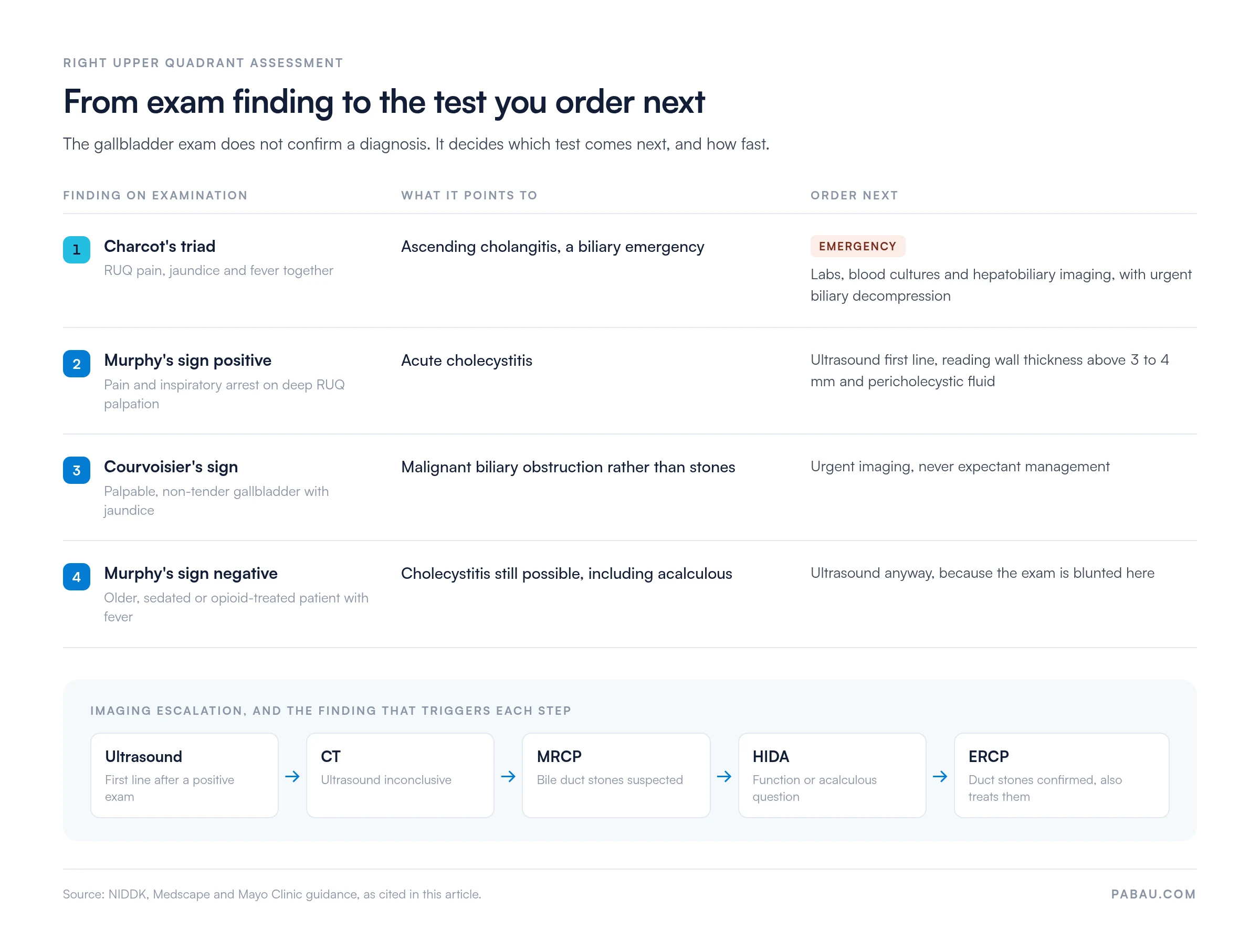
Standard post-exam workup includes:
Record the reasoning behind each test you order. That line is what tells the next clinician why a lipase was sent, and what would have changed if it came back high.
Ultrasound is the first-line imaging test after a positive gallbladder exam. It assesses gallbladder wall thickness, where more than 3 to 4 mm is abnormal. It also shows pericholecystic fluid, gallstones, and the sonographic Murphy’s sign.
If ultrasound is inconclusive, CT of the abdomen and pelvis gives a broader view. Magnetic resonance cholangiopancreatography (MRCP) is indicated when common bile duct stones are suspected.
A hepatobiliary scintigraphy (HIDA) scan measures gallbladder ejection fraction, which matters when acalculous cholecystitis or biliary dyskinesia is the working diagnosis. Endoscopic retrograde cholangiopancreatography (ERCP) both confirms duct stones and removes them.
A well-performed exam recorded as “abdomen soft, no tenderness” gives the next clinician no gallbladder information at all. Structured, specific documentation protects both the patient and the practitioner. It is also what makes a finding reproducible when someone repeats the exam tomorrow.
Use the SOAP structure for clear, reproducible notes. A worked example with the fields already laid out sits in our abdominal exam documentation template.
Our guide to writing safer clinical notes covers the principles that hold across every examination type, including audit and medico-legal requirements.
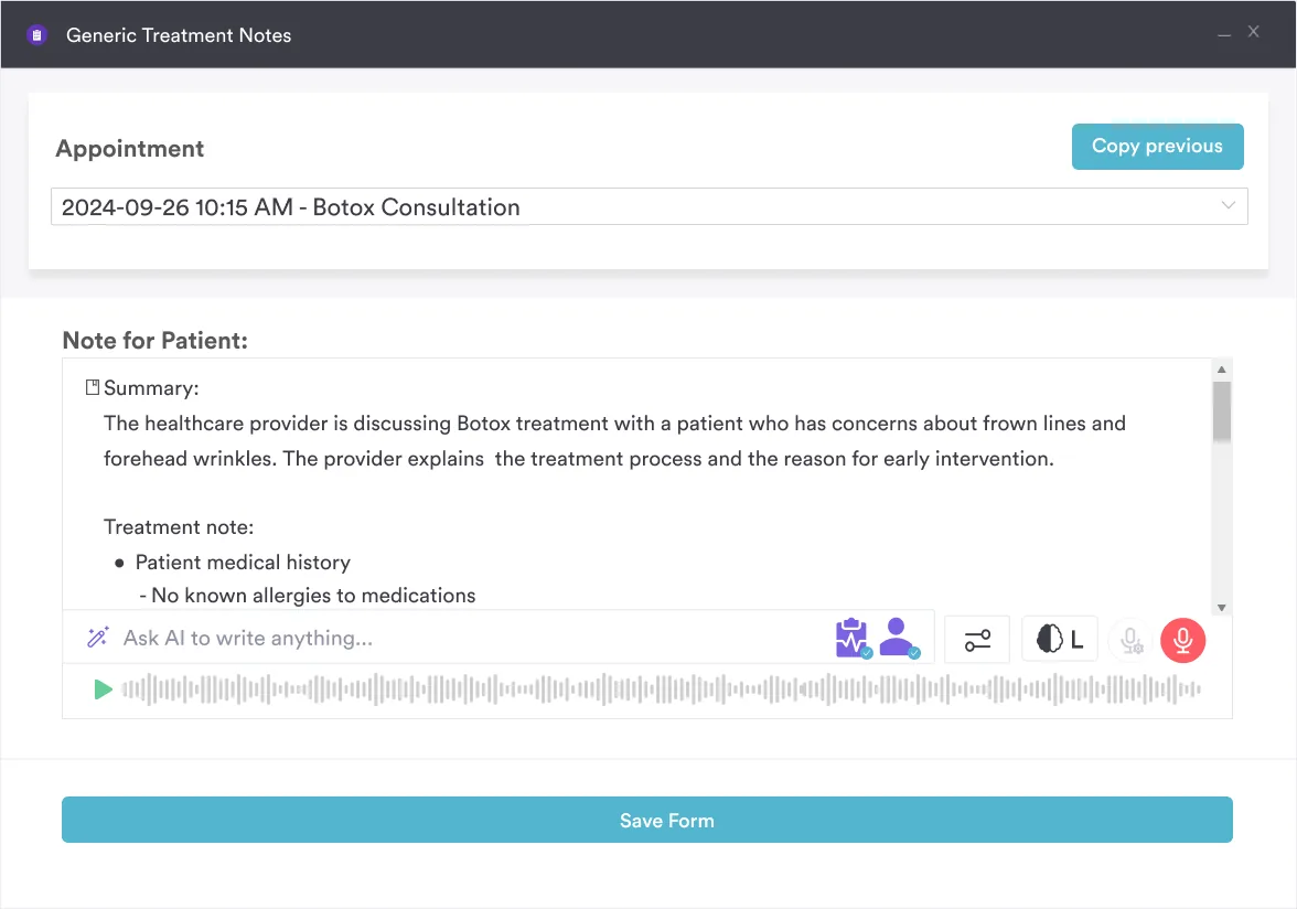
The shift from paper notes to digital clinical forms has made structured examination documentation more consistent. Free-text notes vary by clinician. A structured template prompts for each component of the examination instead.
Whether Murphy’s sign was tested, and what the result was, becomes a checkbox with a free-text field beside it. So does whether rebound tenderness was assessed. The prompt is what stops a maneuver going unrecorded.
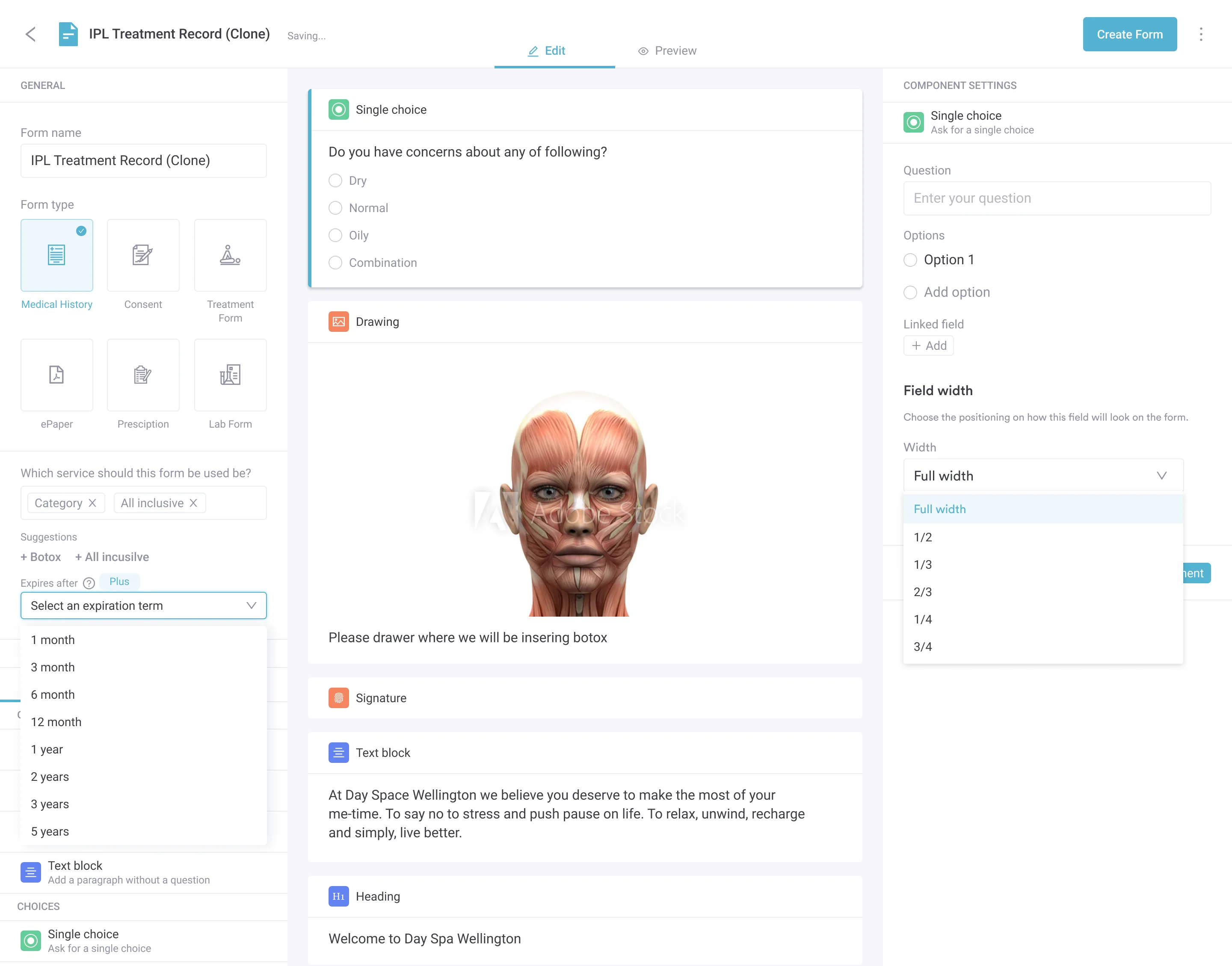
Voice-to-text tools change the time cost of note-writing. For an abdominal exam, though, a dictated finding is only as specific as what the clinician says aloud. Naming each maneuver out loud is what gets it into the record.
In most practices a gallbladder exam ends up in three places. The maneuvers go into a free-text note. The imaging request goes through a separate system, and the report comes back by email. Reconstructing the encounter later means opening all three.
Practice management software like Pabau keeps all three in one client record. A digital form holds the examination fields, so Murphy’s sign, Boas’ sign, guarding, and rebound each have somewhere to land. Pabau Scribe, our AI medical scribe, drafts the note from the consultation itself.
So you can palpate with both hands and read the note back afterwards, instead of typing while the patient holds a breath. The lab results, the imaging report, and the plan you wrote sit against the same encounter. When the patient returns, the previous exam is one click away.

Pabau helps physicians in primary care and private practice capture examination findings, lab results, and imaging requests in one structured client record. That removes the separate systems a gallbladder workup usually spans.

The gallbladder exam earns its place by deciding what happens in the next hour, not by naming a diagnosis. A positive Murphy’s sign sends the patient for an ultrasound. Charcot’s triad sends them somewhere faster.
The trade-off worth remembering is that a negative exam buys you very little. In an older, sedated, or opioid-treated patient, the absence of a sign is weak evidence. Order the imaging anyway.
Whichever way the exam goes, the note is what carries the decision forward. Book a demo to see how Pabau keeps exam findings, labs, and imaging reports against one client record.

Need the finished note, not just the technique? Abdominal exam documentation gives you the fields and worked examples for recording a right upper quadrant assessment.
Working through the wider abdominal assessment? Abdominal physical exam sets out the full four-quadrant sequence the gallbladder exam sits inside.
Another bedside examination to sharpen? Otoscope examination covers technique, findings, and documentation with the same structure used here.
Examining children rather than adults? Pediatric physical examination walks through the age-adjusted sequence and what changes in a young patient.
Want your notes to hold up under audit? Write faster, safer clinical notes covers the documentation principles that apply to every examination type.
A gallbladder physical exam is a targeted abdominal assessment used to detect signs of biliary pathology, including acute cholecystitis, biliary colic, and choledocholithiasis. It includes inspection, auscultation, percussion, and palpation of the right upper quadrant, with Murphy’s sign as the primary diagnostic maneuver.
Hook the fingers of your right hand under the right costal margin at the midclavicular line (Murphy’s point) and ask the patient to inhale deeply. A positive sign is when the patient experiences pain sufficient to cause inspiratory arrest as the inflamed gallbladder descends onto the examining fingers.
The earliest gallbladder symptoms are typically episodic right upper quadrant or epigastric pain occurring 15-60 minutes after fatty meals (biliary colic), accompanied by nausea. As disease progresses, pain becomes constant, fever develops, and patients may notice jaundice, indicating obstruction or acute cholecystitis.
No. A gallbladder physical exam guides the diagnostic workup but cannot confirm gallstones or cholecystitis without imaging. Ultrasound is the required first-line confirmatory test, with laboratory tests adding information about inflammation, obstruction, and organ involvement.
Murphy’s sign is elicited anteriorly by pressing under the right costal margin during inspiration. Boas’ sign is a posterior finding: hyperesthesia (increased skin sensitivity) in the right subscapular region caused by referred pain via phrenic nerve irritation. Both associate with acute cholecystitis but Boas’ sign has less modern validation data.