HIPAA compliant texting: What you can send patients & how to communicate securely
HIPAA compliant texting is possible when a text holds no protected health inform...

Last Updated: Oct 16, 2026
A bradykinesia test is a structured clinical assessment of slowness, shrinking amplitude, and fatigue in repeated voluntary movement. Neurologists and movement disorder specialists use it to detect parkinsonian motor impairment and stage how severe it is.
The standard version scores five MDS-UPDRS Part III tasks (items 3.4 to 3.8) from 0 to 4 on each side. Finger tapping sits at its core. According to the Parkinson’s Foundation, about one million people in the US live with Parkinson’s disease, so this is routine work in neurology. Scored consistently, the test turns a vague sense of “slower than last time” into data you can compare across visits.

A bradykinesia test measures movement slowness, amplitude reduction, and fatiguing decrements across standardized motor tasks.
MDS-UPDRS Part III items 3.4 to 3.8 are the validated scoring backbone for bradykinesia in clinical and research settings.
The finger tapping test is the most widely administered bedside component, scored on a 0 to 4 scale per hand.
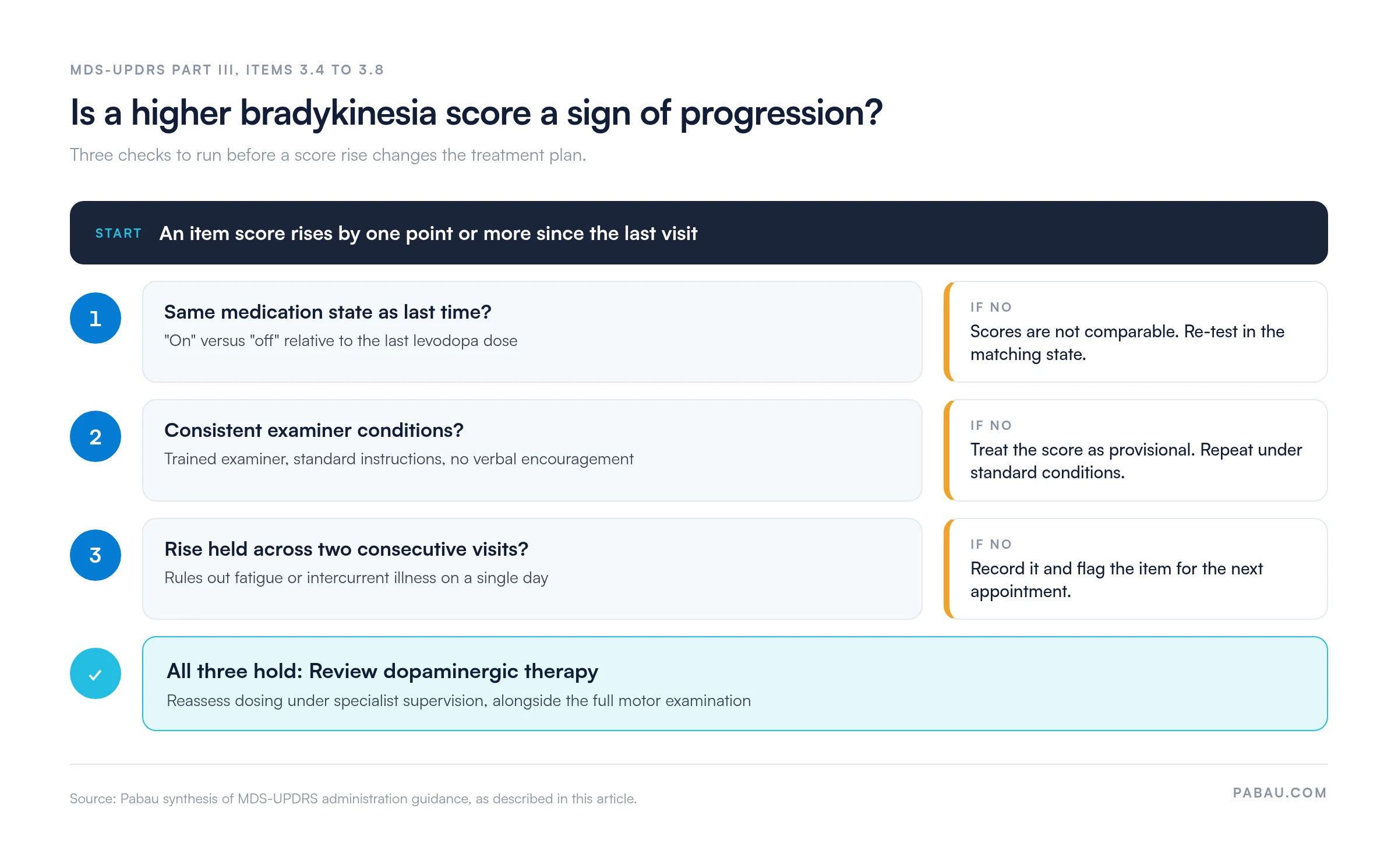
A higher score signals progression only when the medication state and examiner conditions match and the rise holds across two visits.
Structured longitudinal documentation of bradykinesia test scores supports disease staging, medication titration decisions, and medico-legal records.
A bradykinesia test is a motor examination protocol that quantifies three distinct movement deficits. These are speed reduction (bradykinesia proper), amplitude reduction (hypokinesia), and progressive fatiguing across repeated cycles (decrement). Under the Movement Disorder Society’s 2015 clinical diagnostic criteria, bradykinesia is an obligate cardinal feature of Parkinson’s disease (PD). Without it, a PD diagnosis cannot be made.
Dopamine loss in the substantia nigra of the basal ganglia disrupts the motor control signals that regulate movement initiation, speed, and rhythm. This mechanism underlies bradykinesia. It also separates PD-related slowing from the general slowness that comes with aging, depression, or musculoskeletal conditions.
Clinically, the test matters because it produces reproducible, trackable data. A neurologist who scores finger tapping at UPDRS 2 on first presentation and 3 at follow-up has objective evidence of progression. That same data informs levodopa dosing decisions, triggers specialist referrals, and supports medico-legal documentation. Subjective observation cannot serve those functions reliably.
Before formal assessment, bradykinesia shows itself through signs a practitioner can note during history-taking and general observation.
These pre-test observations set clinical context and help examiners focus on the most affected limbs during the motor examination. They also prompt differential thinking: Rigidity, tremor, and postural instability may co-present alongside bradykinesia, each requiring its own examination component. None of these signs alone is sufficient for a clinical bradykinesia test score.
Three categories of tool make up the current assessment landscape. The right one depends on the clinical setting, examiner training, and whether quantitative data or bedside efficiency is the priority.
The rapid finger tapping test is the most widely used component of any bradykinesia test battery. It appears as item 3.4 in MDS-UPDRS Part III and also works as a standalone bedside screen. The protocol below follows the MDS-UPDRS standardized instructions.
Examiners must not give verbal encouragement during the task. Verbal cues artificially improve performance in PD patients (a phenomenon called “paradoxical kinesia”) and invalidate the score. For the same reason, the patient must not watch a clock or visual cue while tapping.
Test the less affected limb first when the patient presents with asymmetric symptoms. Starting with the better side sets a performance baseline and avoids contamination of the affected-side score by warm-up effects.
A complete motor examination for bradykinesia uses several tasks beyond finger tapping. Each targets a distinct aspect of motor control and contributes a separate MDS-UPDRS subscore. Every task also has its own administration method and a defined observation target.
The patient opens and closes the hand repeatedly in a large, rapid movement. The examiner watches for reduced amplitude and progressive slowing over 10 seconds. This tests bradykinesia in a different motor program than the pincer-grip pattern of finger tapping.
The patient alternately pronates and supinates each forearm as rapidly as possible with the arm extended. Asymmetry between hands is clinically significant and often matches the side of the predominant dopaminergic deficit. The examiner watches for rhythm irregularity and decrement over 10 seconds.
Toe tapping mirrors the finger tapping protocol: The patient taps the heel on the floor while lifting the whole foot high and fast. Leg agility asks the patient to stamp the leg up and down alternately. These lower-limb items capture bradykinesia that may be disproportionately severe in patients whose upper-limb scores appear mild.
The pull test (also called the retropulsion test) is not a bradykinesia measure, but it’s always part of the full PD motor examination. The examiner stands behind the patient and delivers a firm, brief backward tug on the shoulders. The patient is warned beforehand to resist falling.
A normal response is one to two corrective steps. Three or more steps, or no recovery without examiner support, scores as postural instability. This test requires specific safety precautions: The examiner must be positioned to catch the patient. Skip the test if the patient has severe balance impairment or recent falls.
Where the pull test is unsafe, the timed up and go test offers a structured fall-risk screen that doesn’t rely on a backward tug.
The MDS-UPDRS (revised 2008) replaces the original UPDRS. Items 3.4 through 3.8 together make up the bradykinesia subscale. Each item is scored independently on a 0 to 4 ordinal scale, and the scores are not summed into a composite without specialist interpretation. Consistent examiner technique is the main determinant of score reliability.
Inter-rater reliability in MDS-UPDRS administration is moderately good in trained examiners. Intraclass correlation coefficients for the bradykinesia items fall in the 0.75 to 0.90 range, per MDS training validation studies. Reliability drops significantly in examiners who have not completed the official MDS-UPDRS training program.
Practices using the scale for research or medico-legal purposes should document examiner certification alongside each score.
The BRAIN (Bradykinesia Akinesia Incoordination) test is a computerized assessment that quantifies motor impairment through timed keystroke sequences on a standard keyboard. It was validated in a peer-reviewed study published in Movement Disorders (PMID 10928573). Bedside UPDRS scoring depends on trained examiner judgment, while the BRAIN test captures objective timing data.
That data includes inter-tap intervals, amplitude proxies from keystroke force, and the coefficient of variation across a tapping sequence. The patient alternately presses two designated keys as rapidly as possible for a defined period. Software then calculates bradykinesia, akinesia (movement initiation delay), and incoordination (rhythm irregularity) scores from the raw timing data.
This removes examiner-specific bias and produces a quantitative output you can compare directly across visits. The BRAIN test is most commonly used in research and specialist movement disorder practices. Its evidence base, while well established in the academic literature, comes from controlled research settings.
Before adopting it in routine practice, confirm it complements your standard UPDRS-based assessment rather than replacing it. Also check that your patient population reflects the cohorts in the original validation research. It is not a diagnostic instrument: Like all bradykinesia tests, it supports clinical assessment rather than replacing specialist evaluation.
Bradykinesia test results support clinical decision-making rather than standing alone as diagnostic conclusions. A single UPDRS subscore or BRAIN output does not diagnose Parkinson’s disease. It informs the clinician’s overall assessment within a full neurological examination, so score in context, compare across visits, and flag outliers for specialist review.
A score of 0 indicates no detectable bradykinesia on that item. Scores of 1 to 2 across multiple items suggest mild to moderate motor impairment consistent with early to mid-stage PD. Scores of 3 to 4 indicate significant functional impairment. They typically signal a need for medication review or dose adjustment under specialist supervision.
The pattern of change across visits carries more clinical weight than any single score. A patient stable at UPDRS 2 who rises to 3 across two consecutive appointments warrants reassessment of their dopaminergic therapy. An isolated rise at a single visit may reflect medication timing (testing during an “off” period), fatigue, or intercurrent illness rather than disease progression.
Before a higher score changes the treatment plan, it should clear the three checks below.
Progressive amplitude decrement across a 10-second tapping sequence is a highly specific sign of basal ganglia dysfunction. It differs from the overall slowness that any fatigued healthy person might show. Arrests are complete stops mid-sequence, sometimes called “freezing” of the limb. They’re scored as a separate qualifier and, when present, push the score toward 3 or 4 regardless of residual speed.
Bradykinesia is not exclusive to PD. Drug-induced parkinsonism, most commonly from antipsychotics and antiemetics acting on dopamine receptors, produces an identical motor picture on bedside tests. Vascular parkinsonism, multiple system atrophy (MSA), and progressive supranuclear palsy (PSP) all show bradykinesia alongside their own distinguishing features.
Cervical myelopathy also slows the hands and makes them clumsy, so patients fumble buttons and drop objects. A cervical myelopathy reflex screen separates cord compression from parkinsonian slowing before the exam settles on PD.
A bradykinesia test result alone cannot tell these conditions apart. The examiner has to integrate the finding with the full clinical picture, imaging, and history.
The bradykinesia test does not stand in isolation. A complete PD neurological examination covers four domains, and each contributes distinct diagnostic and staging information. Physical therapists who document neurological rehabilitation in physical therapy EMR software work across the same four domains.
Together, these four domains determine Hoehn and Yahr stage and whether a patient meets MDS clinical diagnostic criteria for probable or clinically established PD. A positive bradykinesia test alone is necessary but not sufficient for that determination.
Structured longitudinal documentation turns a series of point-in-time scores into a clinically useful progression record. The practical challenge in busy movement disorder practices is getting UPDRS subscores off paper forms and into a format that shows change between visits.
Effective documentation captures UPDRS items 3.4 to 3.8 per side, the medication state at the time of testing, and the visit date. Note the medication state as “on” or “off” relative to the last dose. This allows direct comparison across appointments, even when different clinicians administer the test.
Structured field-based recording outperforms free-text narrative for any scored assessment. Rehab and neurology teams choosing a system for it can compare the leading physical therapy practice software side by side.
Record the patient’s medication state alongside every UPDRS score. A score taken two hours after levodopa (‘on’ state) is not directly comparable to one taken before the morning dose (‘off’ state). Inconsistent recording of medication timing is the most common source of spurious score fluctuation in longitudinal PD data.
Many neurology and movement disorder practices still record UPDRS subscores on paper or in free-text notes. Finding last visit’s finger tapping score then means digging through scanned pages, and the medication state is often missing.
Practice management software like Pabau replaces that with structured fields. With clinical measurement tracking, UPDRS items 3.4 to 3.8 are recorded against each appointment and tracked over time. You can also build a standardized UPDRS form with a required medication-state field that saves straight to the patient record.
The score history is there before the patient sits down. Dosing conversations start from data rather than memory, and a rise that doesn’t clear the three checks above is easy to spot.

Pabau’s measurement tracking and digital forms let movement disorder practices record UPDRS subscores at each appointment. Every score is saved to the patient record, ready to compare at the next visit.

Choose the test that fits the question you’re answering. Finger tapping and the other MDS-UPDRS items suit routine bedside staging. The BRAIN test earns its place where you need examiner-independent timing data.
Then protect the score. Record the medication state, keep examiner conditions consistent, and wait for a rise to hold across two visits before changing therapy. That patience costs a visit, but it keeps an “off”-period score from triggering a dose change the patient didn’t need.
Book a demo to see how Pabau keeps every UPDRS score, medication state, and visit date in one patient record.

Assessing fall risk alongside the pull test? Timed up and go test covers how to administer and score the standard mobility and fall-risk screen.
Need a broader measure of hand function? Dexterity test explains the fine motor assessments that sit alongside finger tapping.
Checking for upper motor neuron signs? Clonus reflex test walks through the technique, grading, and documentation of the reflex exam.
Running a full neurological exam? Cranial nerves test shows how to assess all 12 cranial nerves during the same visit.
Documenting motor function in rehab? Motor assessment scale gives you a scoring guide and a free PDF template for functional motor tasks.
A bradykinesia test is a structured motor examination that quantifies movement slowness, amplitude reduction, and progressive fatiguing of repeated voluntary movements. The most common version uses the MDS-UPDRS Part III scoring system, with the finger tapping test (item 3.4) at its center. Results support clinical diagnosis and disease staging but don’t replace specialist neurological assessment.
Seat the patient with hands on their lap and demonstrate a large-amplitude tap of the index finger to the thumb. Then ask them to tap as big and as fast as possible for 10 seconds. Test each hand separately and watch for speed, amplitude, decrement, and arrests. Score it per MDS-UPDRS item 3.4 on a 0 to 4 scale, and don’t give verbal encouragement during the task.
Bradykinesia is most commonly caused by dopamine deficiency in the substantia nigra of the basal ganglia. This disrupts the neural signals that control movement initiation, speed, and rhythm. Parkinson’s disease is the leading cause. Bradykinesia also occurs with drug-induced parkinsonism (particularly from antipsychotics), vascular parkinsonism, multiple system atrophy, and progressive supranuclear palsy.
MDS-UPDRS scores of 0 indicate no impairment, 1 to 2 reflect mild to moderate impairment, and 3 to 4 indicate significant functional deficit. Change across visits is the most clinically meaningful signal: A score increase of one point across two consecutive appointments warrants review of dopaminergic therapy. That holds only if both tests ran under consistent medication and examiner conditions.
The BRAIN (Bradykinesia Akinesia Incoordination) test is a computerized assessment of keystroke timing, rhythm, and inter-tap intervals. The patient alternately presses two keys as rapidly as possible. Validated in peer-reviewed research (PMID 10928573), it produces objective scores without examiner bias. It’s used mainly in research and specialist movement disorder settings.
The pull test (retropulsion test, MDS-UPDRS item 3.12) assesses postural stability rather than bradykinesia. The examiner gives a firm backward tug on the patient’s shoulders and observes the corrective response. One to two steps is normal. Three or more steps, or failure to recover, indicates postural instability and feeds into Hoehn and Yahr staging and fall-risk assessment.