



GLP-1 diet plan
A GLP-1 diet plan is a structured eating framework for patients taking semagluti...

2
Last Updated: Oct 13, 2026

The clonus reflex test elicits rhythmic, involuntary muscle contractions by applying a sudden passive stretch to a joint.
In adults, clonus is generally pathological. StatPearls classifies more than 10 beats as sustained clonus, the most severe grade.
Clonus is not one of ACOG’s severe features of preeclampsia. SOMANZ and ISSHP guidance treat three or more beats of ankle clonus as a warning sign.
Pabau’s digital forms and structured patient records help practitioners document clonus findings, laterality, and beat counts consistently across neurological assessments.
Most neurological examinations include a clonus reflex test, yet documentation of findings varies widely between clinicians.
Beat counts go unrecorded. Laterality is omitted. Serial changes over time are lost. According to NCBI StatPearls, clonus in an adult is generally pathological, and more than 10 beats is classified as sustained clonus. That grading only earns its place when practitioners record what they found systematically. This guide covers what the clonus reflex test is and how to elicit it at each anatomical site. It also covers how to interpret the beats and how to document findings so they stay usable.
Clonus is a series of rhythmic, involuntary muscle contractions and relaxations triggered by a sudden passive stretch applied to a joint. Unlike a single deep tendon reflex response, clonus sustains itself for multiple beats as long as the stretch stimulus is maintained. It is a clinical sign of an upper motor neuron (UMN) lesion rather than a diagnosis in its own right.
Normal reflex arcs involve a one-time stretch response. In clonus, that response loops. The muscle contracts, the stretch is momentarily relieved, the muscle stretches again, and the cycle repeats. Three anatomical sites are routinely assessed: the ankle, the patella, and the wrist.
The Cleveland Clinic notes that clonus is associated with conditions including multiple sclerosis, stroke, cerebral palsy, spinal cord injury, and amyotrophic lateral sclerosis. It co-occurs with other UMN signs such as spasticity, hyperreflexia, and a positive Babinski reflex. Those are distinct findings and each one should be documented separately.
Upper motor neurons run from the cortex down the corticospinal tract to the spinal cord. They normally exert descending inhibitory control over lower motor neurons and the stretch reflex arc. When a UMN lesion disrupts that inhibitory pathway, the stretch reflex becomes disinhibited and exaggerated.
The gamma motor neuron loop is central to this process. Gamma motor neurons set the sensitivity of muscle spindles. Without adequate descending inhibition from UMN pathways, spindle sensitivity is elevated. A sudden passive stretch fires the spindle, which fires the alpha motor neuron, which contracts the muscle, which then re-stretches the spindle, restarting the cycle. That self-sustaining loop is clonus.
This is also why clonus co-occurs with spasticity and hyperreflexia. All three reflect the same underlying loss of descending inhibition, though each presents differently. Spasticity is velocity-dependent increased tone. Hyperreflexia is an exaggerated tendon jerk.
Clonus is the oscillating loop. Naming the shared mechanism helps when only one of the three signs shows up on examination. Practices using structured patient records in practice management software like Pabau can capture all three findings in one assessment note.
The clonus reflex test requires the patient to be relaxed. Muscle guarding from pain, anxiety, or an awkward position introduces voluntary tone that masks the reflex. Position carefully before applying the stimulus at each site.
The ankle clonus test is the most frequently performed clonus assessment. Use it as the primary site in any standard neurological exam.
A normal response is no oscillation at all. Document the number of beats, which ankle, and whether the beats continued or subsided spontaneously. Clonus is elicited in the same pass as the ankle jerk, so record both together. A structured deep tendon reflex exam keeps the reflex grades and the clonus beat count on one sheet.
Patellar clonus is less commonly assessed but remains part of a comprehensive neurological examination, particularly when ankle findings are equivocal.
Rhythmic oscillation of the patella and quadriceps against maintained pressure is a positive patellar clonus test. Record the exact beat count rather than a bare positive or negative. Document the finding alongside the ankle assessment to establish the pattern and severity of UMN involvement.
Wrist clonus is the least commonly elicited of the three sites. It is most relevant in patients with suspected cervical myelopathy or high spinal cord lesions affecting upper limb function.
Wrist clonus in isolation is uncommon. When present alongside ankle and patellar clonus, it suggests a more widespread UMN lesion affecting both upper and lower limbs.
Beat count is the primary metric for interpreting the clonus reflex test. No universally validated numeric grading scale exists, but the framework below follows NCBI StatPearls and standard clinical teaching. StatPearls is explicit on two points. Clonus in an adult is generally pathological, and sustained clonus means more than 10 beats.
A five-beat cutoff for sustained clonus is still quoted in some teaching material. StatPearls sets the sustained threshold higher, at more than 10 beats, so record the exact count every time. A number travels between clinicians. A label does not.
Two beats of clonus is a commonly asked-about finding. On its own, two beats does not confirm a UMN lesion. It carries weight when it occurs alongside other UMN signs such as hyperreflexia, a positive Babinski, or spasticity. Document the full neurological picture rather than relying on any single finding in isolation.
Practitioners working in a physical therapy EMR often track clonus longitudinally. Serial beat counts help monitor disease progression or treatment response in conditions like multiple sclerosis and spinal cord injury.
Clonus in an adult signals disruption to the upper motor neuron pathway, and sustained clonus makes that signal stronger. It does not identify the specific cause or location of the disruption. It is one piece of a larger diagnostic picture. The conditions below are consistently associated with clonus in clinical practice, including in the lists published by Physiopedia and the Cleveland Clinic.
Clonus confirms a UMN sign is present. It does not localize the lesion or confirm a diagnosis. Imaging, history, and additional neurological findings complete the picture. Occupational therapists monitoring functional recovery in these conditions can benefit from occupational therapy practice software that tracks assessment findings over time.
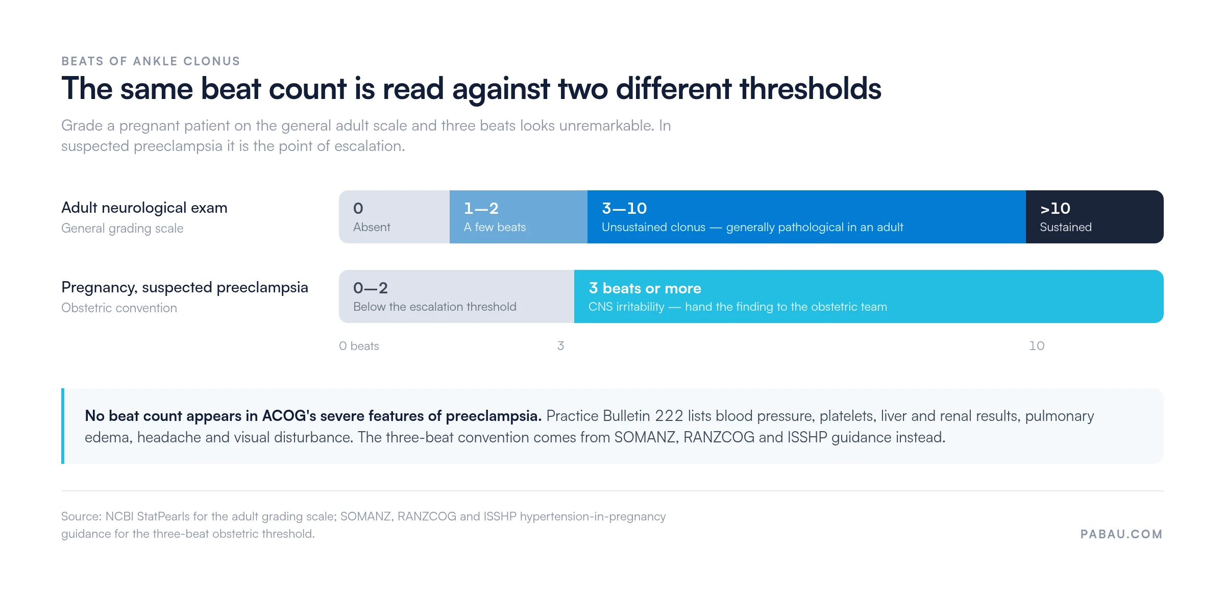
Clonus assessment during pregnancy is not routine neurological testing. It is a targeted check for central nervous system irritability in a patient with suspected preeclampsia, and a positive result demands immediate escalation.
Clonus is not one of ACOG’s severe features of preeclampsia. Practice Bulletin 222 lists blood pressure at or above 160/110 mmHg, thrombocytopenia, impaired liver function, renal insufficiency, pulmonary edema, treatment-resistant headache, and visual disturbances. No beat count appears anywhere in that list.
The three-beat convention is real, but it comes from elsewhere. SOMANZ, RANZCOG, and ISSHP guidance on hypertensive disorders of pregnancy treats three or more beats of ankle clonus as a sign of CNS irritability. Clinicians use it to support a decision on magnesium sulfate prophylaxis, alongside blood pressure, symptoms, and laboratory results. Reading the same beat count against both scales shows why the context matters.
Practitioners working in obstetric or women’s health settings should document clonus findings with timestamps, beat count, laterality, and the clinical context that prompted testing. Pre-built fields for neurological examination in the patient record prevent omissions in high-pressure situations.
Document ankle clonus in obstetric patients with a timestamp, the beat count per ankle, and the clinical trigger for the assessment. Record the trigger itself, such as a systolic blood pressure above 160 mmHg. This supports handover to obstetric or emergency teams and leaves a clear record if the patient later develops eclampsia.
Clonus can be mistaken for tremor or fasciculation during a rapid assessment. The three phenomena are distinct in their mechanism, trigger, and clinical significance. Getting the differential right matters because the management implications differ substantially.
A practical clinical distinction: clonus stops (or markedly reduces) when you release the stretch stimulus. Tremor does not stop because it is not stretch-dependent. Fasciculations are visible but not palpable as rhythmic beats against the examiner’s hand. If you are unsure whether you are feeling clonus or muscle guarding, reposition the patient, reassess after a few minutes, and compare both sides.
Incomplete records are where clonus assessments most commonly lose their diagnostic value. A beat count that sits in a handwritten note and is never reviewed or compared against baseline contributes nothing to the patient’s management. The same applies when the finding never reaches the rest of the care team.
A complete clonus record should capture six elements for every assessment.
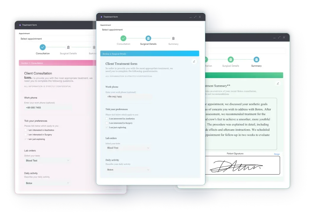
Practitioners writing structured neurological examination notes benefit from templates that prompt for each element. Pabau’s digital intake forms let a practice build custom examination templates with dropdown fields for beat count, laterality checkboxes, and UMN sign co-documentation. Findings then get captured the same way regardless of which clinician performs the assessment.
Serial monitoring in MS or spinal cord injury depends on the first assessment. Record it in a searchable, comparable format, or later comparisons rest on guesswork. Aim for specificity, structure, and completeness at the point of care rather than reconstruction from memory later.
A neurological examination is hard to write up while you are still performing it. Pabau Scribe, our AI scribe, transcribes examination findings in real time. That cuts the cognitive load during a complex assessment. The beat count lands in the patient record instead of an unrecorded verbal handover.
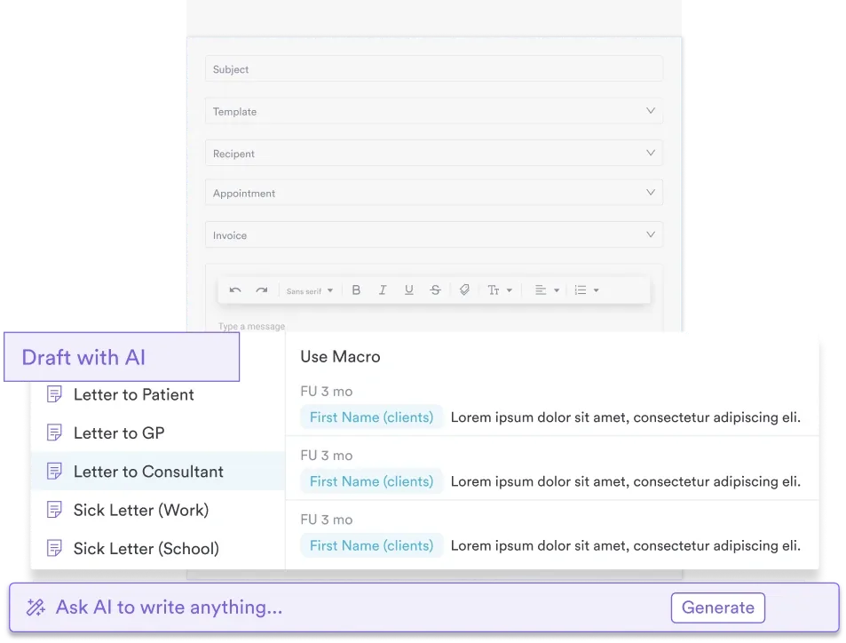
Teams working across neurology, physical therapy, and occupational therapy can start from a ready-made examination template. Adapt it once and every clinician records the same fields in the same order, which is what makes serial comparison possible.
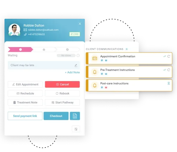
Automated workflow rules can then trigger a follow-up task when a clonus finding is positive. That prompts an imaging referral or specialist escalation without a manual chase. An abnormal beat count no longer sits in a note waiting for someone to notice it.

Pabau gives physical therapy and neurology practices digital examination forms, structured patient records, and AI-assisted documentation. Clonus findings get captured the same way across every assessment and every clinician.

The clonus reflex test is a high-value bedside tool, but its value depends on how findings are elicited, interpreted, and recorded. Clonus in an adult is generally pathological, and more than 10 beats counts as sustained clonus. In a pregnant patient, three or more beats of ankle clonus flags CNS irritability and warrants urgent obstetric review. None of that helps if the beat count, laterality, and clinical context never reach the record.
Pabau’s digital forms and structured patient records give neurology and rehabilitation teams a documentation framework they can reuse. It turns a 30-second bedside test into a data point you can track over time. To see how Pabau supports systematic neurological examination workflows, book a demo.

Need the wider reflex examination this test sits inside? Reflex test covers how to elicit and grade every deep tendon reflex in a standard neurological exam.
Want the reflex grades and the clonus count on one sheet? Deep Tendon Reflex Exam is a ready-made form for recording reflex grades, laterality, and beat counts.
Assessing cranial nerves in the same neurological workup? Cranial Nerve Examination Checklist walks through all twelve nerves in a fixed order so nothing is skipped.
Tracking motor recovery after stroke or spinal cord injury? Fugl-Meyer Assessment Form scores motor function over time, which is where serial clonus counts earn their value.
The clonus reflex test is a bedside neurological assessment that elicits rhythmic involuntary muscle contractions by applying a sudden passive stretch to a joint. Clonus is caused by loss of descending inhibition from an upper motor neuron lesion, which lets the stretch reflex arc self-sustain in an oscillating loop. Common underlying causes include stroke, multiple sclerosis, spinal cord injury, cerebral palsy, and preeclampsia with severe features.
Position the patient supine with the knee slightly flexed. Support the calf with one hand and cup the sole of the foot with the other. Apply a firm, rapid dorsiflexion in a single movement. Maintain the pressure and count the rhythmic beats that follow. In an adult, clonus is generally pathological at any beat count, and more than 10 beats is classified as sustained clonus.
In an adult, clonus is generally pathological at any beat count, according to NCBI StatPearls. StatPearls reserves the term sustained clonus for more than 10 beats, the most severe grade. A few beats can also reflect anxiety, fatigue, or muscle guarding, so interpret a short run alongside other upper motor neuron signs. The five-beat cutoff quoted in older teaching material does not match the StatPearls scale.
Clonus is triggered by passive stretch and stops when the stretch is released. It reflects an upper motor neuron lesion. Tremor is spontaneous and persists regardless of examiner input. It arises from basal ganglia, cerebellar, or essential tremor mechanisms rather than UMN disruption. Clonus produces rhythmic beats felt against the examiner’s hand at the joint being stretched. Tremor oscillates a whole body part during voluntary movement or at rest.
Clonus involves rhythmic contraction of a whole muscle group in response to passive stretch, and it signals an upper motor neuron lesion. Fasciculation is a spontaneous, brief, irregular twitch of a single muscle fascicle visible under the skin. It signals lower motor neuron or anterior horn cell pathology, such as ALS. Fasciculations occur at rest without an examiner-applied stimulus and do not produce the repeating rhythmic beats felt in clonus.
Ankle clonus assessment in pregnancy uses the same technique as the standard test. Apply rapid dorsiflexion, maintain the pressure, and count the beats. Clonus does not appear in ACOG’s severe-feature criteria for preeclampsia. SOMANZ, RANZCOG, and ISSHP guidance treats three or more beats of ankle clonus as a sign of CNS irritability. A positive finding in a pregnant patient with raised blood pressure needs urgent obstetric review.






