Nitrates medication list
A nitrates medication list is a point-of-care reference that sets out the main c...

Last Updated: Oct 6, 2026

A cranial nerves test works through all 12 nerves in a fixed order, from smell (CN I) to tongue movement (CN XII).
Seven bedside items cover the full exam: Penlight, ophthalmoscope, Snellen chart, cotton wisp, tongue depressor, tuning fork, and familiar-scent containers.
Upper motor neuron facial palsy spares the forehead, while a lower motor neuron lesion such as Bell’s palsy affects the whole ipsilateral face.
Document each nerve group by name, with the finding and the time, because vague wording will not support a specialist referral.
Pabau’s digital forms and AI-assisted documentation record structured neuro findings at the point of care.
A cranial nerves test checks all 12 cranial nerves in sequence, one function at a time, to find where a neurological problem sits. It takes 10 to 15 minutes, needs seven pieces of bedside equipment, and localizes lesions that a scan may not explain on its own.
This guide covers the technique for each nerve, the normal finding, and the abnormality that changes your management. It also covers what comes after the exam. That means how to word the note, and how much of the sequence each clinical setting expects.
The cranial nerves test assesses all 12 cranial nerves in sequence to detect deficits in the brain, brainstem, or peripheral nervous system. Standard neurological practice is to work through them in the same order every time, so no nerve gets skipped when the exam is interrupted.
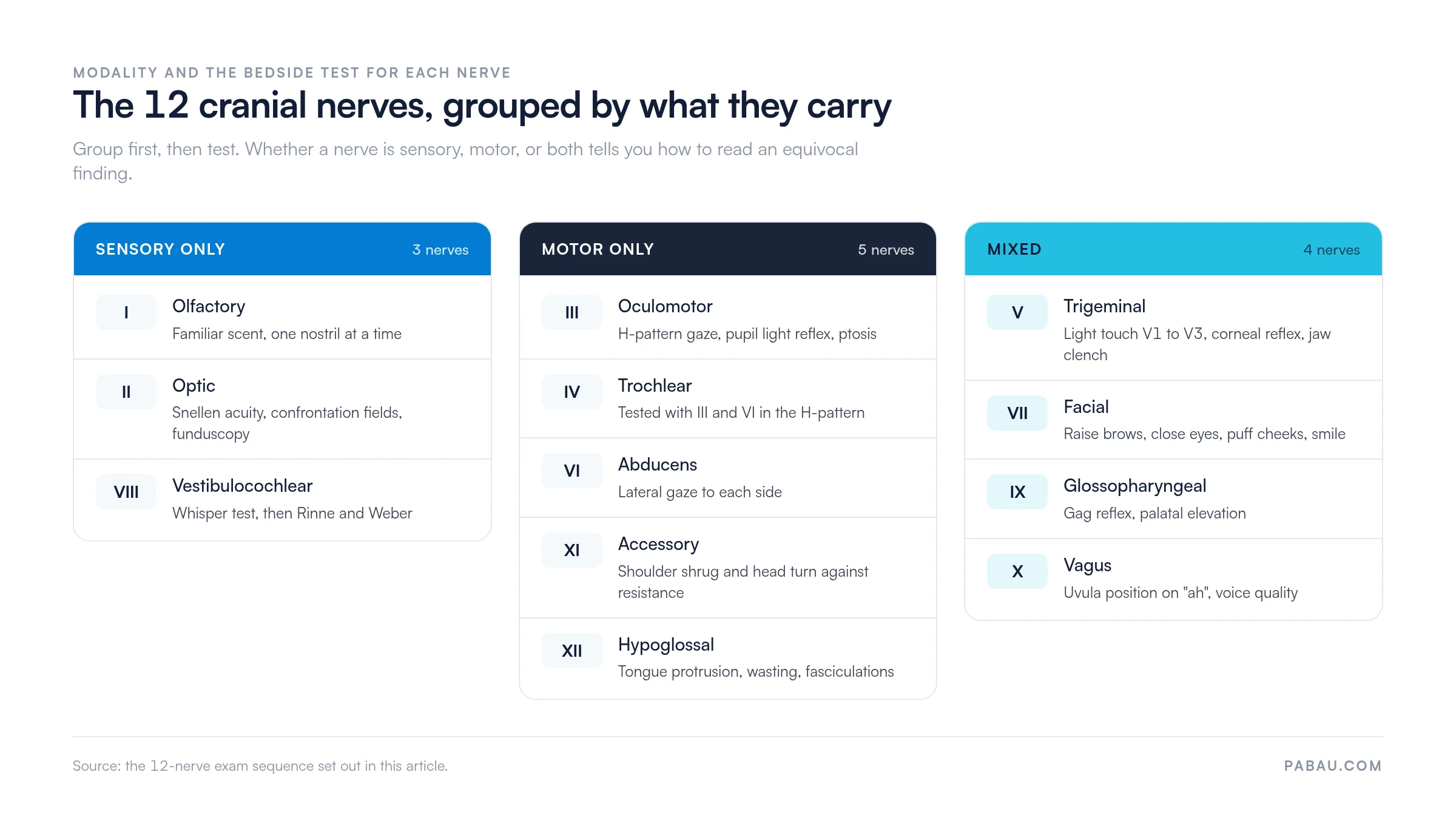
Each nerve has a distinct function: Sensory, motor, or both. Knowing which nerves are purely motor, purely sensory, or mixed tells you how to read a finding that sits on the edge of normal.
The grouping below sets out which nerves fall into each category, with the bedside test each one needs.
The full kit fits in a standard bedside bag. Set it out before the patient arrives so the exam runs without a pause to fetch something.
Position the patient seated upright when possible. Explain each test before you perform it. A relaxed, cooperative patient gives more reliable results, especially for subtle findings like mild facial asymmetry or early nystagmus.
The convention is CN I through CN XII in sequence. In practice, III, IV, and VI are tested together, because they jointly control eye movement. IX and X are also tested together, since their functions overlap. What follows covers technique, normal finding, and key abnormality for each nerve group.
Occlude one nostril. Present a familiar, non-irritant scent (coffee, vanilla, peppermint) to the other. Repeat on the opposite side. The patient should identify the odor correctly with each nostril independently.
Normal finding: Correct identification bilaterally. Abnormal: Anosmia or hyposmia. Unilateral loss may point to an ipsilateral olfactory groove meningioma or a frontal lobe lesion. Bilateral loss after COVID-19 may reflect central olfactory pathway involvement, so it warrants investigation beyond CN I alone.
Test visual acuity with a Snellen chart at 20 feet, or with a near-vision card. Then assess visual fields by confrontation. Cover your own eye, hold fingers in the four quadrants, and ask the patient to report when they appear. Perform funduscopy to examine the disc and retina.
Normal: 20/20 acuity, full fields, pink disc with sharp margins. Abnormal: Field defects localize the lesion, and a homonymous hemianopia suggests post-chiasmal pathology. Papilledema indicates raised intracranial pressure (ICP) and requires urgent referral.
Ask the patient to hold their head still and follow your finger in an H-pattern. Watch for full, conjugate, smooth movement across all six cardinal directions. Assess pupil size, symmetry, and the direct and consensual light reflex. Note any ptosis.
Normal: Full extraocular movements, no nystagmus, no diplopia, pupils equal and reactive to light (PERRL). Abnormal: CN VI (abducens) palsy is the most common isolated cranial nerve palsy, and it produces failure of lateral gaze on the affected side. A CN III palsy with a dilated, unreactive pupil and ipsilateral headache may indicate a posterior communicating artery aneurysm. Treat that as a neurosurgical emergency and escalate immediately.
Test light touch across the three divisions: V1 (ophthalmic, forehead), V2 (maxillary, cheek), and V3 (mandibular, jaw). Use a cotton wisp and ask the patient to report where they feel it with their eyes closed. Test the corneal reflex by touching the cornea lightly from the side, and both eyes should blink. Then ask the patient to clench their teeth while you palpate the masseters.
Normal: Symmetric sensation in all three divisions, an intact corneal reflex bilaterally, and equal jaw muscle bulk. Abnormal: Unilateral facial numbness in a dermatomal pattern localizes the lesion. An absent corneal reflex may indicate CN V sensory loss or CN VII motor loss, the afferent and efferent limbs respectively.
Observe facial symmetry at rest. Then ask the patient to raise their eyebrows, close their eyes tightly against resistance, puff their cheeks, and show their teeth. Asymmetry on any of these tasks points to facial nerve weakness.
The upper motor neuron (UMN) versus lower motor neuron (LMN) distinction matters here. In a UMN lesion such as stroke, the forehead is spared, because it receives bilateral cortical input. In an LMN lesion such as Bell’s palsy, the whole ipsilateral face is affected, forehead included. That single observation sets both the urgency and the direction of your management.
Start with a gross whisper test. Stand behind the patient, occlude one ear, and whisper a two-syllable word. Then perform the Rinne and Weber tests with a 512 Hz tuning fork.
Ask the patient to open their mouth and say “ah.” Observe palatal elevation and uvula position. In unilateral vagus nerve palsy, the uvula deviates away from the side of the lesion, toward the intact side. Test the gag reflex with a tongue depressor. An absent gag reflex is not always pathological, so read it against the rest of the exam.
Assess voice quality and ask about dysphagia. Hoarseness suggests recurrent laryngeal nerve involvement. Dysphagia with hoarseness points to a vagal lesion at or below the jugular foramen.
Test the trapezius by asking the patient to shrug both shoulders against your downward resistance. Test the sternocleidomastoid by asking the patient to rotate their head against your resistance on each side.
Normal: Strong, equal bilateral muscle action. Abnormal: Weakness in shoulder shrug or head rotation on one side indicates an ipsilateral CN XI lesion. Common causes are neck dissection, a jugular foramen tumor, and direct accessory nerve injury.
Ask the patient to protrude their tongue. Inspect for deviation, wasting, or fasciculations. In a lower motor neuron CN XII lesion, the tongue deviates toward the side of the lesion. Fasciculations indicate a lower motor neuron or nuclear lesion.
Normal: Tongue midline on protrusion, no wasting, no fasciculations. Abnormal: Deviation, atrophy, or fasciculations call for further investigation of the brainstem, skull base, or peripheral nerve.
The table below works as a bedside reference for the full cranial nerves test. It consolidates technique, normal findings, and the key abnormal finding for each nerve group.
If you would rather work from a printed form, the cranial nerve nursing assessment covers the same 12 nerves in the same order.
Most abnormal cranial nerve findings are non-urgent and guide outpatient investigation. A handful are emergencies, and telling the two apart matters as much as the technique itself.
When any of these patterns appears, record what you found, when you found it, and against what baseline. Then escalate without delay, because a vague note slows specialist review down.
Name the nerve group, state the finding, and give the time. A note reading “neuro exam unremarkable” creates medico-legal risk and leaves the next clinician guessing which nerves you tested.
For neurological findings, these conventions are widely accepted:
Structured templates with a pre-mapped field for each nerve group remove the risk of missing one. Purpose-built clinical documentation software keeps that field order identical for every clinician. A note written on the ward then reads the same way in outpatient neurology.
Build a cranial nerve exam template with one field per nerve group and a dropdown for normal, abnormal, or not assessed. It takes a minute or two at the bedside and gives you a specialist-readable note every time. Pabau’s digital forms make the template reusable at every encounter.
No two settings demand the same depth of cranial nerve assessment. What counts is knowing which abbreviated version is clinically defensible where you work.
Telehealth limits what you can test by hand, so lean on observation and patient self-report. Bring the patient in for an in-person exam as soon as you see asymmetry.
On a ward, the value sits in the trend. Record the same fields at every assessment and compare each one against the documented baseline.
Most practices still write cranial nerve findings as free text, into a paper chart or a blank note field. That leaves the order of the nerves, the wording, and the level of detail up to whoever is holding the pen.
Practice management software like Pabau replaces that blank field with a form you build once. Our digital forms for practitioners let you map one field per nerve group and set the response options. You then reuse the same template at every encounter.
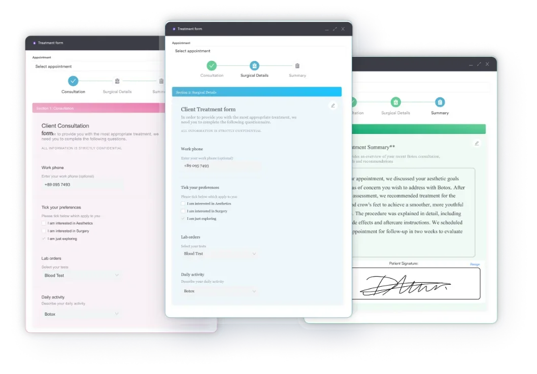
Completed forms write straight into the patient’s structured client records, so the specialist and the nurse on the next shift read the same note.
Pabau Scribe, our AI scribe, adds AI-assisted clinical documentation on top. Dictate findings as you work through the sequence and they land in those same fields. That cuts the delay between examining and recording, which is where recall errors creep in.
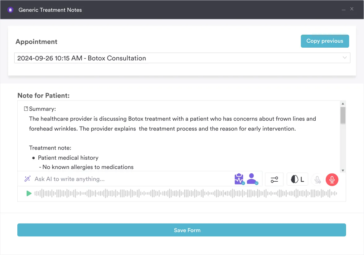

Pabau gives clinical teams structured note templates and AI-assisted documentation that capture cranial nerve findings in the format specialists read. Spend less time writing and more time assessing.

The exam itself is the part you can drill. The sequence needs seven pieces of equipment and 10 to 15 minutes, and you can rehearse it until it runs identically every time.
What decides whether the exam was worth doing is the note that comes out of it. A finding nobody can read six weeks later did not change anybody’s management, and that is where a fixed field order earns its place.
So pick your abbreviated version for each setting, write it into a template, and use the same one every time. Book a demo to see how Pabau structures cranial nerve documentation across your practice.

Need a printable form for the exam itself? Cranial nerve nursing assessment gives you a field for each of the 12 nerves, ready to complete at the bedside.
Assessing a patient who is not fully alert? Level of consciousness assessment sets out how to record responsiveness before you attempt the nerve sequence.
Need a faster neuro check in an emergency? AVPU scale covers the four-point responsiveness screen used when there is no time for a full exam.
Comparing tools for writing clinical notes? Clinical notes software walks through what to look for when notes have to satisfy a specialist reviewer.
Documenting a full systems review as well? 12-point review of systems lists the systems and the wording a complete review needs.
A cranial nerves test is a bedside neurological assessment of all 12 cranial nerves. It checks their sensory, motor, and reflex functions to find deficits in the brain, brainstem, or peripheral nervous system. It forms a core part of any full neurological examination.
Each nerve has its own technique. Smell identification covers CN I, Snellen acuity and funduscopy cover CN II, and the H-pattern with pupil responses covers CN III, IV, and VI. Light touch and the corneal reflex test CN V, facial expression tasks test CN VII, and Rinne and Weber test CN VIII. Palatal elevation and the gag reflex cover CN IX and X, shoulder shrug covers CN XI, and tongue protrusion covers CN XII.
Abnormal findings localize the pathology. Multiple simultaneous palsies suggest a brainstem or skull base lesion. A dilated, unreactive pupil with CN III palsy may indicate a posterior communicating artery aneurysm. Unilateral CN VII weakness with forehead sparing points to an upper motor neuron lesion such as stroke. Each pattern carries a different urgency and a different investigation pathway.
The H-pattern test assesses the three nerves that control eye movement: CN III (oculomotor), CN IV (trochlear), and CN VI (abducens). The patient tracks a finger moving in an H shape through all six cardinal directions of gaze. Any failure of smooth conjugate movement, nystagmus, or diplopia in a given direction identifies which nerve is affected.
Escalate immediately for a CN III palsy with a dilated, unreactive pupil and headache, which may be an aneurysm. Also escalate for sudden bilateral CN VI palsy, bilateral facial weakness, papilledema on funduscopy, or multiple simultaneous palsies. These findings need urgent imaging and specialist review rather than watchful waiting.
A focused screen of CN II, III, VII, and XII is appropriate in three situations. Those are emergency triage for acute stroke, and a telehealth consultation where physical testing is limited. The third is a patient too unwell to tolerate the full sequence. Complete the remaining nerves as soon as the patient’s condition allows.