Bear hug test: Procedure, interpretation and free template
The bear hug test is a quick shoulder special test that screens for subscapulari...

2
Last Updated: Oct 23, 2026

The prone knee bend test, also called the reverse Lasègue test, screens the L2 to L4 nerve roots.
It works by tensioning the femoral nerve, so a positive result is anterior thigh or groin pain.
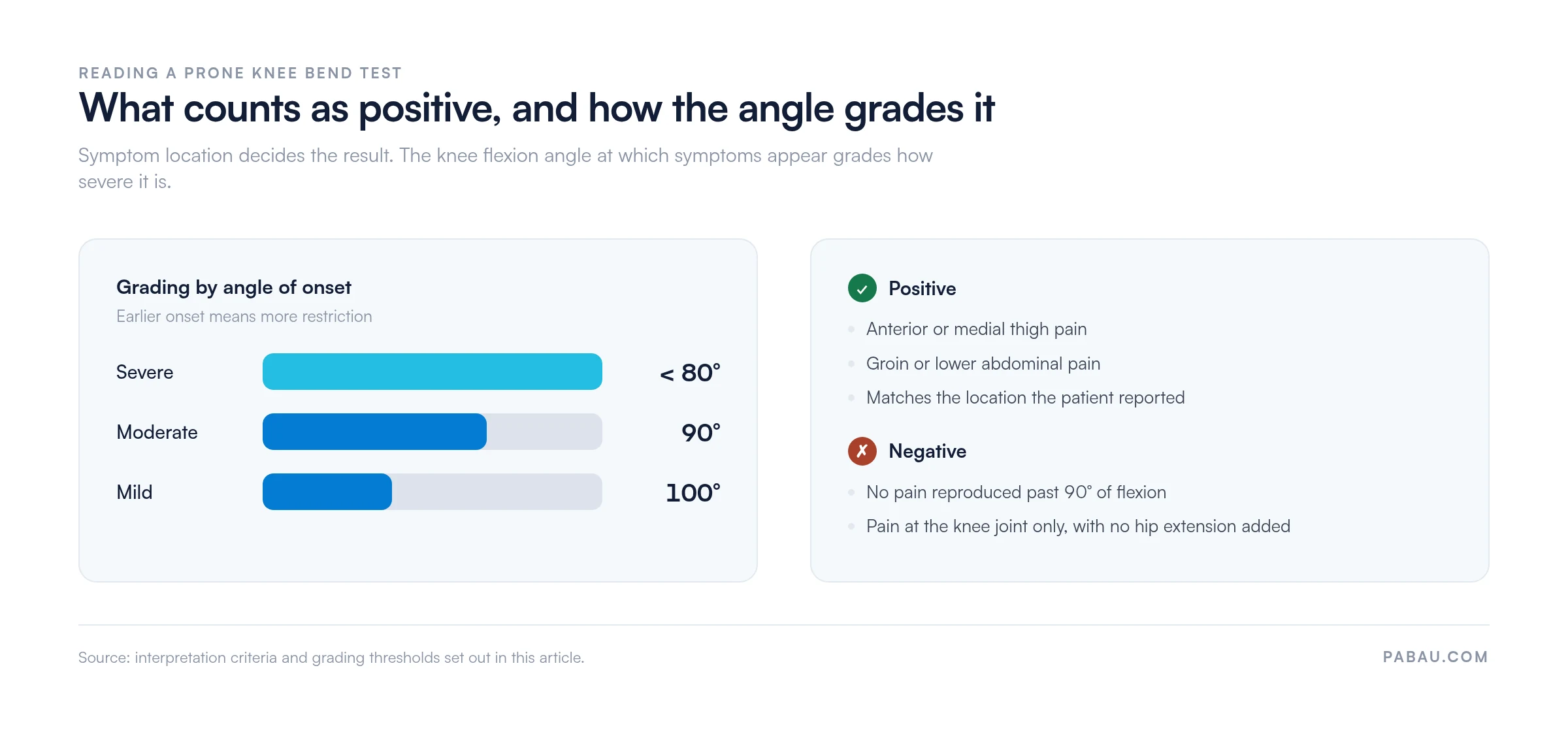
Grade the finding by the angle of onset: mild near 100°, moderate near 90°, severe below 80°.
Published sensitivity and specificity vary by study, so read the result next to the history and other tests.
Practice management software like Pabau keeps the form, the angles, and the treatment note on one record.
A one-page assessment form with fields for patient positioning, flexion angle by side, symptom location, and pain score. It closes with a SOAP note section you can copy straight into the patient record.
Download templateThe prone knee bend test screens the upper lumbar nerve roots, L2 to L4, by putting the femoral nerve under tension. The patient lies prone, you flex the knee passively, and anterior thigh or groin pain counts as a positive finding.
This page covers the procedure, the thresholds that grade a positive result, the contraindications, and the wording to put in the note. The form above gives you the same fields to complete at the treatment table.
It is a special test for femoral nerve tension, also known as the reverse Lasègue test or the femoral nerve stretch test. Physical therapists, sports medicine physicians, chiropractors, and osteopaths all use it in a lower limb neurological exam.
Passive knee flexion in prone applies longitudinal tension along the femoral nerve and stretches the quadriceps group. Pain in the anterior thigh or groin points to nerve root involvement or muscle restriction at L2 to L4.
The straight leg raise targets the lower lumbar and sacral roots, L4 to S2. This test isolates the upper lumbar structures instead, which is what makes it useful when anterior thigh pain is the main complaint.
The femoral nerve comes off the lumbar plexus at L2 to L4. It runs through the psoas major and under the inguinal ligament before it divides into motor and sensory branches.
The motor branches supply the quadriceps, sartorius, pectineus, and iliopsoas. The sensory branches cover the anterior and medial thigh, the medial lower leg, and the medial foot.
Passive knee flexion in prone loads that whole course at once. An upper lumbar disc herniation, nerve root compression, or foraminal stenosis at L2, L3, or L4 turns the tension into familiar pain.
That anatomy also sets the limits of the test. Pain below the knee or in the lateral leg points somewhere else, usually L5 to S1 pathology that the straight leg raise picks up.
Use it when a patient presents with anterior thigh pain, suspected femoral nerve compression, or upper lumbar radiculopathy. Common scenarios include:
Sports medicine and physical therapy teams lean on it for a fast differential before imaging is ordered. A clear result also tells you whether nerve tension is limiting a staged plan, such as a return to running protocol.
Not every patient is suitable for it. Do not perform this test if any of the following apply:
Relative precautions: high-dose anticoagulation, osteoporosis, and suspected vertebral fracture all call for gentle handling. Explain what the test involves and take consent before you start.
Follow a standardized sequence so the result stays comparable between sessions and between clinicians.
Capture the flexion angle reached, symptom reproduction and its location, pain intensity out of 10, and any guarding or compensation you saw.
Positive test: pain in the anterior or medial thigh, the groin, or the lower abdomen, reproduced during passive knee flexion. The location should match what the patient described in the history.
Negative test: no pain reproduction at all. Pain confined to the knee joint also reads as negative, once flexion passes 90° with no hip extension added.
Grading: many clinicians grade the finding by the angle at which symptoms start. Mild sits near 100° of flexion, moderate near 90°, and severe below 80°. Recording that angle is what makes the next session comparable.
Hip flexor length is the most common confounder here, so measure it separately rather than inferring it. The Thomas test gives you a cleaner read on iliopsoas and rectus femoris restriction.
Published accuracy data comes mostly from patients with lumbar disc herniation and nerve root compression. The figures shift with the reference standard used, whether that is imaging, surgical findings, or clinical diagnosis.
Those figures support moderate to good utility once you read them next to the history. No single special test confirms or excludes pathology on its own. Combine the result with a neurological screen, imaging where it is indicated, and tests such as the slump test.
Several variations exist, each changing the neural tension or the mechanical constraint applied:
Pick the version that matches your clinical question and the patient’s tolerance. Record which one you used, so the next clinician repeats it the same way.
This is one tool in a wider examination. Combine it with the tests below to narrow the differential:
A positive straight leg raise with a negative prone knee bend points to lower lumbar pathology at L4 to S1. The reverse pattern points upward instead, to L2 or L3.
The crossed straight leg raise is worth adding when a disc herniation is the working hypothesis. It trades sensitivity for specificity, so a positive result there carries more weight.
None of this helps the differential unless the findings sit side by side in the record. A structured physical therapy intake form keeps the history, the range of motion figures, and every special test in one place.
Contemporaneous notes protect continuity of care, and they are what a medicolegal review reads first. Record the following:
A sample SOAP note entry: “Prone knee bend test: positive bilaterally. Right anterior thigh pain at 85° knee flexion, 7/10. Left at 95°, 5/10. Suggests upper lumbar nerve irritation, likely L3 root involvement on the dermatomal pattern. Correlates with MRI findings of an L3-L4 disc bulge. Plan: neural mobilization, postural education, re-test in one week.”
Paper forms lose the angle and the side within a week of being filed. Practices running software for physical therapists keep those fields on the patient record instead, where the next re-test can sit beside them.
Most practices write this test up as a line of free text, or on a paper form that gets scanned into the file. The angle, the side, and the pain score end up buried in a paragraph, so nobody compares them at the re-test.
Practice management software like Pabau treats them as structured data instead. You build the fields once as a digital form: flexion angle, side, symptom location, and pain score. The completed form attaches to the patient record, and the values carry into the treatment note.
Pabau Scribe, our AI scribe, writes the surrounding note from what you say during the assessment. So the reasoning behind the grade gets recorded while the patient is still on the table, not at 7pm.
The payoff arrives at the follow-up. Two assessments sit side by side with the same fields, so progress becomes a comparison rather than a memory. Each entry carries who recorded it and when, which is what an audit asks for.

Pabau records flexion angle, side, symptom location, and pain score against the patient record, then carries them into the SOAP note. Every re-test lines up with the last one, so progress is easy to read.

The prone knee bend test earns its place as a fast screen, not as a diagnosis. It tells you whether upper lumbar nerve tension belongs in the differential, and the angle of onset tells you how much.
What decides whether the test was worth doing is the record it leaves. An angle, a side, a location, and a score turn the next session into a comparison. A note that says “positive on the right” leaves the next clinician guessing.
Download the form above, use the same fields at every re-test, and the trend does the reasoning for you. Book a demo to see how Pabau keeps special test findings, digital forms, and treatment notes on one patient record.

Need another upper lumbar screen? The passive lumbar extension test covers a prone provocation test for lumbar instability, including how to grade what you find.
Ruling out a muscular cause first? The hip flexor strain test walks through separating a strain from neural tension in the anterior thigh.
Want the patient to map the pain first? Back pain location charts give you a printable diagram for the history, before any hands-on testing.
Building out the wider lower limb exam? Our guide to the hip examination sets out the sequence to follow and what each stage rules out.
Anterior thigh or groin pain reproduced during passive knee flexion. It points to femoral nerve tension or upper lumbar (L2 to L4) nerve root irritation, often from a disc herniation or foraminal stenosis.
The prone knee bend test targets the upper lumbar roots, L2 to L4, through femoral nerve tension. The straight leg raise targets L4 to S2 through supine hip flexion. A full lower limb screen uses both.
L2, L3, and L4, through tension on the femoral nerve. Where the pain sits on the anterior thigh hints at which root is involved, but imaging is needed to confirm it.
Some clinicians use sacroiliac variants, but the evidence for them is thin. The thigh thrust test is better established for sacroiliac dysfunction. Do not rely on this test alone for that question.
Skip the test after acute knee injury or recent knee surgery, and in severe lumbar instability. Skip it too with acute neurological compromise, an intolerance of lying prone, or significant hip pathology.
Estridge et al. (1982) reported sensitivity around 84% and specificity around 89% for lumbar disc herniation. Later figures vary with the population and the reference standard, so treat the test as one input among several.