



GLP-1 diet plan
A GLP-1 diet plan is a structured eating framework for patients taking semagluti...

2
Last Updated: Oct 13, 2026

The crossed straight leg raise test reproduces sciatica in the painful leg while you raise the opposite, pain-free leg.
A positive result is a strong rule-in sign for lumbar disc herniation, with specificity of roughly 85 to 90 percent.
Sensitivity sits near 25 to 30 percent, so a negative crossed SLR never rules a herniation out.
Call the test positive only when radicular pain appears in the opposite leg at 30 to 70 degrees of hip flexion.
Practice management software like Pabau stores the angle, the side raised, and the symptom detail in one structured note.
Lift a patient’s pain-free leg, and the sciatica fires down the other side. That crossover response is the crossed straight leg raise test. Very few bedside findings point this firmly at a lumbar disc herniation pressing on a nerve root.
The test is also easy to perform badly. Raise the leg too fast, let the knee bend, or accept hamstring tightness as a positive, and the result tells you nothing. Get the technique right and you gain a finding that shapes your next decision, from the imaging referral to the wording of the note.
It tells you that tension on one side of the spinal canal is provoking pain on the other. That crossover only happens when something sizeable sits close to the midline and irritates the opposite nerve root. Usually, that means a lumbar disc herniation.
The same maneuver travels under several names, which makes older papers and referral letters harder to follow.
One difference separates it from the standard straight leg raise. Here you lift the asymptomatic leg, and the pain has to appear in the symptomatic one. The standard test lifts the painful leg directly. Because each version loads the nerve roots from a different direction, their accuracy profiles look nothing alike.
Both nerve roots share one dural sleeve, so tension applied on one side pulls on the other. Lifting a straight leg drags the sciatic nerve and its roots downward. That pull travels across the dural sac and reaches the root on the far side.
The sciatic nerve is formed by the L4 through S3 nerve roots. In disc-related sciatica, though, the levels that matter most are L4, L5, and S1, because a lumbar herniation usually compresses one of those. Displaced nuclear material presses on the root, and the pain follows that root’s dermatome.
Position explains the rest. A medial or large central herniation sits close to the midline, so rising dural tension can reach the root on the opposite side.
Lateral herniations behave differently. They compress their own root locally and rarely trouble the far side. That single anatomical point drives both the high specificity and the low sensitivity below.
Technique decides whether the result means anything. Limb position, speed, and the questions you ask all change the answer. So work through the same six steps every time.
Adding ankle dorsiflexion at the angle of onset is a useful confirmation step. If symptoms sharpen, the finding is neural rather than muscular. Practitioners working in physical therapy and musculoskeletal settings should capture both the angle and the response. Referral letters carry far more weight with that detail attached.
Three things must line up before you call it positive. Pain on its own is not enough, and this is where examiners most often disagree with each other.
A handful of judgment calls come up on almost every patient. Here is how each one lands.
Think of the crossed SLR as a rule-in test rather than a screen. Surgical cohorts and systematic reviews put its specificity high and its sensitivity low. The table below shows how that compares with the standard version.
A positive likelihood ratio near 3.4 to 4.0 moves the odds of disc herniation up by a worthwhile amount. No bedside test clears a ratio of 10, so pair the finding with the history and the rest of the neurological exam. StatPearls reports the same pattern across published studies.
The sign has held up for decades, too. A 1979 study described the crossover response as a diagnostic sign of herniated disc, and later reviews have not overturned that finding.
Run the standard test first, then the crossed one. Order matters here, because each test answers a different question. Used interchangeably, they waste each other.
Practitioners in chiropractic and sports medicine settings and sports medicine settings tend to record both results side by side. Written together, the angle, the symptom response, and the test order tell a specialist far more than a bare positive.
Always perform and document the standard SLR before the crossed SLR. The standard test gives you the sensitivity screen. The crossed test adds the specificity. Recording both angles and both symptom responses in the SOAP note gives the receiving specialist the full picture instead of a binary result.
A positive crossed SLR changes what you do next, not only what you write down. The test rarely fires without a herniation behind it, so the finding carries weight in imaging and referral decisions.
Guidelines point the same way. The American College of Physicians treats clinical examination as the basis for initial triage in low back pain. NICE supports imaging once findings suggest meaningful neural compromise.
Red flags still outrank the test. Recent trauma, osteoporosis, or long-term steroid use raises the chance of a vertebral fracture, and no neurodynamic test rules that out. Those patients go for imaging on history alone, and the diagnosis may land on a fracture code such as S22.049K.
It sits in the middle of the battery, after the standard SLR and before you commit to a level. On its own it localizes nothing. Combined with the tests below, it helps you name the segment.
Segment-specific tests round the picture out. The passive lumbar extension test screens for instability that neurodynamic testing cannot show. A written neurological exam checklist keeps the sensory and reflex work in the same order every time.
Running a battery this long from memory invites omissions. Structured digital assessment forms that prompt for each test, angle, and response cut transcription errors and make referral letters quicker to write.
Six mistakes account for most unreliable results, and every one of them is avoidable.
Hamstring tightness causes the most trouble. Above 70 degrees it produces posterior thigh pain that mimics root pain closely enough to fool a rushed examiner. Stopping at onset and asking the patient to compare the pain with their usual sciatica sorts most of those cases out.
Before you rely on a positive result, run through this checklist.
One caution sits behind all of it. Reported agreement between examiners is inconsistent, so a single positive from a single clinician is a strong hint rather than a verdict. Repeat the test at the next visit before you build a plan on it.
The same naming precision applies elsewhere: distinguishing a positive Eichhoff’s test from the related Finkelstein maneuver keeps wrist tendon findings just as unambiguous.
Write the finding the way the next reader needs to read it. That means the side raised, the angle, the symptoms, and the side that answered. A bare “SLR positive” tells a surgeon almost nothing, and it tells a reviewer even less.
Six elements make up a complete entry.
Written out, that becomes two lines under Objective. “Crossed SLR positive. Right leg raised to 45 degrees, reproducing shooting pain from left buttock to lateral calf, matching presenting complaint. Dorsiflexion at 45 degrees increased the pain.”
The note also has to support whatever diagnosis code follows. Confirmed lumbar disc disease with root pain sits under M51.16, while sciatica without a proven disc lesion sits in the M54.3 family, as in M54.31.
A template does the remembering for you. The SOAP note framework puts examination findings squarely under Objective, and a saved form keeps the fields consistent between clinicians. It works just as well for a snapping scapula syndrome test or any other special test you run often.
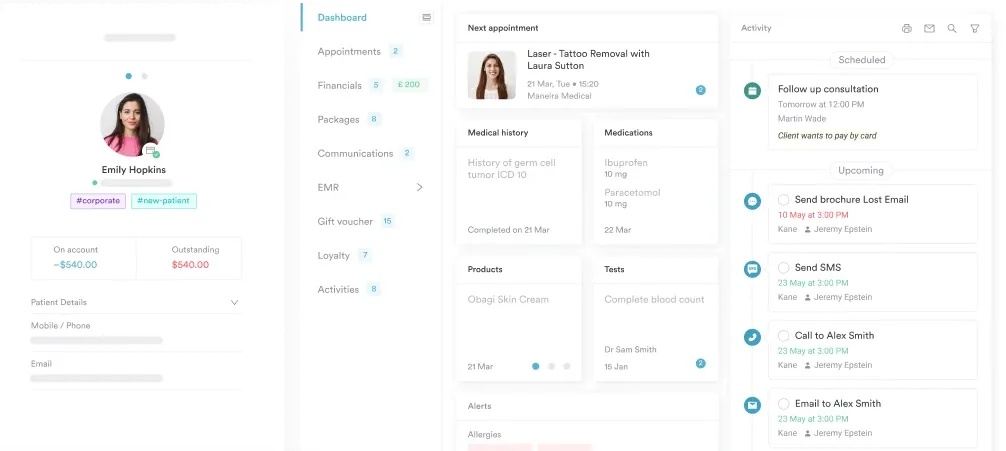
Most musculoskeletal practices split a single examination across three places. The angle lands on a paper proforma, the plan goes in the notes, and the referral letter gets typed from scratch a week later. Detail leaks out at every handover.
Practice management software like Pabau keeps all three in one client file. You can build a lumbar assessment form in Pabau’s capture forms software that prompts for every field. The side raised, the angle, and the symptom response each get their own box, so nothing is left blank by accident.
Completed forms drop straight into the patient’s client records, alongside the history, the imaging request, and the correspondence. When you re-test at review, last visit’s angle is already on screen. Writing the referral then becomes a matter of pulling the entry rather than rebuilding it.

Pabau gives physical therapists, osteopaths, and chiropractors structured assessment forms, SOAP notes, and referral letters in one client file. Examination findings like the crossed SLR stay linked to the patient’s history instead of sitting on paper.

Treat the crossed straight leg raise as a confirmation step, never as a screen. Its worth comes from what a positive result rules in, and that worth disappears the moment technique slips. So aim for a slow lift, a locked knee, and contralateral pain between 30 and 70 degrees.
The trade-off to remember is the low sensitivity. Roughly seven in ten patients with a genuine herniation will still test negative, so a negative crossed SLR should never end the conversation. Keep the standard SLR as your screen and let the crossed test do the confirming.
Whatever you find, the note is what travels. Record the side, the angle, and the symptom match while the patient is still on the table. Book a demo to see how Pabau keeps musculoskeletal exam findings, notes, and referrals in one client record.

Testing for nerve compression higher up the body? Wright test walks through the shoulder and arm version of provocation testing, including the positions that produce false positives.
Need a prone test for anterior thigh pain? Ely’s test covers the technique, the interpretation, and how it fits alongside femoral nerve testing.
Standardizing how your team measures movement? Range of motion assessment sets out the measurement methods and the recording conventions worth agreeing on across a practice.
Treating horizontal canal BPPV? BBQ roll maneuver walks through the four rotations, hold times, and how to pick the affected ear.
Planning rehabilitation after the diagnosis lands? Return to running protocol shows how staged criteria turn examination findings into a progression plan.
Yes, and it is a warning sign. Bilateral positives suggest a large central herniation squeezing roots on both sides. Check saddle sensation, bladder function, and bowel function straight away, then arrange same-day assessment if any of them are altered.
No. It supports a herniation diagnosis, but surgical decisions rest on neurological deficit, pain severity, and response to conservative care over time. The test contributes one piece of evidence toward a referral, not the referral outcome itself.
Use the slump test instead. Seated testing loads the same neural structures through neck flexion and knee extension, so it suits patients who cannot tolerate supine positioning. Record which version you used, because the two are not interchangeable in notes.
Always. A documented negative shows you checked, and it gives you a baseline for comparison at review. If the test turns positive later, that change carries real clinical meaning, and it only exists if the first result was written down.
Not really. The crossed SLR looks for reproduction of existing radicular symptoms, so it has little to offer in back pain without any leg involvement. Spend the time on movement testing and red flag screening instead.






