
 Anja Dodevska

Values exploration worksheet
Download your free values exploration worksheet A two-page printable form that o...

2
Last Updated: Nov 6, 2026
Free PDF 5 pages, print-ready Clinician-reviewed
A foot diagram pain chart links each zone of the foot, from heel to toes, to the conditions most often behind pain there. Podiatrists, physical therapists and primary care teams use it to narrow their questions before a full assessment. The chart guides triage, and examination confirms the cause. Numbness, burning or a wound on a diabetic foot calls for a structured exam instead. This guide pairs the chart with a free diabetic foot examination form. It records symptoms, monofilament sensation, foot findings and a risk category in one place.
A five-page form covering diabetes history, foot symptoms, a structured foot exam and monofilament sensation at four sites on each foot. It closes with a risk category, a referral and education plan, and two signature blocks.
Start from scratch, or upload this PDF or any form you already use, and Pabau's AI Form rebuilds it with tick boxes, text fields and a signature. Attach it to the treatment once, and every patient gets it on time.
AI Form rebuilds the consent, history and aftercare forms you already use. You review each one, then publish.
Patients complete forms on their phone before they arrive, so the front desk stops retyping paper.
Every signed form is versioned and attached to the treatment record, ready when you need it.
Set how long a consent stays valid. Returning patients skip forms they have already signed.
Supports HIPAA and GDPR compliance AES-256 encryption 4.8 on G2 4.7 on Capterra Trusted by 4,000+ practices worldwide

A foot diagram pain chart narrows the likely causes of foot pain, and examination confirms the diagnosis.
Each foot zone points to a different set of conditions, from plantar fasciitis at the heel to Morton’s neuroma at the ball.
Numbness, burning or a wound on a diabetic foot moves the visit from the chart to a structured foot exam.
The free download is a diabetic foot examination form, with monofilament sensation checks and a 0 to 3 risk category.
Digital intake forms can capture the pain zone at the first visit, so it sits in the patient record.
A foot diagram pain chart is a labeled outline of the foot, with each zone linked to the conditions most often associated with pain there. Clinicians use it for triage and documentation at the first visit.
Its value is structure. Rather than asking a patient to describe foot pain from scratch, the clinician points to a zone and works through the conditions listed for it.
The chart covers the heel, arch, ball of the foot, top of the foot, outer edge, toes and sole. It also flags the numbness and burning that can follow nerve involvement. The diagram below numbers each zone and the conditions to rule out first.
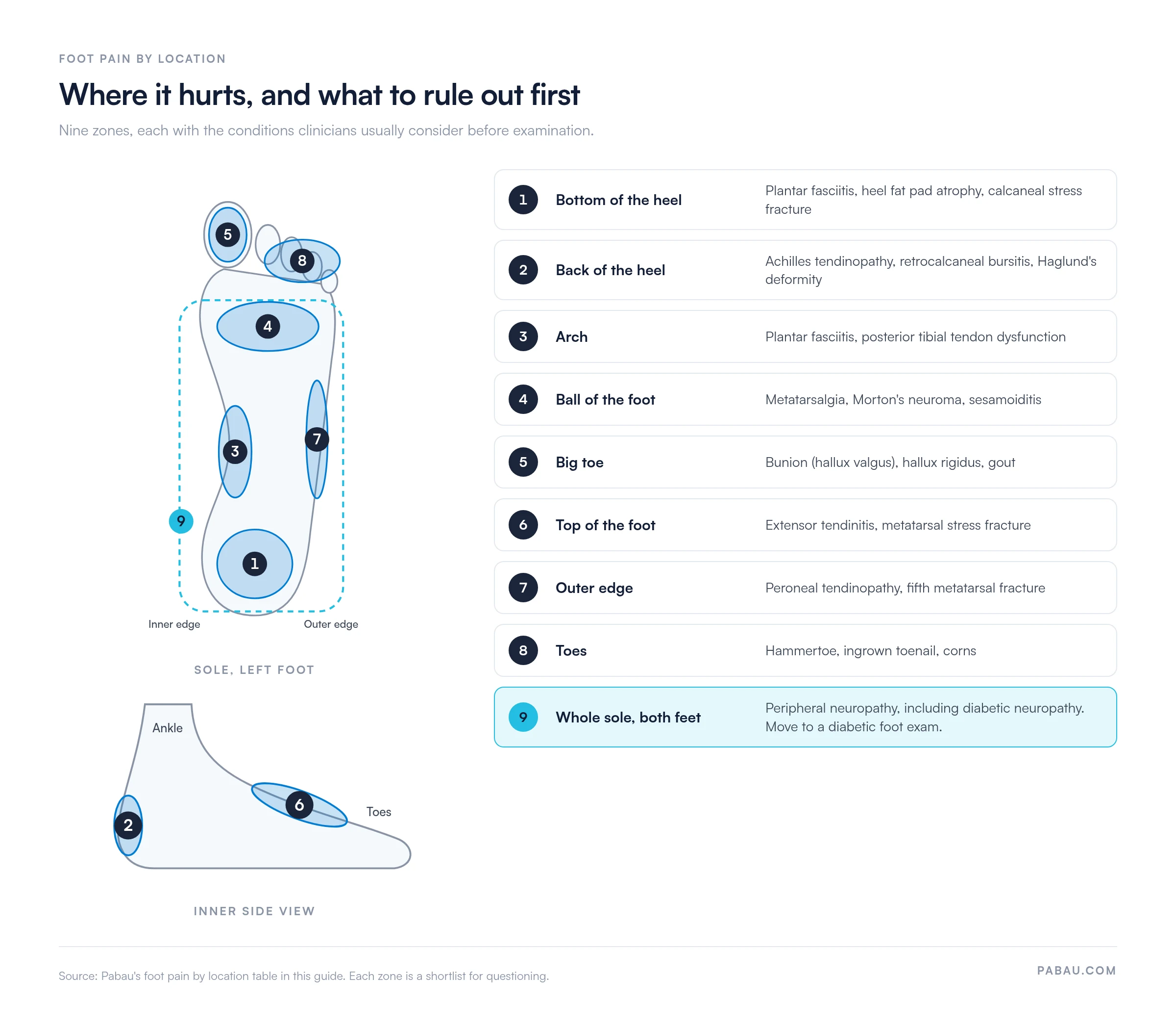
Each zone is a starting point for questions, examination and, where needed, imaging. For pain that spreads beyond the foot and ankle, a body pain chart applies the same zone approach to the whole body.
The table below maps each zone of the foot to the conditions clinicians most often consider first. Treat it as a shortlist for questioning, since other causes remain possible in each zone.
Plantar fasciitis is one of the most common causes of heel pain, and the NHS plantar fasciitis guidance is a useful patient-facing companion. For pain under the heel, the Windlass test helps separate plantar fascia involvement from other causes.
Use the foot diagram pain chart in five steps: locate, match, review, screen and record.
The chart narrows the list of possibilities but cannot confirm a condition. Examination and, where indicated, imaging make the diagnosis.
Foot pain in a patient with diabetes needs a full foot exam, because nerve damage can mask how serious a problem is. A patient with reduced sensation may feel little pain from an ulcer, a fracture or a tight shoe.
Move from the pain chart to a structured exam when any of these appear:
The NICE guideline on diabetic foot problems recommends a foot assessment at least once a year for adults with diabetes. That assessment includes a 10 g monofilament test for loss of protective sensation.
The download with this guide is a five-page diabetic foot examination form. It does not include a blank pain diagram. It gives you a structured record of the exam that the pain chart leads into.
The chart and the form work as a pair. The chart tells you where the pain sits, and the form records what the full exam finds.
Keep each completed form with the patient’s record. Comparing risk categories year over year shows whether protective sensation is changing.
Many practices still print foot exam forms, fill them out by hand and scan them into the file later. The pain location ends up in one note and the monofilament results in another. Last year’s risk category then takes a search to find.
Pabau, our practice management and EHR software, turns a paper exam like this one into a digital form that patients and clinicians complete on screen. Patients answer the history and symptom questions before the visit. The clinician adds exam findings and sensation results in the same record, next to treatment notes and photos.
Each signed form is stored with the patient, so this year’s findings sit beside last year’s for a quick comparison. Rehab teams get the same setup in a time-saving physical therapy EMR, with exercise plans and progress notes in the same record.

See how Pabau keeps intake answers, foot exam findings and follow-up in one patient record, so each year’s results are easy to compare.

Start with the foot diagram pain chart when a patient says their foot hurts. It gives the first conversation a shape and a shortlist of causes to test.
Once numbness, a wound or diabetes enters the picture, stop relying on the chart alone. Run the full exam on the form, and the findings will hold up at next year’s review.
Book a demo to see how Pabau keeps foot exam findings, forms and follow-up together for your practice.

Seeing pain at the back of the heel? Achilles tendon rupture treatment guidelines handout gives patients clear recovery guidance after a tendon injury.
Checking foot posture behind arch or heel pain? Foot Posture Index FPI-6 gives you a six-item standing assessment with a free scoring template.
Suspect plantar fascia involvement? Plantar fascia rupture test explains how to assess a suspected tear and record the result.
Tracking ankle mobility alongside foot pain? Ankle dorsiflexion test shows how to measure and track ankle range of motion between visits.
Choosing software for a foot care practice? Practice management software for podiatry compares the systems podiatry teams use to run bookings, notes and forms.
Pain in each part of the foot points to a different set of causes. Bottom-of-heel pain often means plantar fasciitis, and ball-of-foot pain suggests metatarsalgia or Morton’s neuroma. Outer-edge pain can follow a peroneal tendon problem or fracture. A clinician confirms the cause by examination.
No. A foot diagram pain chart narrows the likely causes and guides questioning, but it cannot confirm a diagnosis. Examination, and imaging where indicated, makes the diagnosis.
The free download is a five-page diabetic foot examination form. It records diabetes history, foot symptoms and a structured foot exam. It also captures monofilament sensation at four sites per foot, a 0 to 3 risk category and the care plan.
Run a diabetic foot exam when a patient with diabetes reports foot pain, numbness, burning or a slow-healing wound. Adults with diabetes should also have a foot assessment at least once a year, even without symptoms.
The 10 g monofilament test checks for loss of protective sensation. The clinician presses the filament at each marked site and records a plus if the patient feels it and a minus if they do not.