Head to toe assessment cheat sheet: Printable nursing guide
A head to toe assessment cheat sheet gives you one fixed order at the bedside, s...

2
Last Updated: Oct 23, 2026

Capillary refill test (CRT): Press a nail bed firmly for 5 seconds, release, and count how long color takes to return.
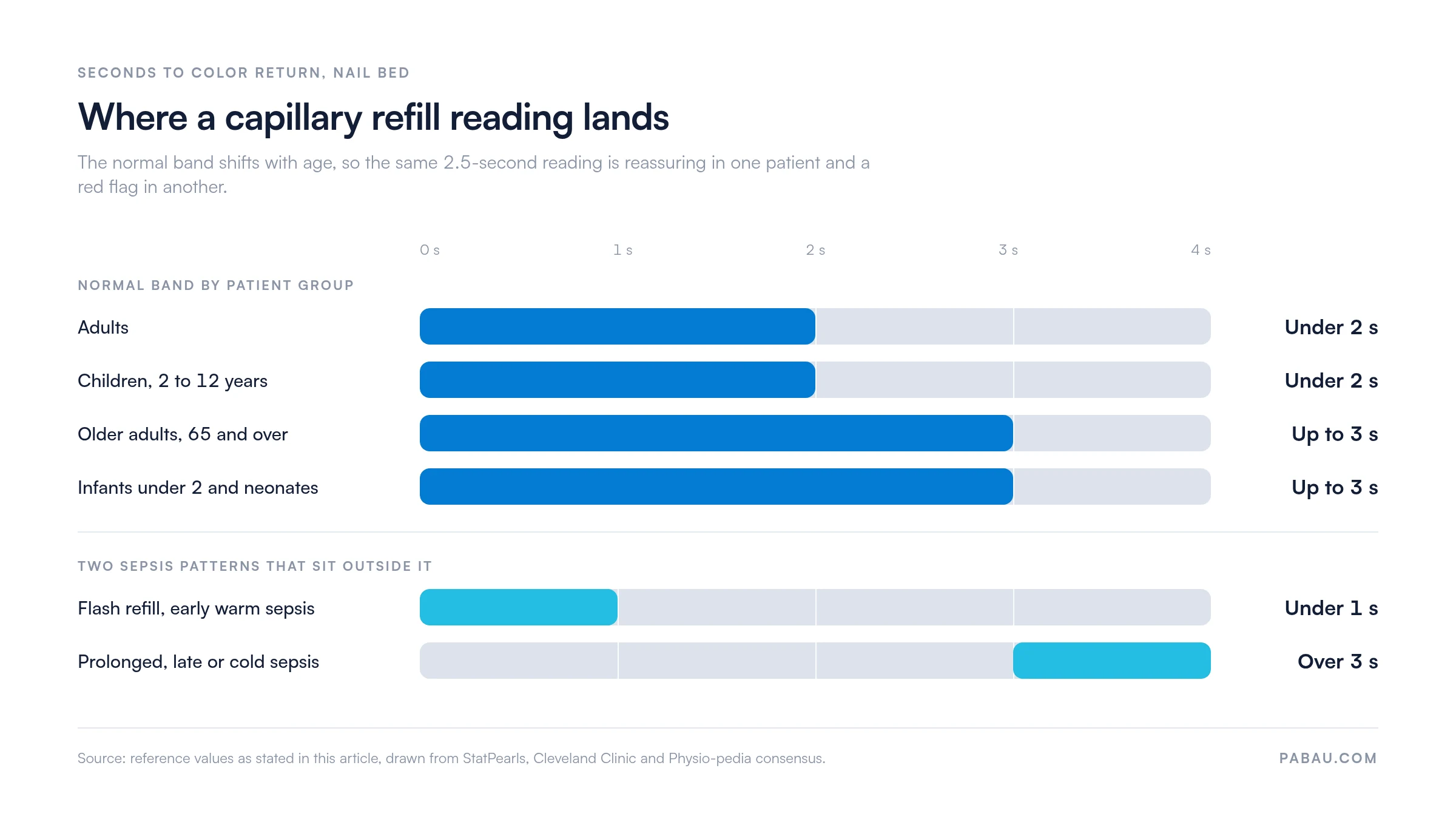
Normal CRT is under 2 seconds for adults, while infants under 2 years may reach 3 seconds normally.
Prolonged CRT in an adult, meaning over 2 seconds, points to reduced peripheral perfusion from shock, dehydration, or vascular disease.
Practice management software like Pabau lets you log the CRT result, the assessment site, and ambient conditions in one patient record.
A capillary refill test takes under ten seconds and costs nothing. Yet it can flag dehydration, early shock, or compromised peripheral circulation before other vital signs shift. That speed is why emergency nurses, paramedics, physical therapists, and primary care teams reach for it during a rapid perfusion assessment.
This guide covers how to perform the test correctly, how to read results across patient groups, and what causes a prolonged reading. It also sets out a five-point documentation format that turns single readings into a usable trend. It is written for healthcare professionals who perform or supervise bedside assessments.
The capillary refill test (CRT) is a bedside assessment of peripheral perfusion. Pressure temporarily forces blood out of the capillary bed beneath a nail, so the skin blanches white. When you release, the time color takes to return reflects how efficiently blood re-enters the peripheral circulation. No equipment is needed beyond a light source and a timer.
CRT sits alongside pulse rate, blood pressure, and skin turgor in the rapid assessment of circulatory status. It earns its place in prehospital and emergency settings, where patients cannot always report symptoms. It also matters in pediatric assessment, where blood pressure readings are often less reliable early in compromise.
According to NCBI StatPearls, CRT is a fast, simple assessment of peripheral perfusion and circulatory status. It is used across emergency medicine, critical care, and allied health settings. Its value lies in speed, not in replacing more definitive investigations.
Technique consistency matters, because small variations in pressure, duration, and site all move the result. Follow these steps for the standard nail bed method used in adults and children.
Practitioners in physical therapy practices and other allied health settings often perform CRT as part of a broader limb assessment. In those contexts, comparing left and right hand readings adds value when peripheral vascular disease or trauma is suspected.
A single threshold does not fit all patients. Age and peripheral circulation status both shift the normal reference range. Use the table below as your clinical reference, drawn from StatPearls, Cleveland Clinic, and Physio-pedia consensus values.
These values are well established in the literature, but CRT is not a standalone diagnostic. A reading of 2.5 seconds in a warm, well-hydrated adult carries different weight than the same reading in a cold trauma patient. Always interpret with the full clinical context.
Plotted on one scale, the age bands overlap more than the table suggests, and the two sepsis patterns sit clearly outside them.
A prolonged CRT tells you blood is returning to the peripheral capillary bed more slowly than expected. That delay reflects reduced cardiac output, peripheral vasoconstriction, or obstructed local circulation. The reading does not pinpoint the cause. It flags impaired perfusion and prompts further assessment.
When CRT exceeds the threshold for the patient’s age group, consider these categories of cause:
Whatever the cause, the value of a reading depends on what happens to it next. A structured record of each CRT finding makes the trend visible instead of leaving it scattered across handwritten notes.
Separating acute from chronic causes shapes the clinical response. An acute change in a previously normal patient is urgent. A persistently prolonged CRT in established peripheral arterial disease calls for different management than new-onset prolongation after surgery.
One localized cause is worth naming separately. Extravasation or poor perfusion around an IV site can produce a prolonged CRT in that limb alone. Comparing the access limb against the opposite hand separates a local access problem from a systemic one.
Sepsis assessment relies on several perfusion markers, and CRT has a specific place in international guidelines. The Surviving Sepsis Campaign recognizes peripheral perfusion assessment, including CRT, as a resuscitation endpoint alongside lactate and mean arterial pressure. The picture in sepsis is more nuanced than in straightforward dehydration.
In early, warm sepsis, capillaries are often maximally dilated. CRT can fall under 1 second, a flash refill, with warm and flushed extremities. That pattern can falsely reassure a clinician about perfusion status. In later or cold sepsis, cardiac output falls and vasoconstriction develops, so CRT extends beyond 3 seconds.
A 2019 trial published in JAMA, the ANDROMEDA-SHOCK study, examined CRT-guided resuscitation in septic shock. It found that targeting CRT normalization was at least as effective as lactate-guided resuscitation for 28-day mortality. That result established CRT as a credible sepsis endpoint.
Clinicians using CRT in sepsis should record both the reading and the peripheral temperature of the extremity. The combination separates the warm and cold patterns, which the number alone cannot do.
Pediatric CRT assessment uses the same technique as in adults, with different thresholds and a different clinical framework. In children aged 2 and over, the adult threshold of under 2 seconds applies. In infants under 2 years and neonates, up to 3 seconds can be normal.
CRT is a core component of the Pediatric Assessment Triangle (PAT), a widely used rapid evaluation framework. Within the PAT, CRT contributes to the circulation to skin domain alongside skin color and temperature. A prolonged reading in a child matters partly because children compensate far longer than adults before decompensating.
Several confounders can produce a falsely prolonged or falsely normal CRT. Recognizing them matters as much as knowing the thresholds, because acting on a bad reading carries risk.
Practitioners in sports medicine practices assess CRT after musculoskeletal injuries to check distal limb perfusion. Cold pitches, compression garments, and baseline athletic vascular adaptation all complicate interpretation in that setting.
CRT has clinical utility, but it is not a precise diagnostic instrument. Understanding its limitations prevents both over-reliance and under-reliance.
Inter-rater variability is the most frequently cited limitation. Studies show meaningful disagreement between clinicians measuring CRT on the same patient at the same time, particularly near the 2-second threshold.
The disagreement comes from differences in applied pressure, timing, and judgment of the color endpoint. Agreeing a pressure and a timer method within the team reduces this variability without eliminating it.
Sensitivity and specificity are moderate. CRT misses some cases of compromised perfusion and occasionally flags well-perfused patients as abnormal. That is why Advanced Trauma Life Support and PALS embed CRT within multi-sign assessments rather than treating it as standalone.
Structured decision rules make the same point. The Ottawa Ankle Rules outperform any single physical sign used on its own, and CRT is no exception to that pattern.
When not to use CRT as a primary guide: In known peripheral vascular disease, Raynaud’s phenomenon, or severe hypothermia, CRT says little about systemic perfusion. A central site such as the sternum or forehead is more reliable in those patients.
Where the question is positional vascular compression rather than systemic perfusion, a provocative maneuver answers it better. Adson’s test examines whether shoulder position compresses the vessels feeding the arm.
Standardize your team’s CRT technique by agreeing on three points. Always use the index finger at heart level. Count with a clock rather than in your head. Record the ambient temperature whenever a result is borderline. This protocol cuts inter-rater variability without any extra equipment.
Documentation is where CRT earns its keep. A single reading is useful. A series showing CRT slide from 1.5 seconds to 3.5 seconds over four hours describes a deteriorating patient. That series only exists if every reading is recorded the same way.
Best-practice CRT documentation captures five data points every time:
Structured digital forms keep those five points consistent across every practitioner in the practice. Assessment templates inside patient record software can carry CRT alongside other perfusion markers, so each reading joins a timeline rather than sitting alone.
Most practices record CRT wherever a field is nearest, whether that is a free-text observation box, a paper chart, or a handover note. The reading survives, but the trend does not, and the next clinician has to reconstruct it.
Practice management software like Pabau replaces that with a structured assessment template. Your team builds the CRT field set once, covering site, result, ambient conditions, and action taken. Every practitioner then completes the same fields, and each entry timestamps itself into the patient record.
The outcome is a readable perfusion timeline instead of scattered notes. A clinician picking up the patient at hour four can see hour one. A slide from 1.5 to 3.5 seconds becomes obvious rather than buried.

Automated workflows can also prompt for the CRT field at set intervals inside a monitoring protocol. Nobody has to remember when the next reading is due.


Pabau lets your team build assessment templates that capture CRT, perfusion observations, and clinical context in one timestamped patient record. Every reading lands in the same place, for every practitioner.

CRT is one of the most information-dense bedside assessments available. Five seconds of pressure and a two-second count can surface shock, dehydration, vascular compromise, or sepsis before other vital signs move. Its value depends on consistent technique, the right threshold for the patient in front of you, and honesty about the confounders.
The trade-off worth remembering is precision. CRT will never match an arterial line or a lactate, and it does not need to. It buys you a perfusion signal in ten seconds, at the bedside, with no equipment at all.
Pick one technique, agree it across the team, and record all five data points every time. Book a demo to see how Pabau turns single CRT readings into a perfusion timeline your whole team can read.

Assessing upper limb circulation from another angle? Wright’s test checks whether shoulder position compresses the vessels feeding the arm.
Need a bedside sensory screen to sit alongside CRT? Two-point discrimination test covers the technique, the thresholds, and what a poor result tells you.
Want a worked example of a structured decision rule? Ottawa Ankle Rules calculator shows how a rule outperforms any single physical sign.
Documenting a positional vascular test as well? Adson’s test comes with a template you can hand straight to your team.
Tracking another cardiovascular reference range? Heart rate recovery chart by age gives you the age-banded values in a printable format.
A capillary refill test is a bedside assessment of peripheral perfusion. The clinician presses a nail bed firmly for 5 seconds, then releases and times the return of color. Normal return in adults is under 2 seconds. The test is used across emergency medicine, critical care, and allied health settings.
Normal capillary refill time is under 2 seconds in adults and in children aged 2 and over. In infants under 2 years and neonates, up to 3 seconds is normal. Older adults may sit closer to 3 seconds because of age-related peripheral vascular changes. Always read the result alongside ambient temperature and the wider clinical picture.
A delayed capillary refill time, meaning over 2 seconds in an adult, signals reduced peripheral perfusion. Common causes include hypovolemic shock, dehydration, peripheral vascular disease, cold or late sepsis, and hypothermia. It is not a diagnosis on its own. It tells you that further assessment is needed.
No. Reliability drops in patients with pre-existing peripheral vascular disease, older adults, and anyone assessed in a cold environment. Nail polish and artificial nails obscure the nail bed entirely. Inter-rater variability between clinicians is a recognized limitation. Use CRT as one component of a multi-sign assessment, never as a standalone test.
In sepsis, CRT works as a peripheral perfusion endpoint alongside lactate and mean arterial pressure. The Surviving Sepsis Campaign supports targeting CRT normalization during resuscitation. Early warm sepsis can produce flash refill under 1 second, while late cold sepsis prolongs it. Document the peripheral temperature of the extremity alongside the reading.
Children aged 2 and over use the adult threshold, so under 2 seconds is normal. Infants under 2 years may reach 3 seconds normally. CRT forms part of the Pediatric Assessment Triangle. It carries extra weight because children compensate for circulatory compromise longer, then decompensate rapidly.