


How to Start a Pelvic Health Practice: A Step-by-Step Guide
Skilled pelvic floor therapists often stay tethered to employer-owned clinics lo...
July 21, 2026
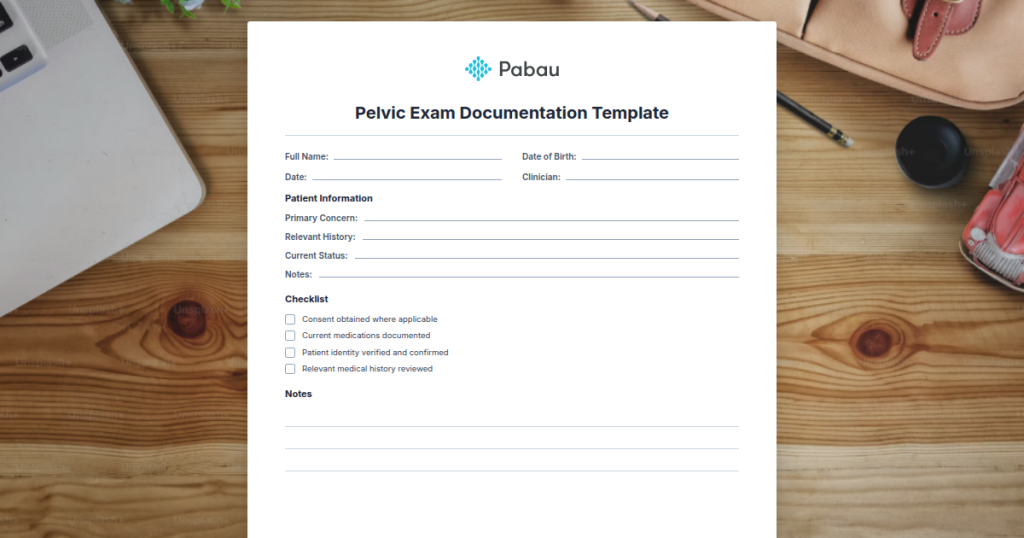
A pelvic exam documentation template standardises the recording of all examination components: external, speculum, bimanual, and rectovaginal findings.
CMS and HIPAA require explicit chaperone documentation, anatomical terminology, and clear normal vs abnormal findings to meet audit and compliance standards.
Accurate documentation directly links to correct ICD-10 coding (Z01.419 for routine exam), reducing billing denials and supporting E/M level assignment.
Pabau’s digital forms and Pabau Scribe documentation tools automate template population and integrate pelvic exam records directly into clinical workflows.
A pelvic exam documentation template is a structured clinical form used to record the findings of a comprehensive gynaecological examination. It captures anatomical observations across four distinct examination phases: inspection of external genitalia, speculum visualisation of the cervix and vaginal vault, bimanual assessment of uterine and adnexal structures, and rectovaginal examination when indicated.
The primary purpose of this pelvic exam documentation template is to ensure consistent, legally defensible clinical records that meet regulatory standards. HIPAA requires explicit documentation of patient consent and chaperone presence. ACOG clinical guidelines specify that pelvic examination findings must be recorded in anatomical detail, not generic summaries. CMS coding audits verify that examination documentation supports the reported E/M level-without specific findings documented, claims face denial risk.
This template serves gynaecology practices, family medicine clinics, women’s health centres, and any pelvic health clinic conducting routine screening or symptomatic pelvic evaluations. It bridges the gap between clinical assessment and accurate coding, reducing compliance burden.
A standardised clinical record capturing external genitalia inspection, speculum exam, bimanual examination, rectovaginal findings, chaperone presence, and patient consent details. Print or digital use.
Download templateComplete the pelvic exam documentation template in a logical, sequential order that mirrors the actual examination workflow. This ensures no findings are missed and the record reads naturally for auditors and referral clinicians.
Once findings are complete, link them to the appropriate CMS ICD-10 coding guidelines for accurate billing and establish structured medical forms workflows to reduce documentation gaps.
Gynaecology and women’s health practices: Specialists conducting routine annual exams, follow-up visits for abnormal findings, or symptomatic evaluations depend on this template to capture the depth of assessment required for diagnosis and coding.
Fertility clinics: These practices conduct pelvic exams as part of initial fertility assessment and treatment monitoring. Documenting baseline and interval changes supports clinical decision-making for ovulation induction and embryo transfer timing.
Family medicine and primary care clinics: Primary care providers offering well-woman exams or addressing gynaecological complaints benefit from a structured approach to ensure completeness and appropriate coding under E/M guidelines, the same approach that supports other focused screens they perform daily, such as a PERRLA eye exam form for rapid neurological checks.
Sexual health clinics and post-menopausal care centres: These settings require clear documentation of atrophic changes, differential diagnosis consideration, and contraceptive counselling aligned with examination findings.
Compliance and legal protection: Explicit chaperone documentation and anatomical detail meet CMS audit standards and GMC/CQC requirements in the UK. Medicolegal defensibility increases when findings are specific rather than vague.
Accurate E/M and diagnostic coding: Detailed pelvic examination findings link directly to ICD-10 codes and CPT visit levels. Documented findings justify higher E/M levels (99213 vs 99214) and reduce claim denials tied to insufficient documentation.
Workflow efficiency: A standardised template ensures nothing is missed during the exam, reducing the need for follow-up documentation and second visits due to incomplete records.
Continuity of care: When examination findings are clearly documented, other clinicians (specialists, locums, covering physicians) understand the baseline status and detect interval changes reliably. This is critical for monitoring fibroids, ovarian cysts, or cervical abnormalities over time.
Document chaperone presence by name and specific time of presence within the examination (e.g., ‘Present during speculum and bimanual exam, left prior to rectovaginal’). Generic ‘chaperone present’ notes create audit liability if the clinician’s recollection differs from the patient’s account.
Beyond structure, the quality of pelvic exam documentation depends on precise anatomical language and differential thinking. Clinical documentation safety practices emphasise using standardised terminology rather than abbreviations that may be misinterpreted. “RLQ ovarian mass” is clearer than “R adnexal palpable”-the first indicates the specific anatomical location, the second is ambiguous.
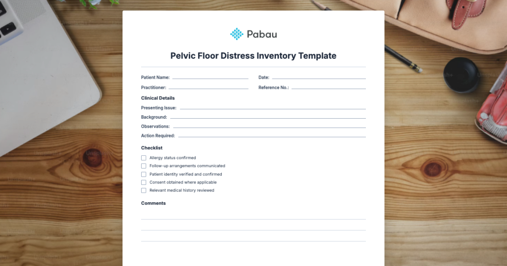
Distinguish between routine screening findings and symptomatic exam detail. A routine well-woman visit requires less exhaustive documentation of normal findings than an exam triggered by pelvic pain, abnormal bleeding, or post-menopausal symptoms. Document the indication for the exam and how findings either reassure or raise concern. When the indication is pelvic floor symptoms such as prolapse or incontinence, pairing the objective findings with a patient-reported Pelvic Floor Distress Inventory captures the symptom burden the exam alone does not quantify.
When using digital clinical documentation tools like Pabau Scribe, verify that auto-populated pelvic exam templates include your specific findings rather than generic defaults. Auditors and insurers recognise boilerplate language and may question claim legitimacy if documentation appears templated without customisation.
Pabau's digital form templates and Pabau Scribe automate documentation while maintaining clinical accuracy and compliance standards.

Digital forms in practice management systems enable pelvic exam templates to populate directly into the patient record, auto-fill from previous visits for comparison, and trigger ICD-10 coding suggestions based on documented findings. This integration reduces transcription errors and ensures no examination step is overlooked.
Many HIPAA-compliant clinic software systems allow customisation of pelvic exam templates to match your practice’s protocols. Configure fields for your specific examination sequence, add branching logic that reveals additional fields based on initial findings (e.g., if “mass palpated,” expand detail fields), and ensure consent language is prominently displayed before exam fields are unlocked.
A pelvic exam documentation template ensures that every gynaecological examination is recorded with the anatomical precision, legal completeness, and coding accuracy that auditors, regulators, and patient safety demand. By standardising the structure of documentation and integrating it into SOAP note and clinical documentation workflows, clinics reduce compliance risk and improve care continuity.
Pabau’s digital forms and automated documentation tools eliminate manual charting delays and ensure your pelvic exam records meet both clinical standards and billing requirements. Book a demo to see how your clinic can streamline pelvic exam documentation while staying audit-ready.
Standard sections include patient consent and chaperone presence, external genitalia inspection, speculum examination of the cervix and vaginal vault, bimanual assessment of uterus and adnexa, and rectovaginal examination when clinically indicated. Include fields for abnormal findings, differential diagnosis reasoning, and E/M coding level justification.
For a normal exam, use concise anatomical language: “External genitalia: normal. Speculum exam: cervix pink, midline, no discharge. Bimanual: cervix mobile, non-tender, uterus normal size and position, ovaries palpable bilaterally, no masses. Rectovaginal: no masses or tenderness.” Avoid vague shorthand like “WNL” (within normal limits)-specificity prevents coding disputes.
ICD-10-CM code Z01.419 (Encounter for gynecological examination without abnormal findings) applies to asymptomatic well-woman visits. If abnormal findings are present (e.g., fibroid, ovarian cyst), code the specific condition (e.g., D25.9 for leiomyoma). Always document the exam indication and findings to support the assigned diagnosis code.
State cervical mobility and tenderness, uterine size (normal or weeks if enlarged), contour and position, and adnexal findings for both sides. Example: “Uterus: normal size, anteverted, firm, mobile, non-tender. Right ovary: palpable, non-tender. Left ovary: palpable, non-tender.” Avoid “no masses” without specific ovarian palpation documentation.
Explicit chaperone documentation protects clinician and patient from allegations of impropriety. GMC guidance and CQC standards in the UK require this. In the US, malpractice insurers expect chaperone notation. Include the chaperone’s name, role, and the specific portion of the exam during which they were present.