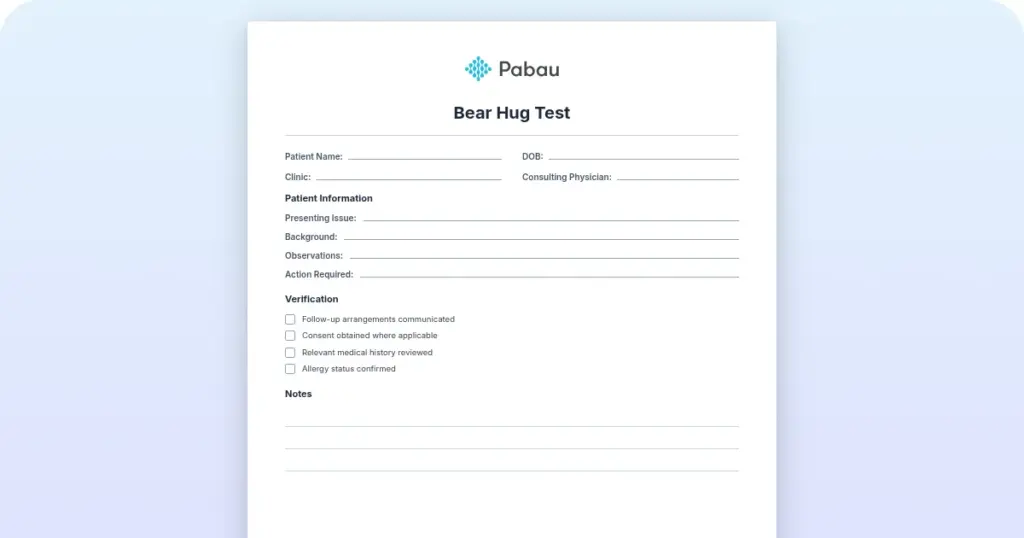
Bear hug test: Procedure, interpretation and free template
The bear hug test is a quick shoulder special test that screens for subscapulari...

3
Last Updated: Oct 23, 2026

A peroneal tendon tear is a split or a rupture in one of the two tendons running behind the outer ankle bone.
No single maneuver confirms a tear, so the exam runs palpation, resisted eversion, a single-leg heel rise, and a subluxation check.
MRI confirms the diagnosis and sizes the tear, while dynamic ultrasound is better at catching tendons that slip inside the sheath.
Sobel grades I to III usually start with conservative care, and a grade IV rupture earns a surgical opinion.
Digital assessment forms in practice management software like Pabau keep every exam finding in the client record.
A clinical assessment form for a suspected peroneal tendon injury, with fields for symptom history, palpation landmarks, and each provocative test result. It also carries the interpretation guide and a treatment planning section.
Download templatePeroneal tendon injuries are a common cause of lateral ankle pain in athletes and active patients. The peroneal tendon tear test is how you separate a tear from tendonitis, subluxation, or a ligament sprain. It follows the examination sequence used in the peer-reviewed orthopedic literature.
This guide covers the exam in order, how to read each positive finding, and when imaging is worth ordering. Whether you work in sports medicine, physical therapy, or an orthopedic practice, every finding below has a matching field on the downloadable form.
A peroneal tendon tear test is a structured exam of the two tendons that run behind the outer ankle. Those tendons are the peroneus brevis and the peroneus longus. They stabilize the ankle and drive eversion, which turns the sole of the foot outward. A tear brings lateral ankle pain, swelling, and a feeling of instability.
No single maneuver settles the question. The exam combines provocative maneuvers with careful palpation and a functional test. You are trying to reproduce the pain, find weakness, and rule out the other causes of lateral ankle pain. Recording each result in digital assessment forms keeps the note defensible.
Which tendon tore changes what you find on the exam, so start with the anatomy.
Both tendons sit in a groove behind the fibula and are held there by the superior peroneal retinaculum, known as the SPR. A lax SPR lets the tendons slip. A tear causes pain and a loss of strength. That difference decides the treatment plan.
Take the history before you touch the ankle. Patients usually describe an inversion injury, long-standing lateral instability, or pain that built up under repetitive load.
Acute tears arrive with sudden pain and swelling. Chronic tears build slowly and get mistaken for peroneal tendonitis. Your client record should carry the onset, the mechanism, and what the patient can no longer do.
Work in the same order every time. Inspection comes first, then palpation, then range of motion, then the provocative maneuvers. A physical therapy EMR keeps that order on screen, so no step gets skipped under time pressure.
No maneuver here reaches 100% sensitivity or specificity. Read the findings together with the history and, where you have it, the imaging.
Two named systems get used here, and they answer different questions. The Sobel grading system scores how badly the peroneus brevis is split, running from grade I to grade IV.
Grade I is a flattened or attrited tendon with no split. Grade II is a split shorter than 1 cm, and grade III a split of 1 to 2 cm. Grade IV is a complete rupture.
Raikin’s classification describes something different. It covers intrasheath subluxation, where the tendons move inside a sheath that is still intact. Type A is two intact tendons swapping position. In type B, the peroneus longus slips through a split in the brevis.
The grade matters because it points at a route. Grades I to III usually start with conservative care, while a grade IV rupture earns a surgical opinion.
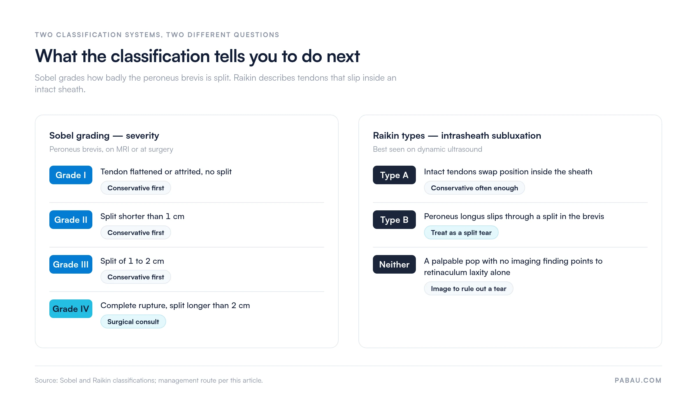
Record the grade, and the subluxation type where it applies, so the rest of the care team plans against the same pathology. Ankle injuries collect named systems like this. The Danis-Weber classification does the same job for fibular fractures.
Imaging confirms what the exam suspects. MRI is the standard for soft-tissue detail, tear size, and location. Ultrasound adds a moving picture and catches subluxation that a static scan can miss. Cleveland Clinic’s guidance treats the two as complementary.
Treatment splits at this fork, so it is worth being sure. Tendonitis is inflammation with the tendon still intact. A tear is a break in continuity.
A palpable defect higher up the calf points at the Achilles instead, which is where the Thompson test earns its place. Once imaging is booked, patient scheduling software ties the scan and the review appointment to the same episode of care.
Severity, chronicity, activity level, and the response to a conservative trial decide the route.
Set the recovery window with the patient before they leave the room. A six-week boot and a four-month post-operative course are two different conversations, and the second one lands better early.
The template gives you a checklist for the exam and somewhere to put each result. It carries fields for symptom history, palpation landmarks, provocative test results, imaging recommendations, and treatment planning.
Filling the same fields every time makes findings comparable between clinicians and between visits. It also means the note holds up if the case gets reviewed months later.
Pair it with automated workflow software to send the post-exam summary, book the imaging appointment, and check in during rehab. The same layout carries to other regions, so our quadriceps strain test form documents the thigh the same way.
Most practices run this exam on paper and type a summary into the record hours later. The palpation map ends up in a drawer, the imaging request sits in an inbox, and the rehab notes live somewhere else again. Comparing week one with week six then means opening three systems.
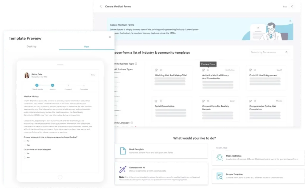
Practice management software like Pabau keeps the episode together. The assessment form is a digital form the clinician fills in the room, so the findings save straight to the client record. Imaging requests, review appointments, and rehab measurements attach to that same client.
So when the patient comes back at week six, you have one place to look. You also have one export to hand over if a report is ever requested. Nothing gets retyped, and no finding sits in a drawer waiting to be filed.

Pabau’s digital forms capture peroneal tendon findings in the room and store them alongside the client’s imaging, appointments, and rehab progress. Your whole team works from one record.

A peroneal tendon tear starts as a clinical suspicion that imaging then confirms, and the exam is what earns the scan. Run the same sequence every time and the pattern gets easy to spot.
What is worth remembering is that this exam narrows the possibilities without closing them. A palpable defect with weakness on resisted eversion still goes to MRI before anyone discusses surgery.
Standardizing the documentation is the cheap part of this, and it pays off at the review visit. A client portal also keeps the patient looking at their own rehab plan between appointments.
Book a demo to see how Pabau holds assessment findings, imaging, and rehab progress in one client record.

Assessing a knee instead? Apley’s test template walks through the grind and distraction sequence for a suspected meniscal tear.
Need the same structure for the thigh? Quadriceps strain test covers resisted extension, palpation landmarks, and grading.
Checking reflexes in the same workup? Deep tendon reflex exam gives you a grading scale and a place to record each side.
Measuring joint movement? Hip range of motion test shows how to log degrees consistently across visits.
Writing up the rehab visits? Progress note template keeps each session comparable to the one before it.
The core tests are resisted eversion, a single-leg heel rise, palpation along the fibular groove, and a subluxation check for SPR laxity. No one test is definitive. Diagnosis needs the clinical picture plus imaging confirmation on MRI or ultrasound.
Yes, many patients can walk with a peroneal tendon tear, though it usually hurts and the ankle can feel unstable. Tolerance depends on how severe and how recent the tear is. Acute complete ruptures often need crutches at first. Ask a qualified orthopedic professional before you progress weight-bearing.
Conservative care typically runs six to twelve weeks of structured physical therapy, with weight-bearing and strengthening built up in stages. Recovery after surgical repair spans four to six months. A full return to sport can take six to twelve months. Your clinician should set that timeline against your tear grade.
A tear is a break in the tendon, while tendonitis is inflammation without rupture. Tears bring pain and clear weakness on resisted eversion. Tendonitis hurts but rarely causes weakness. MRI or ultrasound settles it, showing visible disruption in a tear and thickening in tendonitis.
MRI is the standard for full soft-tissue assessment and for planning surgery. Ultrasound is cheaper, more available, and better at catching subluxation, because it shows the tendons moving. Many clinicians use ultrasound first and add MRI if surgery is on the table.
Not always. Partial tears and many complete tears respond to immobilization, bracing, and physical therapy when treatment starts early. Surgery gets considered for complete ruptures, for tears with retraction, and after a conservative trial fails at six to twelve weeks. High-demand athletes may choose it sooner.