CRM for chiropractors: how to manage patients, bookings and marketing
A CRM for chiropractors is a patient relationship management system built around...

Last Updated: Oct 13, 2026

Hip range of motion testing measures six movement planes with a goniometer: flexion, extension, abduction, adduction, internal rotation, and external rotation.
Normal values are flexion 0-120°, extension 0-30°, abduction 0-45°, adduction 0-30°, and 0-45° for both internal and external rotation.
Active range of motion sits 5-10° below passive range. A larger difference points to weakness or pain inhibition rather than stiffness.
Manual goniometry overstates true hip motion, so compare a patient against their own baseline rather than against another clinician’s readings.
Practice management software like Pabau records bilateral degrees, end feel, and notes in the chart, so every follow-up starts from the last measurement.
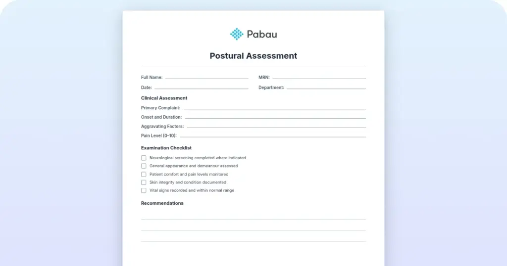
A standardized assessment form covering all six movement planes, with bilateral recording fields, goniometer entries, end feel notation, and room for interpretation. Print it or fill it in on screen at the treatment table.
Download templateA hip range of motion test measures how far the hip moves in six directions, recorded in degrees using a goniometer. Normal figures run from 0-120° in flexion to 0-30° in adduction. Both hips are always measured, because the difference between sides often matters more than either number on its own.
This guide covers the testing procedure for active and passive motion, goniometer placement, the normal reference values, and how to read your findings. It also explains what manual goniometry can and cannot tell you, which decides how much a 5° change is worth.
The free form above gives you somewhere to record it all.
A hip range of motion test measures the angular movement available at the hip joint across six planes. Three things get recorded at each plane:
Standard goniometry is the reference method for all three. A 12-inch goniometer sits with its axis at the center of the hip joint, one arm along the pelvis and the other along the femur. The angle between the two arms, in degrees, is the recorded measurement.
Reference values come from the standardized testing protocols published by the American Academy of Orthopaedic Surgeons (AAOS) and repeated across the clinical literature. The table below sets out the accepted ranges for all six planes.
Measure against these and the restricted planes show up immediately, along with how far off they are. That is usually enough to point your clinical reasoning at a tissue or a structure.
AROM shows what the patient can produce under their own muscular control. Five steps keep the measurement repeatable:
AROM usually runs 5-10° below PROM, because it depends on force production. A difference wider than 10° points to muscle weakness or pain inhibition holding back voluntary movement.
In software built for occupational therapy practices, scores from scales like this feed straight into progress notes.
PROM shows structural mobility independent of the patient’s strength. It is what separates contractile tissue, meaning muscle, from inert tissue such as capsule and ligament.
PROM that barely exceeds AROM usually means muscle tightness. PROM well above AROM points the other way, toward weakness or inhibition limiting what the patient can do voluntarily.
Rotation is the plane that goes first in hip dysfunction, so it deserves a careful setup. Test it prone, with the knee flexed to 90°, which isolates the hip and stops the lumbar spine from helping.
A 10-15° difference between sides turns up often in athletes and other active patients.
Recording both rotation values at every visit is what lets you show the correction later.
Accurate goniometry comes down to tool choice and consistent landmarks. Use a 12-inch instrument for the hip, since shorter arms wobble against the length of the femur.
It is worth knowing what those degrees represent. A manual goniometer reads the angle between two body segments, not the angle inside the joint itself. It therefore overstates true hip motion, and the size of that error changes with the plane you are testing.
One reliability study in patients with hip impingement found strong agreement for abduction and internal rotation. Adduction was the weakest plane of the six. Repeat readings by the same examiner held up well across all of them.
Two practical consequences follow. Compare a patient against their own earlier readings rather than a colleague’s, and keep the same examiner where the schedule allows. Treat a 3-5° shift between visits as noise, and look for 10° or more before you call it progress.
Consistency is easier to hold when the recording sheet asks for the same fields every time.

Test and record both hips every time. An asymmetry is a finding in its own right, and it often shapes the treatment plan more than the absolute numbers do.
That level of detail only helps if you can find it again months later.

ROM values point your reasoning at a structure. These are the patterns that come up most often:
When the findings line up with what the patient complains about, you have an outcome measure you can retest.
Reliable baselines depend on the same form being used every time.
Paper is still the usual route. Degrees get written on a sheet at the table, then summarized into the notes that evening. Six weeks later, nobody can say whether flexion started at 95° or 105°.
Practice management software like Pabau replaces the sheet with a form on the screen in front of you. You build the hip ROM assessment once, with bilateral fields for each plane and an end feel selector beside them. It opens straight from the patient’s chart, already carrying their name, the date, and yours. Recording measurements the same way each visit is what makes progress visible, and physical therapy EMR software enforces it.
In practice the workflow runs like this:
The practical result is fewer repeat measurements and no transcription errors between sheet and chart. You also get an objective record you can hand to a payer or a referring physician.

Pabau’s digital assessment forms capture bilateral degrees and end feel at the treatment table, then store them in the patient record. Every follow-up opens with the previous measurement already on screen.

Hip ROM testing earns its place because it turns a vague complaint into a number you can retest. The technique itself is not hard. Holding it steady between visits, and between clinicians, is what decides whether the number means anything.
So set the protocol once, use the same form every time, and record both sides at every visit. Do that and a 15° improvement counts as evidence rather than an impression. Book a demo to see how Pabau stores hip ROM measurements and surfaces the last reading at the follow-up.

Need another lower-limb screening test? Crossed straight leg raise test walks through performing and interpreting a test that points to nerve root involvement.
Assessing the upper limb as well? Wright test covers the hyperabduction maneuver used to screen for thoracic outlet syndrome.
Screening an older patient for fall risk? 4 stage balance test template gives a checklist, timing guidance, scoring table, and fall-risk interpretation.
Rehabbing a patient after a groin pull? Adductor strain exercises handout stages isometric, concentric, and eccentric work through return-to-sport progressions.
Building a first-time exercise routine for a client? Beginner workout plan for women sets a four-week structure with frequency, exercises, and progression for beginners.
Normal values are flexion 0-120°, extension 0-30°, abduction 0-45°, adduction 0-30°, internal rotation 0-45°, and external rotation 0-45°. These are the reference standards used to identify restrictions.
Active range of motion is movement the patient produces with their own muscle strength. Passive range of motion is movement you apply for them, with no effort on their part. Passive usually reads 5-10° higher. A wider difference suggests weakness or pain limiting voluntary movement.
Place the axis at the hip joint center, one arm along the pelvis midline and the other along the femur midline. Read the angle between the arms and record it in degrees. A 12-inch goniometer is the standard tool for the hip.
It is reliable enough to track one patient over time, and less reliable as an absolute figure. A goniometer reads the angle between body segments, so it overstates true hip motion. Agreement is strongest for abduction and internal rotation, and weakest for adduction.
Treat a limitation as pathological when it falls more than 15° below the normal value. Pain, a clear asymmetry between sides, or functional impairment also count. Loss of internal rotation is often the first sign of hip joint pathology.
Yes. Test both hips at every assessment and record the sides separately. Asymmetries between sides are clinically significant and guide the treatment plan.
End feel indicates which tissue is limiting the movement. Soft suggests muscle tightness, firm indicates capsular stretch, and hard can mean a bony block. That distinction separates contractile from inert tissue and shapes the treatment approach.