Bear hug test: Procedure, interpretation and free template
The bear hug test is a quick shoulder special test that screens for subscapulari...

2
Last Updated: Oct 23, 2026

Apley’s test is a prone-position knee exam that assesses meniscal integrity through compression and rotation.
Pain on compression-rotation suggests meniscal pathology, while relief on distraction points to a ligamentous cause.
External rotation stresses the medial meniscus, and internal rotation stresses the lateral meniscus.
Reported sensitivity clusters around 60 to 61 percent and specificity around 70 to 76 percent, so confirm with MRI.
Practice management software like Pabau lets you record compression and distraction findings in structured SOAP notes.
A ready-to-use assessment form covering patient positioning, the compression and distraction sequence, positive sign criteria, and differential diagnosis fields. Print it for the exam room or attach it to the patient record.
Download templateApley’s test is the prone compression-and-distraction maneuver clinicians use to screen a painful knee for meniscal injury. This page covers the technique, how to read each phase, and what the published accuracy data actually supports.
The reference card above gives you a documentation form to fill in during the exam. The sections below are written for the clinician performing the test, not for the patient reading about it.
Apley’s test is an orthopedic examination technique that assesses the integrity of the knee meniscus. Alan Graham Apley described it in 1947, and it remains a standard screening maneuver for suspected meniscal tears.
The patient lies prone with the knee flexed to 90 degrees. The examiner applies compressive force and tibial rotation while watching for pain or clicking.
The test runs in two phases. Compression loads and stresses the meniscus. Distraction unloads it and helps separate ligamentous pain from meniscal pain. A painful response on compression-rotation points toward meniscal involvement, while relief on distraction raises suspicion of a ligamentous cause.
The meniscus is a crescent-shaped fibrocartilage that sits between the femur and tibia in each knee. Each knee holds two of them. The medial meniscus sits toward the body’s midline, and the lateral meniscus sits toward the outer edge. Both absorb shock, distribute load, and stabilize the joint through movement.
Apley’s test stresses these structures by driving axial compression through the foot while the tibia rotates. Internal rotation stresses the lateral meniscus, and external rotation stresses the medial meniscus.
Pain in one rotational direction hints at which meniscus is involved, though MRI confirmation is still required before a definitive diagnosis.
Perform the test when you suspect meniscal pathology and want a quick screen inside a full knee examination. Mechanical symptoms such as catching, locking, or giving way are the usual trigger.
Avoid the test when the patient has an acute fracture, severe acute pain, or suspected ligamentous instability. Skip it in the immediate post-injury phase as well. Ask about trauma and screen with Lachman and anterior drawer before you proceed. Where a fracture is plausible, the patellar-pubic percussion test screens for one before you load the joint.
Follow these steps to run the maneuver correctly and safely:
Physical therapy practice software lets you log compression-phase pain, distraction-phase findings, and mechanical symptoms in the same note. The assessment record then builds itself as you work.
Positive result: the patient reports pain, catching, or clicking during the compression-rotation phase. This points to possible meniscal pathology, whether a tear, degeneration, or meniscal-capsular separation.
Negative result: no pain on compression-rotation. Meniscal pathology becomes less likely, though the absence of pain does not rule out a tear.
Compression against distraction: pain that eases or resolves during distraction suggests ligamentous involvement rather than pure meniscal pathology. The ACL, PCL, or joint capsule may be the source. This is why Apley’s protocol carries both phases rather than one.
The test is low-cost and patient-friendly, but its diagnostic accuracy is modest. Published figures vary with the study population, the operator’s experience, and the type of tear.
Across the three most cited estimates, sensitivity lands in a narrow band while specificity sits noticeably higher.
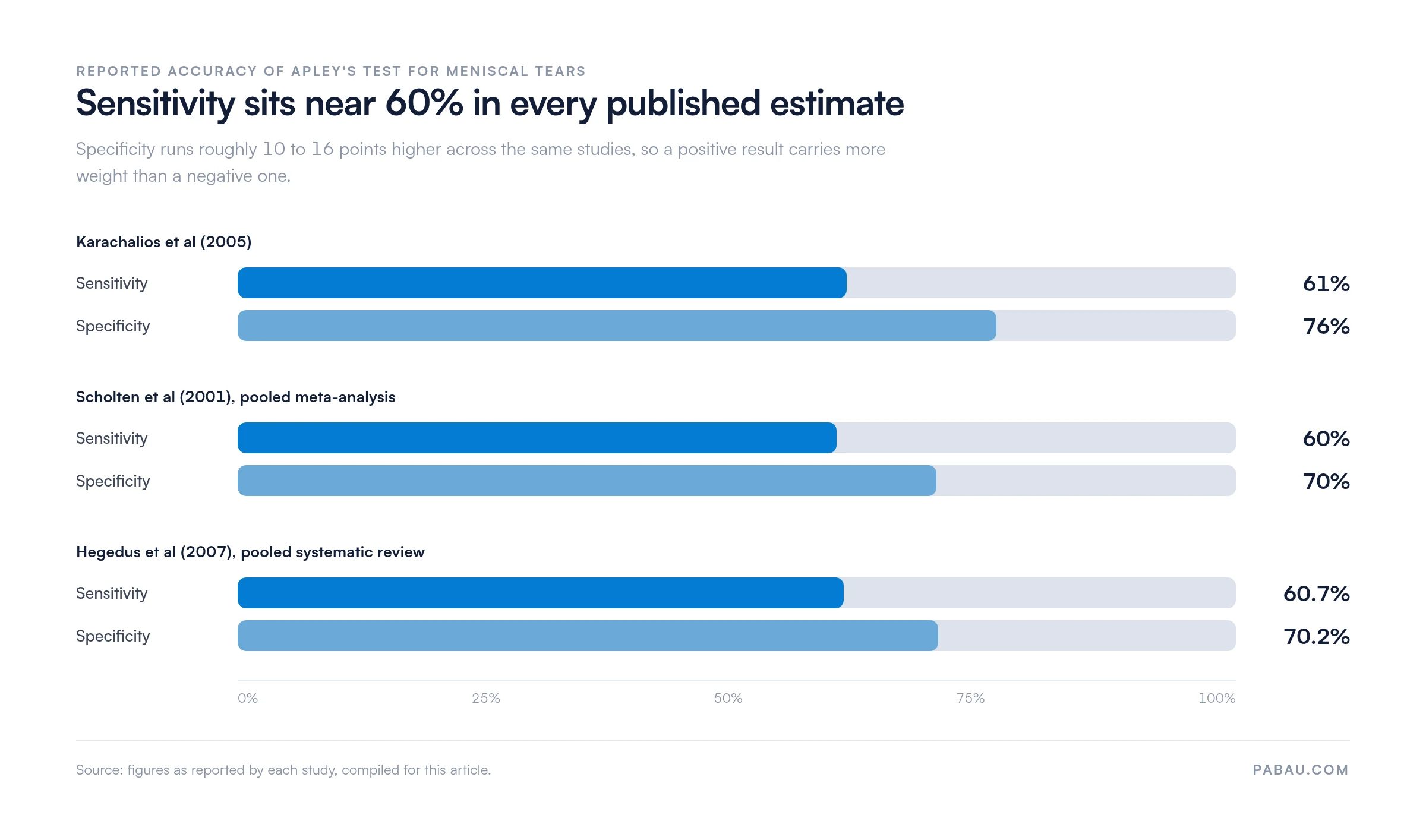
In practice: never use Apley’s test on its own. Pair it with other knee tests and imaging before you commit to a diagnosis. The modest sensitivity means a negative test does not exclude meniscal pathology. The moderate specificity means a positive test in the right clinical context earns further investigation.
Three meniscal tests dominate clinical practice, and each stresses the joint differently. Knowing where they differ helps you build a battery rather than lean on a single result.
Explain each maneuver before you perform it. A guarded patient tenses the hamstrings and muddies the reading, so a few words of warning improve the result you get. Talking the patient through the plan is also one of the cheapest ways to improve patient engagement.
A positive finding should trigger a structured response. Do not diagnose meniscal pathology on this test alone.
In a sports medicine practice, that referral loop runs most weeks, so the handoff is worth templating rather than improvising each time. Where conservative management wins out, a baseline measure such as a proprioception test gives you something to re-check at discharge.
Accurate documentation protects the patient and the practice. A structured SOAP entry keeps the record defensible and easy to re-read months later.
Subjective: the symptoms the patient reported before the test, including mechanical complaints and how the problem started.
Objective: the findings themselves, in a repeatable form. For example, “Apley’s performed prone, knee at 90° flexion. Compression with internal rotation: [pain or no pain, and where]. Compression with external rotation: [pain or no pain]. Distraction phase: [pain, relief, or no change]. Clicks: [none noted].”
Precision matters here more than prose. Structured record fields keep the wording consistent between clinicians and make the entry retrievable later. A shared progress note template stops each colleague inventing their own phrasing.
Assessment: your interpretation. For example, “Positive Apley’s suggesting possible meniscal involvement; distraction phase did not reproduce the pain.”
Plan: the next steps. For example, “MRI referral raised, McMurray and Thessaly planned for the next visit, documentation filed per practice compliance standards.”
File the entry dated and legible in the patient’s permanent record within 24 hours of the assessment.
A knee assessment usually lands in the record as free text. Wording drifts between clinicians, the distraction phase often goes unrecorded, and six months later the note cannot answer whether traction eased the pain.
Pabau, our practice management software, replaces that free text with structured clinical note templates. You build one Apley’s block with fields for position, compression findings by rotation direction, distraction findings, and mechanical symptoms. Every clinician then fills the same fields in the same order.
The record carries the same shape for every knee you assess. Auditing your own accuracy becomes possible, and referral letters pull straight from the note. A colleague reading the file months later sees the detail you saw on the day.
Attach the reference card above as a patient-facing form and the completed version files itself against the appointment. Nothing gets scanned, and no loose paper sits in a drawer waiting to be matched to a patient.

Pabau’s structured clinical notes capture compression and distraction findings in named fields, so every knee assessment reads the same. Referral letters and audits pull straight from the record.

Apley’s test earns its place because of the distraction phase, not the compression phase. Plenty of maneuvers load the meniscus. Few give you a same-position way to ask whether the pain is coming from ligament instead.
Treat the result as one input among several. Sensitivity near 60% means a clean negative buys you very little. A patient with mechanical symptoms still needs the rest of the battery and, in most cases, an MRI.
The part worth fixing this week is the note. An entry that records both phases separately is what makes the test useful to the next person who opens the file. Where you also manage the rehab, a documented baseline such as a return to running protocol gives the discharge decision something to sit on.
Book a demo to see how Pabau structures knee assessment notes for physical therapy and sports medicine practices.

Tightening up your note format? Safer clinical notes covers the SOAP structure and compliance standards behind a defensible assessment entry.
Need a format for follow-up visits? PIRP note template gives you a problem, intervention, response and plan structure for ongoing care.
Coordinating imaging and referrals? Patient care management keeps assessment findings, imaging orders and specialist referrals in one place.
Apley’s test screens for meniscal tears and injuries in the knee. It combines compression and rotation to stress the meniscus while the patient lies prone. Pain on compression-rotation suggests possible meniscal pathology and warrants MRI imaging plus orthopedic evaluation.
Diagnostic accuracy is modest. Reported sensitivity clusters around 60 to 61 percent and specificity around 70 to 76 percent. That makes it useful inside a full assessment but unreliable on its own. Pair it with McMurray and Thessaly, then confirm with imaging before you diagnose.
Compression loads the meniscus with downward force plus rotation, so pain in that phase suggests meniscal involvement. Distraction unloads the meniscus with gentle traction plus rotation. Pain that eases during distraction points to a ligamentous cause instead. That contrast is the whole point of the protocol.
Apley’s runs prone with the knee at 90 degrees, while McMurray runs supine. Apley’s leads with compression-rotation and adds a distraction phase for differentiation. McMurray uses extension with rotation to clamp the meniscus at the joint line. McMurray carries higher specificity, so most clinicians run both.
Document the findings precisely, run the rest of the knee battery, and refer for MRI if meniscal pathology still looks likely. Never diagnose on this test alone. Coordinate with an orthopedic specialist or physical therapist to decide between conservative rehabilitation and surgery.
Avoid the maneuver in the immediate post-injury phase or where the patient reports severe acute pain. Screen for fracture and ligamentous instability first with palpation, Lachman and anterior drawer. The test suits subacute or chronic knee symptoms where trauma has already been ruled out.