Treatment summary
A treatment summary is a written record of what care was delivered during an epi...

2
Last Updated: Oct 16, 2026

The Danis-Weber classification categorizes ankle fractures by fibular fracture level relative to the distal tibiofibular syndesmosis, using three types: A, B, and C.
Weber A fractures are infrasyndesmotic and generally stable; Weber C fractures are suprasyndesmotic with obligate syndesmotic disruption requiring surgical fixation in most cases.
Weber B fractures are the most common and clinically challenging: stability is variable and requires stress radiographs or clinical assessment before deciding on operative vs. nonoperative management.
Pabau supports orthopaedic and musculoskeletal practices with structured clinical documentation, follow-up scheduling, and digital forms to streamline fracture management workflows.
Ankle fractures are among the most common orthopaedic injuries seen in emergency departments and outpatient practices. Most are misclassified at first contact. The Danis-Weber classification gives clinicians a fast, radiograph-based framework to characterize fibular fractures and guide initial management decisions, and it takes roughly ten seconds to apply once you know the anatomy.
This guide covers each Weber type in detail, explains how to apply the classification on plain radiographs, compares it with the Lauge-Hansen system, and outlines the clinical decision thresholds that separate conservative from operative management. It is aimed at orthopaedic trainees, emergency medicine practitioners, radiologists, and musculoskeletal physiotherapists involved in physiotherapy practice management.
The Danis-Weber classification, also called simply the Weber classification, organizes fibular fractures by their position relative to the distal tibiofibular syndesmosis. The core question is simple: where on the fibula did the fracture occur in relation to that joint?
Robert Danis, a Belgian surgeon, is credited with the original description circa 1949. Bernhard Georg Weber later refined and popularized the system, which is why both names appear. The AO Foundation subsequently incorporated Weber’s framework into its own alphanumeric classification (AO/OTA 44), making the two systems largely interchangeable in modern orthopaedic literature.
Three types exist. Each carries distinct implications for syndesmotic integrity, fracture stability, and treatment pathway.
A Weber A fracture sits below the level of the syndesmosis, typically at or below the tip of the fibula. Because the syndesmosis is intact and the ankle mortise is unaffected, these injuries are almost always stable.
Weber A is the least concerning type clinically, but a clean fracture below the syndesmosis does not automatically exclude medial-sided injury. Check the deltoid ligament and the medial malleolus on every radiograph before signing off on conservative management.
Weber B fractures occur at the level of the syndesmosis and represent the majority of ankle fractures presenting clinically. They are the most challenging type within the Weber classification because stability varies: the syndesmosis may be partially torn, fully intact, or frankly disrupted.
The Ottawa ankle rules can help triage Weber B patients toward imaging, but they do not determine stability. That assessment is clinical and radiographic. When in doubt, stress radiographs taken with the patient weight-bearing or under fluoroscopy are the gold standard.
Recovery time for conservatively managed Weber B fractures is typically 6-8 weeks to full weight-bearing. Individual variation based on patient age, bone density, and rehabilitation compliance means that timeline can extend to 12 weeks or more.
Weber C fractures occur above the level of the syndesmosis. The higher the fibular fracture, the more energy was involved and the more extensive the syndesmotic injury. This is the most surgically significant type in the Weber classification.
Always examine the proximal fibula on X-ray when a Weber C pattern is suspected at the ankle. A Maisonneuve fracture, a fibular neck fracture with ankle mortise disruption, is easily missed if the knee is not imaged.
Unrecognized syndesmotic instability is widely recognized in the orthopaedic literature as a leading cause of poor ankle fracture outcomes. That is why proximal fibular imaging matters whenever a high fracture pattern is suspected.
Post-operative wound care at the ORIF site typically involves a secured dressing, billed under A4463, changed at scheduled follow-up visits.
The table below consolidates the key features of each type in the Weber classification for quick clinical reference.
Applying the Weber classification begins with two plain radiographic views: anteroposterior (AP) and mortise. The mortise view (AP with 15-20 degrees of internal rotation) is essential because it shows the ankle mortise space without fibular overlap, allowing accurate assessment of medial clear space, tibiotalar congruence, and the syndesmotic interval.
Three measurements matter most on the mortise view:
For Weber B fractures specifically, a single static mortise view can miss instability. If syndesmotic injury is suspected, weight-bearing mortise radiographs or fluoroscopic stress views, such as the cotton test or an external rotation stress test, are needed to establish whether the mortise widens under load.
Textbooks rarely give this step the weight it deserves, and busy emergency departments can skip it under time pressure. Structured clinical documentation, such as using safer clinical notes frameworks, helps ensure stress testing findings are consistently recorded and available for the treating orthopaedic surgeon.
The Weber classification and the Lauge-Hansen classification both describe ankle fractures, but they approach the problem from different angles. Understanding when to use each system is a practical skill for any orthopaedic or emergency clinician.
The Lauge-Hansen system, with its four patterns of supination-adduction, supination-external rotation, pronation-abduction, and pronation-external rotation, is the preferred system for surgical planning because it maps fracture morphology to the sequence of ligamentous injuries. However, it requires accurate injury mechanism history and is more time-consuming to apply in an emergency setting.
Most practitioners use the Weber classification for initial triage and the Lauge-Hansen system when preparing for operative intervention.
The Weber type narrows the decision tree, but it does not make the decision for you. Stability assessment drives operative vs. nonoperative management, and stability is not always determined by Weber type alone, particularly for Weber B.
A practical decision framework based on Weber type:
Clinicians managing post-operative ankle fracture patients in outpatient settings benefit from structured follow-up scheduling and documented outcome tracking. Sports medicine workflows involving complex fractures require clear documentation at each visit and reliable recall systems to avoid patients being lost to follow-up before full recovery. Recovery has a psychological side too: some patients develop anxiety after injury, and panic disorder treatment combines CBT, medication, and self-help when symptoms escalate.

From structured clinical documentation to automated follow-up scheduling, Pabau helps orthopaedic and musculoskeletal practices keep every patient on track from injury through recovery.

No classification system is perfect, and the Weber classification has well-documented limitations that every clinician should know.
For complex cases, Weber classification should be supplemented by the Lauge-Hansen system or the AO/OTA classification. The AO Foundation’s 44-series codes map onto Weber types but add alphanumeric staging for medial and posterior injuries, making them more comprehensive for surgical documentation.
When reviewing a Weber B ankle fracture in the emergency department, always document medial clear space measurement from the mortise view in millimetres. A written measurement in the notes is far more defensible than ‘appears normal’ and provides the treating orthopaedic surgeon with a baseline for comparison if displacement occurs during conservative management.
For orthopaedic, sports medicine, and physiotherapy practices managing ankle fracture patients post-acutely, the administrative burden is often as challenging as the clinical one. Coordinating follow-up appointments, tracking weight-bearing progression, and ensuring documentation meets medicolegal standards all require reliable systems.
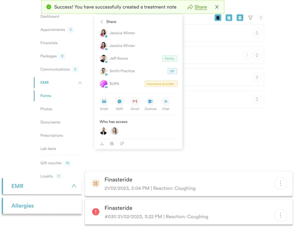
Pabau provides clinical documentation tools designed for musculoskeletal workflows, including structured SOAP notes, body charting, and injury-specific form templates. For post-fracture follow-up, automated follow-up workflows can be configured to trigger at the 1-week, 2-week, and 6-week milestones that Weber B conservative management requires, reducing the risk of patients being discharged from follow-up before fracture consolidation is confirmed on radiograph.
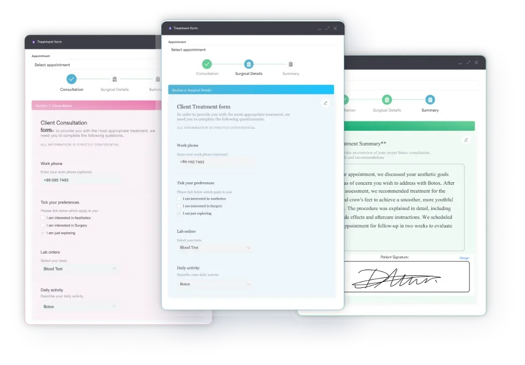
Practices managing a high volume of ankle fracture referrals also use Pabau’s digital intake forms to capture injury mechanism, initial Weber classification, surgical history, and rehabilitation goals before the first appointment, keeping consultation time focused on clinical assessment rather than administrative data entry. Physical therapy practice management built on structured documentation supports better continuity of care across the injury-to-recovery pathway.
For practices with appointment scheduling demands tied to post-operative protocols, such as Weber C patients requiring syndesmotic screw removal at 8-12 weeks, Pabau’s scheduling tools help teams build and maintain structured recall pathways without manual tracking.
The Weber classification remains the most widely used system for initial ankle fracture categorization because it is fast, radiograph-based, and clinically actionable. Weber A is stable and conservative. Weber C is unstable and surgical. Weber B is the system’s most nuanced type and the one most likely to generate diagnostic and management errors without rigorous stability assessment.
Supplementing Weber with Lauge-Hansen for surgical planning and using structured documentation to capture measurements, stress testing results, and follow-up milestones gives clinicians and their patients the best outcomes. If your orthopaedic or musculoskeletal practice needs better tools for structured clinical notes, return-to-running protocols, and automated fracture follow-up workflows, book a demo to see how Pabau handles it end to end.

Need a fast bedside test for a suspected foot fracture? Foot stress fracture test covers the clinical tests, home checks, and imaging pathways used to confirm a diagnosis.
Assessing ankle injuries in older or high-risk patients? Bernese ankle rules offers a validated alternative decision tool for imaging referral.
Need a fast way to document imaging findings? Ankle radiograph results template gives practices a structured format for recording mortise and AP view measurements.
The Danis-Weber classification is a system for categorizing ankle fractures based on the level of the fibular fracture relative to the distal tibiofibular syndesmosis. It has three types: Weber A (below the syndesmosis, stable), Weber B (at the level of the syndesmosis, variable stability), and Weber C (above the syndesmosis, unstable). Originally described by Robert Danis and later popularized by Bernhard Georg Weber, it is now the most commonly used ankle fracture classification system worldwide.
Weber A fractures are infrasyndesmotic, leave the syndesmosis intact, and are almost always stable and managed conservatively. Weber B fractures are transsyndesmotic, may partially involve the syndesmosis, and require stability testing to determine management. Weber C fractures are suprasyndesmotic, disrupt the syndesmosis in all cases, and are unstable, typically requiring surgical fixation with ORIF and syndesmotic repair.
Weber B fractures are neither consistently stable nor consistently unstable: stability must be assessed individually. A mortise radiograph showing medial clear space greater than 4 mm, or a positive stress test under fluoroscopy, indicates instability requiring surgical management. When the mortise is congruent and the deltoid ligament is intact, conservative management in a cast or functional boot is appropriate.
In virtually all cases, yes. Weber C fractures involve obligate syndesmotic disruption and produce an unstable ankle mortise that cannot be maintained without surgical fixation. Standard treatment is ORIF of the fibula combined with syndesmotic stabilization using screws or a suture-button device. Non-operative management of Weber C injuries is associated with poor outcomes due to progressive mortise widening and post-traumatic arthritis.
The Danis-Weber classification is anatomical, based on fibular fracture level on radiograph. The Lauge-Hansen classification is mechanistic, based on foot position and direction of deforming force, and describes sequential ligamentous injury patterns. Weber is faster to apply from plain radiographs; Lauge-Hansen provides more detail for surgical planning. Most clinicians use Weber for initial triage and Lauge-Hansen when planning operative intervention.
Weber fracture types map to ICD-10-CM codes within the S82 category (fracture of lower leg, including ankle). The specific code depends on laterality, displacement status, and whether the fracture is open or closed, rather than Weber type directly. For example, S82.61 covers fracture of lateral malleolus of right fibula, with further characters specifying displacement and encounter type. Clinical coders should select based on documented fracture characteristics rather than Weber type alone; verifying against current CMS ICD-10-CM tables is recommended.