Sulcus Test: Shoulder Instability Assessment
The sulcus test is an orthopedic special test that checks for inferior glenohume...

3
Last Updated: Oct 22, 2026
The inverted supinator test is a quick reflex check for cervical myelopathy, which is compression of the spinal cord in the neck. You tap the brachioradialis tendon near the wrist and watch the forearm and fingers together.
A positive result pairs a weak or absent forearm jerk with an unexpected flick of the fingers. That pattern points to an upper motor neuron lesion at or above C5-C6, rather than a pinched nerve root. A 2024 case report in Annals of Internal Medicine: Clinical Cases describes the sign.
Miss it, and a patient with a compressed cord may leave with a neck-stretch handout instead of an MRI request. Below, we cover the anatomy, technique, grading, documentation and referral.

A positive inverted supinator test pairs an absent or reduced brachioradialis reflex with reflex finger flexion.
The pattern suggests an upper motor neuron lesion at or above C5-C6, which separates cord compression from a C6 root problem.
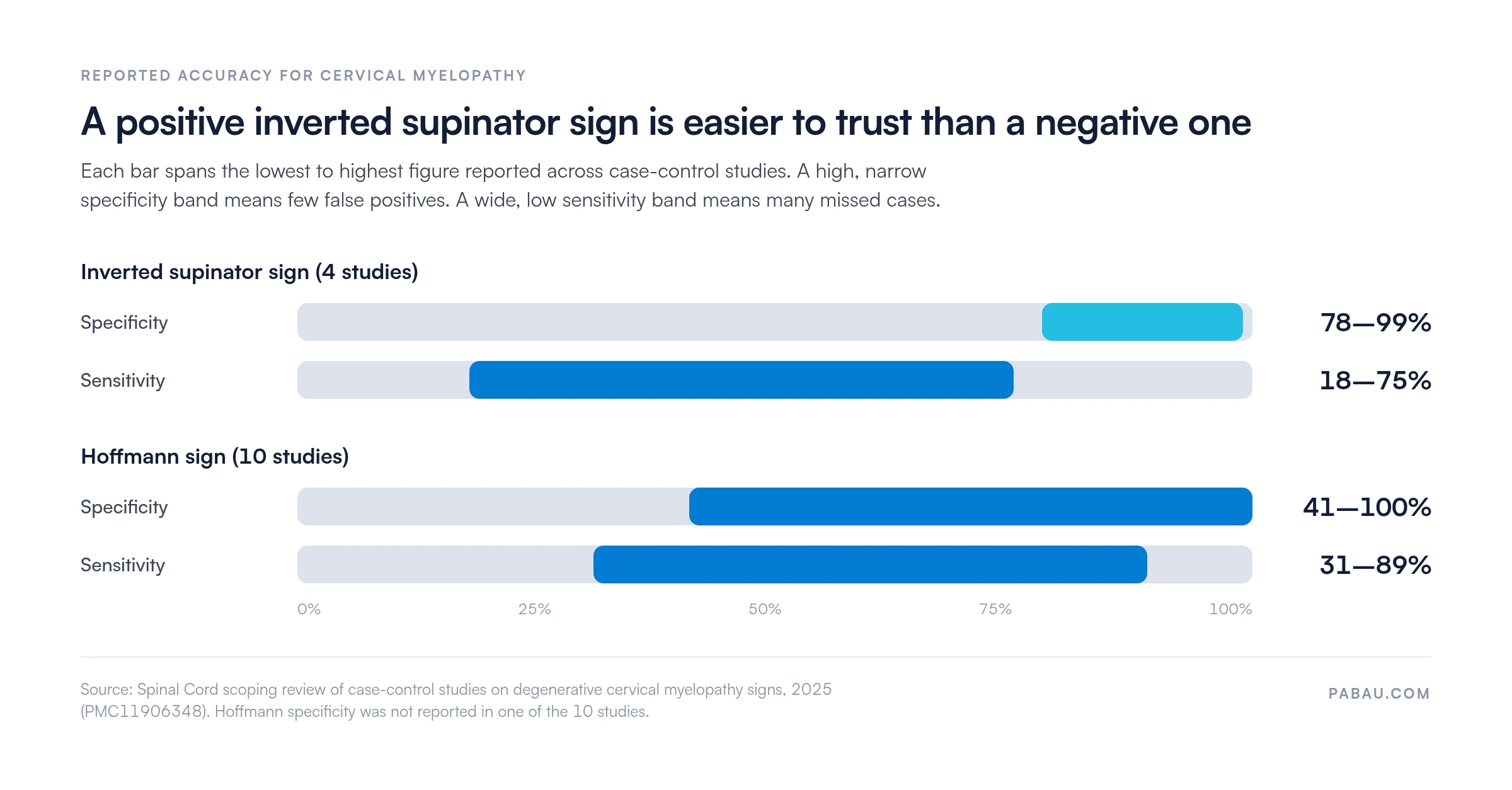
Reported specificity is high (78-99%) but sensitivity is low and variable, so a negative test never rules out myelopathy.
Grade the brachioradialis on the 0-4+ scale and record finger flexion separately for each side, so serial exams stay comparable.
Practice software such as Pabau keeps customizable assessment forms and structured client records together for physical therapy and musculoskeletal practices.
The inverted supinator test is a deep tendon reflex exam that looks for cervical cord compression. One tap on the brachioradialis tendon checks two things. First, it tests the C5-C6 reflex arc that bends the elbow and turns the forearm. Second, it shows whether the finger flexors below that level have lost their normal brake.
You’ll also see it called the inverted supinator sign, the inverted radial reflex or the inverted brachioradialis reflex. All of these names describe the same finding. It belongs in the same toolkit as the Hoffmann sign, the Babinski sign and clonus.
A normal tap makes the forearm supinate and the elbow flex slightly. In a positive test, that movement fades or disappears, and the fingers flex instead. You need both halves of the pattern, because either one alone means something different.
The brachioradialis muscle drives the supinator reflex. Its sensory signal enters the cord through the C5 and C6 roots. The motor signal travels back out through the radial nerve and bends the elbow.
Compression of the cord at C5-C6 disrupts this loop in two ways. Damage at that segment weakens or abolishes the local reflex, so the brachioradialis barely responds. The same lesion also interrupts the descending corticospinal pathways that normally damp down reflexes below it.
The finger flexors, supplied mainly by C7 and C8, become overactive as a result. The vibration from the tap is enough to trigger them, so the fingers flex.
That dual pattern is why the sign is highly specific for cord-level disease at or above C5-C6. A C6 radiculopathy can also dampen the brachioradialis reflex. However, the descending tracts stay intact, so the fingers stay still. A Spurling’s test and a dermatomal sensory check help confirm a root problem instead.
A positive inverted supinator sign suggests an upper motor neuron lesion at or above C5-C6. The most common cause is degenerative cervical myelopathy, often called cervical spondylotic myelopathy.
Suspect it in patients over 50 who report clumsy hands, dropped objects, trouble with buttons or an unsteady walk. Other causes of cord compression at this level include ossification of the posterior longitudinal ligament and trauma. Less often, demyelinating disease or a cord tumor is responsible.
The sign doesn’t tell you which cause you’re dealing with. What it does tell you is that the lesion sits in the cord, and that the patient needs imaging.
Short answer: trust a positive result more than a negative one. A 2025 scoping review in Spinal Cord compared case-control studies of myelopathy signs. Across four studies, the inverted supinator sign showed specificity of 78-99%. Its sensitivity ranged from just 18% to 75%.
A 2022 Neurology teaching case illustrates the inverted radial reflex and the Wartenberg thumb sign in a patient with cervical myelopathy. In practice, a positive result justifies prompt imaging. A negative result does not exclude early myelopathy, so keep testing the other signs.
The chart below shows how much tighter the specificity band is than the sensitivity band, with the Hoffmann sign for comparison.
Poor positioning is the easiest way to miss a positive result or create a false one. Set the arm up carefully, then work through the steps on both sides.
Seat the patient with the forearm resting on their lap or your hand, and the elbow flexed about 90 degrees. Hold the forearm midway between pronation and supination. Then ask them to let the arm go heavy and not help the movement.
You’ll see one of three responses:
A one-sided positive result still counts, especially when it matches the patient’s symptoms. Bilateral positivity, though, raises the urgency considerably.
Read the result from what the tap produces in both places, not just what disappears. Use this reference when you record it.
Myelopathy is often monitored over serial exams before anyone decides on surgery. A note that only says “positive” can’t show whether the sign is getting worse.
Grade the brachioradialis on the standard 0-4+ deep tendon reflex scale. Then record finger flexion separately, as present or absent, for each side.
If several clinicians see the same patient, a shared deep tendon reflex exam template keeps the grading consistent.
A typical positive entry might read: “Left BR reflex 1+/4+, finger flexion present. Right BR reflex 2+/4+, no finger flexion. Repeat in 6 weeks or earlier if symptoms progress.” Kept in structured patient records, that entry lets the next clinician judge the trend without starting over.

Most errors with this test come from technique rather than interpretation. These are the ones to watch for.
Screen both sides on every inverted supinator test and record laterality. Bilateral positivity suggests more extensive cord involvement and raises the urgency of referral and imaging. Never rely on one sign alone. Build the picture from the full upper motor neuron exam before you decide how urgent the case is.
Both are upper motor neuron signs used in myelopathy screening, but you elicit them differently. The Hoffmann sign is often spelled Hoffman’s sign.
To run the Hoffman’s sign test, flick the distal phalanx of the middle finger and watch the thumb and index finger. Flexion shows finger flexor hyperreflexia, but no segmental arc is tested. The inverted supinator test checks the C5-C6 arc and the finger flexors with one tap.
Neither sign proves a level on its own. A positive inverted supinator sign suggests a lesion at or above C5-C6. Across studies, the Hoffmann sign tends to pick up more cases, while the inverted supinator sign is more consistently specific.
A positive Hoffmann sign alone can appear in people with naturally brisk reflexes. When the inverted supinator sign is positive too, a normal variant becomes much harder to argue.
No single sign confirms cervical myelopathy. Build a composite picture from several tests, and read them as a cluster.
When the cluster stays negative and symptoms persist, a mechanical source at the cervicothoracic junction is worth ruling out. The rib spring test checks first rib mobility, where restriction is linked to same-side neck pain and thoracic outlet symptoms.
Refer urgently and arrange a cervical spine MRI when a positive test comes with any of these red flags. Escalate the same day for rapidly progressive deficits or sphincter symptoms.
Without red flags, referral is still needed, for example for isolated hand clumsiness with a stable one-sided sign. Timing then follows your local pathways. Record the functional impact alongside the sign, and review within four to six weeks, or sooner if symptoms progress.
The usual next test is MRI of the cervical spine without contrast. Contrast may be added if a tumor or demyelination is suspected. X-rays and CT show bony narrowing, but they miss changes inside the cord.
Your note should capture the finding and the reasoning behind the referral decision. Our guide to physical therapy compliance covers the record-keeping rules for US and UK practices.
Many physical therapy and chiropractic practices still record neuro exams as free text or on paper. Grades end up in different formats, and the second side often goes unrecorded. Comparing this month’s exam with last month’s then means digging through old notes.
Pabau lets you build customizable assessment forms with fields for each side, the 0-4+ grade and finger flexion. Completed forms save to the patient’s Client record, next to appointments and treatment notes. Our physical therapy EMR brings those records together with scheduling and billing.
The result is a reflex history you can read at a glance. You can also book the six-week recheck before the patient leaves, so the follow-up doesn’t depend on anyone’s memory.

Pabau gives physical therapy and musculoskeletal practices customizable assessment forms and structured client records. Reflex grades, serial findings and referral notes stay in one place for the whole team.

Make the inverted supinator test part of every exam for neck pain or a clumsy hand. It takes seconds, and a positive result changes what happens next for the patient.
The trade-off is sensitivity. A negative test reassures far less than a positive one should alarm you, so pair it with Hoffmann, Babinski, clonus and gait testing. Grade both sides every time, and write the result down in a form the next clinician can compare.
Consistent notes are what make serial testing worth the effort. Book a demo to see how Pabau’s assessment forms keep reflex grades and referral notes together for your team.

Want a ready-made grading form? Deep tendon reflex exam gives you a template for recording reflex grades on both sides.
Testing for upper motor neuron signs in the hand? Hoffman’s sign test walks through technique, interpretation and referral.
Checking the lower limbs next? Clonus reflex test explains technique, grading and documentation.
Does neck flexion trigger electric shocks? Lhermitte sign test covers how to perform it and act on the findings.
Suspect a nerve root rather than the cord? Spurling’s test covers the procedure, interpretation and clinical accuracy.
Reported specificity is high (about 78-99%) but sensitivity is modest (about 18-75%), so a negative result does not exclude myelopathy. Confirm a positive result with other upper motor neuron signs and a cervical spine MRI.
Yes. Inverted radial reflex, inverted supinator reflex and inverted brachioradialis reflex all name the same finding. The “supinator” name dates from when the brachioradialis was called the supinator longus.
It’s uncommon. People with naturally brisk reflexes may flex their fingers slightly on the tap, but their brachioradialis jerk stays normal. The sign only counts as inverted when that jerk is reduced or absent.
Physical therapists, chiropractors, primary care physicians, neurologists and spine surgeons use it during an upper-limb neurological exam. It suits any visit for neck pain or arm symptoms.
No. MRI shows cord compression, but many older adults have narrowing on scans without symptoms. The bedside exam shows whether the cord is affected clinically, which helps you interpret the scan.