Pabau GO app

The new Pabau GO is heredownload on the App Store

Download on the App Store
Book a demo Book a demo
Practice Management Tips

Risk for infection nursing care plan

Key takeaways

Key takeaways

Risk for infection is a NANDA-I nursing diagnosis for patients who are susceptible to pathogens but show no infection yet.

Because no signs are present, the statement uses the two-part PE format, so it carries no as-evidenced-by clause.

Write one specific etiology into the statement, because the etiology decides which interventions you choose.

Core interventions are hand hygiene, sterile technique, daily wound and device assessment, vital sign trending, and patient education.

Practice management software like Pabau turns the plan into a fillable form on the client record, so each entry is timestamped.

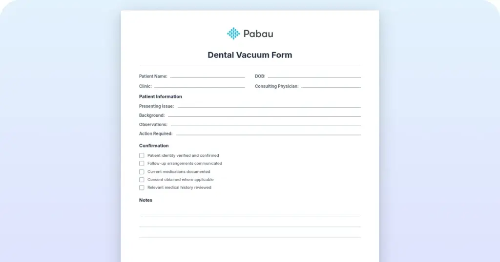
Download your free risk for infection nursing care plan

A printable, blank form covering patient information, medical history, and subjective and objective assessment. It then carries fields for the nursing diagnosis, goals and outcomes, interventions, rationale, evaluation, and nurse sign-off. Fill it in by hand or use it as the field list for a digital form.

Download template

Risk for infection is a prevention diagnosis. You write it when a patient carries risk factors but shows no clinical signs of infection yet. Nurses in surgical units, oncology, and community settings reach for it most days.

This guide covers the NANDA-I definition, the statement format, the five assessment domains, measurable goals, and interventions with rationale. It closes with three sample plans and an escalation panel for the point where infection risk becomes a sepsis call.

What is risk for infection?

Risk for infection is a NANDA-I nursing diagnosis describing a patient’s susceptibility to invasion and multiplication of pathogenic organisms. It focuses on prevention rather than treatment. The patient has not developed clinical signs yet, so they sit in a risk state that needs nursing vigilance.

That distinction changes what you do. A patient with an indwelling urinary catheter has risk factors but no fever, dysuria, or positive culture. The plan activates preventive work: daily catheter assessment, sterile technique during care, and patient education on fluid intake.

Compare that with a patient carrying an actual infection diagnosis, such as pneumonia confirmed on chest X-ray and sputum culture. That patient needs antimicrobial monitoring, symptom management, and an escalation protocol instead.

NANDA-I definition and how to write the statement

Official NANDA-I definition (2024-2026): “Susceptible to invasion and multiplication of pathogenic organisms, which may compromise health.”

The statement follows a standardized format. Risk diagnoses use the two-part PE format (problem and etiology). They drop the “as evidenced by” (AEB) clause, because no defining characteristics are clinically present yet:

Risk for Infection related to [specific risk factor]

  • Post-surgical patient: Risk for Infection related to disruption of skin integrity from a surgical incision
  • Immunocompromised patient: Risk for Infection related to suppressed immune response secondary to chemotherapy
  • Catheterized patient: Risk for Infection related to an invasive urinary catheter and potential bacterial colonization

Note the shape: problem, then “related to”, then one specific etiology. Naming one etiology points your interventions at the cause rather than at generic infection precautions.

This diagnosis applies across very different patient groups. Which risk factors your patient carries decides how deep the assessment goes and how intensive the interventions are. The CDC infection control guidelines group the common ones like this:

Risk factor category Examples At-risk populations
Invasive procedures Indwelling catheters, central lines, intubation, surgical incisions Post-surgical, ICU, and long-term care patients
Compromised immunity Immunosuppressive medication, chemotherapy, HIV/AIDS, organ transplant Oncology, transplant, and rheumatology patients
Impaired skin or tissue integrity Wounds, burns, dermatological conditions, pressure ulcers Wound care, burn unit, and bariatric patients
Nutritional deficiency Low protein, vitamin C, or zinc intake, which slows wound healing Elderly, malnourished, and post-surgical patients
Altered circulation or oxygenation Diabetes, vascular disease, heart failure, respiratory compromise Cardiology, endocrinology, and pulmonary patients

One post-surgical patient can sit in three categories at once. The incision is invasive, pain limits mobility and circulation, and pre-operative NPO status affects nutrition. Each layer raises the risk, so each layer needs its own intervention.

Where a wound has already reached bone, prevention is no longer the job. The osteomyelitis nursing care plan covers that patient, with antimicrobial and mobility goals this form does not carry.

What to assess before you write the plan

Assessment drives the rest of the plan. Without solid subjective and objective data you cannot write a measurable goal or choose the right intervention. Five domains cover it:

  • Vital signs: baseline temperature, fever pattern, heart rate, and respiratory rate. Sepsis often shows as tachycardia before any fever appears.
  • Skin and wound status: color, warmth, edema, drainage character, odor, edge approximation, and suture or staple integrity.
  • Laboratory values: WBC count (4.5 to 11.0 K/uL is normal), bands, and cultures where indicated. Bands are immature neutrophils and point to acute infection.
  • Patient and family report: chills, malaise, confusion, pain at the surgical site, difficulty with self-care, and grasp of prevention.
  • Risk factor inventory: chart review for invasive devices, antimicrobial use, recent procedures, comorbidities, and nutritional status.

A running vital signs record matters more here than any single set of numbers. One reading of 37.6°C means little. The same patient climbing 0.3°C every four hours means something.

Write findings down as you take them, because the assessment is what justifies the diagnosis. Fever with an elevated WBC and purulent drainage points to an infection already underway. An incision with normal vitals, a normal WBC, and a dry dressing calls for prevention only.

Nursing goals and expected outcomes (NOC)

NOC (Nursing Outcomes Classification) outcomes for this diagnosis all aim at stopping infection before it starts. Write SMART goals, meaning specific, measurable, achievable, relevant, and time-bound. A goal like “patient will not get an infection” cannot be measured or evaluated.

  • Infection prevention: patient will keep skin and mucous membranes intact throughout the hospital stay.
  • Wound healing: the surgical incision will show primary intention healing by post-operative day 5, with edges approximated and no drainage or erythema.
  • Knowledge: patient will name three prevention strategies before discharge, covering hand hygiene, catheter care, and fever reporting.
  • Immunity: white blood cell count will stay between 4.5 and 11.0 K/uL through the treatment course.
  • Vital signs: temperature will stay below 38°C (100.4°F), with 48 afebrile hours before discharge.

Each goal traces back to the etiology in your statement. A central line makes exit-site integrity the wound goal. Immunosuppression makes immune markers and symptom absence the goals to track.

Nursing interventions and rationale (NIC)

NIC (Nursing Interventions Classification) interventions work by closing pathogen routes and supporting immune function. Every intervention needs its rationale, grounded in microbiology, physiology, or clinical evidence. The core set, grouped by mechanism:

Intervention Rationale
Perform hand hygiene before and after patient contact, before aseptic procedures, and after body fluid exposure Hands are the most common transmission route for hospital-acquired pathogens. Hand hygiene remains the single most effective prevention measure. The WHO 5 Moments for Hand Hygiene sets out when to do it.
Use sterile technique for invasive procedures, including catheter insertion, wound care, and dressing changes Sterile technique keeps skin flora and environmental pathogens out of sterile body cavities and wounds, which closes a primary infection pathway.
Assess and document wound appearance daily, covering color, warmth, swelling, drainage character, and odor Catching erythema, purulent drainage, or wound separation early allows intervention before the infection turns systemic.
Monitor vital signs every 4 hours or per protocol, and report a temperature at or above 38°C, sustained tachycardia, or tachypnea Fever, heart rate, and respiratory rate move before the WBC rises or a culture turns positive. Trending them shows infection progressing.
Give prophylactic or therapeutic antimicrobials on schedule and watch for adverse effects Prophylactic antibiotics cut surgical site infection risk when the timing is right. Watching adherence and side effects protects both efficacy and safety.
Teach the patient and family hand hygiene, incision care, the signs to report, fluid intake, and nutrition Most of the prevention window happens after discharge, where only the patient and family are present. Teaching reduces post-discharge complications and readmission.

Record each intervention as you perform it. Include the frequency (“every 4 hours”), the specific observation (“no erythema noted”), and how the patient responded.

Sample care plans for three patient types

These three worked examples show the NANDA framework, assessment findings, goals, and interventions applied to real scenarios. Adapt them to your patient population and your institution’s protocols.

Care plan 1: Post-surgical patient after abdominal surgery

Component Detail
Nursing diagnosis Risk for Infection related to disruption of skin integrity from an abdominal surgical incision
Assessment findings
  • Post-op day 2, midline abdominal incision closed with staples.
  • Minimal serous drainage, dressing clean and intact.
  • T 37.2°C, HR 88, RR 18.
  • WBC 12.2 K/uL, slightly high and expected after surgery.
  • Mild incisional pain, no chills or malaise.
Goal (NOC) Patient will keep the incision free of erythema, purulent drainage, and dehiscence through discharge
Nursing interventions (NIC)
  • Assess the incision every 8 hours for erythema, warmth, drainage, and separation.
  • Change the dressing per protocol using sterile technique.
  • Check temperature every 4 hours and report 38.5°C or above.
  • Give the prophylactic cephalosporin on schedule, watching for rash or anaphylaxis.
  • Teach the patient to keep the incision dry and untouched.
  • Encourage ambulation as tolerated, which supports circulation and immune function.
Evaluation, post-op day 5
  • Incision edges approximated, no erythema, staples intact.
  • T 37.0°C for 48 hours, WBC down to 10.1 K/uL.
  • Patient ambulating independently.
  • Discharged with written incision care instructions and a number to call.

Care plan 2: Immunocompromised patient on chemotherapy

Component Detail
Nursing diagnosis Risk for Infection related to suppressed immune response secondary to chemotherapy, at day 10 post-cycle 2
Assessment findings
  • Nadir phase, when the white cell count reaches its lowest point.
  • WBC 2.1 K/uL, ANC 800/mm³.
  • T 36.9°C, HR 92, RR 20.
  • Mucous membranes intact but pale.
  • Fatigue and mild nausea reported.
  • No wounds or skin breakdown, and lives at home with family support.
Goal (NOC) Patient will stay free of infection through the nadir phase and the rest of the chemotherapy course
Nursing interventions (NIC)
  • Apply neutropenia precautions, including a private room if the ANC falls below 500.
  • Ask every visitor to perform hand hygiene on entry.
  • Check temperature every 2 to 4 hours and report any spike at once.
  • Give prophylactic antibiotics per protocol and reinforce adherence.
  • Teach home precautions: no raw foods, live plants, unwashed produce, or crowds.
  • Teach meticulous oral hygiene and a daily shower.
  • Name the signs that need same-day review: fever, chills, cough, dysuria, rectal pain.
  • Assess nutritional intake and encourage protein to support immune recovery.
Evaluation, day 21 post-cycle 2
  • WBC recovered to 4.8 K/uL.
  • Afebrile throughout the nadir, with no infection-related admission.
  • Patient described the home precautions accurately and reported following them.
  • No mucositis and no break in skin integrity.

Care plan 3: Patient with an indwelling urinary catheter

Component Detail
Nursing diagnosis Risk for Infection related to an indwelling urinary catheter and disruption of normal urinary tract flora
Assessment findings
  • Day 3 after hip replacement, 18 Fr Foley placed during surgery.
  • Urine clear yellow with no foul odor.
  • Catheter site clean and dry, no erythema or purulent drainage.
  • Output around 1,200 mL in 24 hours.
  • T 37.3°C, no dysuria, suprapubic discomfort, or bladder spasm.
Goal (NOC) Patient will stay free of catheter-associated urinary tract infection (CAUTI) signs while catheterized and after discharge
Nursing interventions (NIC)
  • Give daily perineal care with soap and water, then assess and document the site.
  • Keep the drainage system closed and never disconnect it without cause.
  • Use aseptic technique for any urine sample.
  • Keep the bag below bladder level at all times to prevent reflux.
  • Empty the bag every 8 hours using clean technique.
  • Check urine color, clarity, and odor, and report any change.
  • Track intake and output, aiming for 1,500 mL a day.
  • Report fever, dysuria, suprapubic pain, or cloudy urine at once.
  • Plan removal as soon as the patient can void, usually day 3 or 4.
Evaluation, post-op day 4
  • Catheter removed once the patient voided spontaneously, post-void residual under 100 mL.
  • Urine stayed clear with no foul odor.
  • No fever, dysuria, or other CAUTI signs.
  • Discharged with the symptoms to watch for and advice to keep drinking.

How to write the plan, step by step

Building your own plan follows the nursing process. Clear nursing documentation at each step is what keeps care continuous across shifts. Here is the working order:

  1. Assess. Review the chart for risk factors such as a surgical incision, immunosuppression, or an invasive device. Take vitals, inspect skin and device sites, palpate for warmth and swelling, and note drainage. Ask the patient about chills, fatigue, pain, and nausea, then record both the subjective and objective findings.
  2. Diagnose. Risk factors with no current signs give you “Risk for Infection related to [etiology]”. Signs already present, such as fever, purulent drainage, or a positive culture, give you an actual infection diagnosis instead. Match your institution’s wording.
  3. Plan. Write SMART goals aimed at preventing infection. Each goal says what the patient will do or demonstrate, and by when. “Patient will keep skin and incision intact through the hospital stay” is measurable every time you assess.
  4. Implement. Carry out each intervention at the frequency you wrote down. Record the action, the observation, and the response as you go. Write “incision assessed 08:00, no erythema, edges approximated, minimal clear drainage, staples intact” rather than “incision looks good”.
  5. Evaluate. At the end of the shift, check each goal against the data. Was the patient afebrile? Was the incision clean? Could they name the prevention steps? Mark the goal met, partially met, or not met, and revise the plan where it was not met.

The cycle repeats as the patient progresses. Digital forms in your patient intake software let you enter findings at the bedside, which cuts transcription errors and keeps the next nurse current.

Pabau digital forms builder showing a template library and a patient form on a tablet
Pabau’s form builder turns the care plan into a fillable record, so each assessment and intervention is timestamped as you chart it.

When infection risk progresses to sepsis

Unrecognized infection risk can escalate to sepsis, a life-threatening systemic inflammatory response. The panel below sets the three rungs side by side, because the same patient can move between them inside one shift.
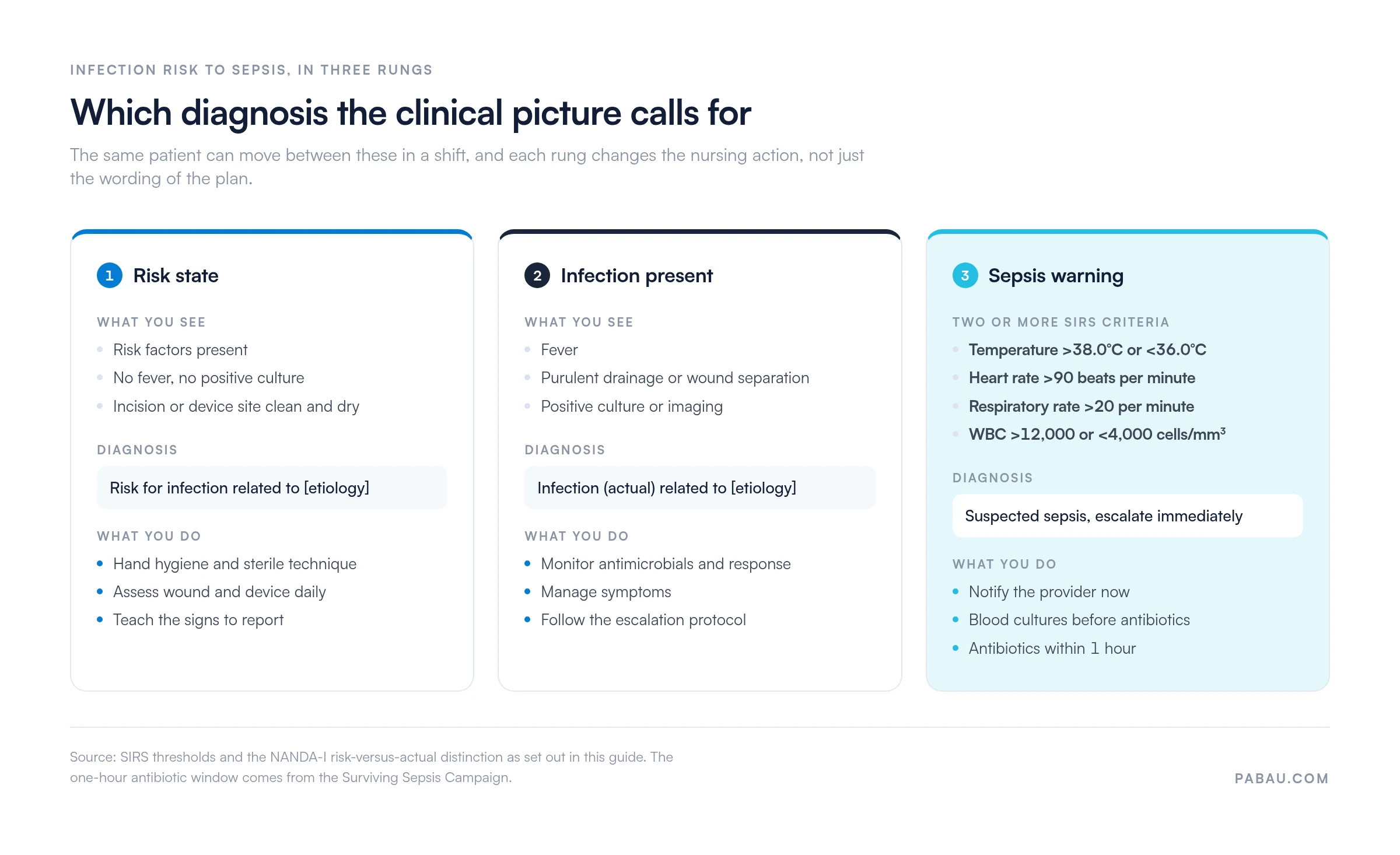
Escalation ladder from infection risk to sepsis warning across three severity rungs
Each rung changes the nursing action, not only the wording of the plan. Thresholds are the SIRS criteria set out below, with antibiotic timing from the Surviving Sepsis Campaign.

Two or more SIRS (Systemic Inflammatory Response Syndrome) criteria point to possible sepsis:

  • Temperature: >38°C (100.4°F) or <36°C (96.8°F)
  • Heart rate: >90 beats per minute
  • Respiratory rate: >20 breaths per minute, or PaCO₂ <32 mmHg
  • WBC: >12,000 or <4,000 cells/mm³, or more than 10% immature bands

Fever plus one more criterion means you notify the provider now. Obtain blood cultures before antibiotics start, then prepare for antibiotic administration and possible ICU transfer. The Surviving Sepsis Campaign’s Hour-1 bundle calls for antibiotics within one hour of recognition, and that speed is what changes survival.

Your job here is continuous surveillance. A patient on this plan with fever, tachycardia, and a raised respiratory rate is showing sepsis warning signs. Escalate rather than watch another set of vitals.

Once a fever is established, prevention stops being the goal and treatment takes over. The fever nursing care plan covers that ground, with cooling measures and antipyretic timing this form does not include.

How Pabau keeps a care plan current at the bedside

Most care plans still start on a paper form or a shared document. The nurse notes findings on one sheet, charts interventions somewhere else, and copies both into the record at the end of the shift.

Detail goes missing in that transfer. The next nurse inherits a plan that is already a shift out of date. That is a problem when the whole diagnosis rests on catching a change early.

Pabau replaces the paper with a fillable form attached to the client record. You build the plan once, with fields for assessment, diagnosis, goals, interventions, rationale, and evaluation. Every entry carries a timestamp and the name of the staff member who made it.

The whole team then reads the same plan, so a temperature logged at 2 AM is visible on the morning round. Reporting across records also shows which prevention steps get documented and which get skipped.

Keep every care plan current at the bedside

Pabau’s digital forms attach the care plan to the client record, so assessment findings and interventions are documented as they happen.

Pabau practice management platform

Conclusion

The value of this diagnosis sits in the etiology, not the label. Name the catheter, the incision, or the suppressed immune response, and the interventions choose themselves.

Two habits separate a plan that works from one that only satisfies an audit. Assess at the frequency you wrote down, and record what you saw rather than that you looked.

Download the form, adapt whichever sample plan matches your patient, and keep the escalation thresholds where your team can see them. Book a demo to see how Pabau keeps care plan documentation attached to the client record.

Continue your research

Continue your research

Preventing surgical site infection before the incision? WHO surgical safety checklist runs the pre-incision checks, including antibiotic prophylaxis timing.

Caring for a patient through chemotherapy? Cancer nursing care plan covers the wider oncology plan that neutropenia sits inside.

Unsure how to word the evaluation step? Evaluation nursing care plan shows how to judge a goal met, partially met, or not met.

Handing infection risk over at shift change? Bedside shift report structures the handoff so risk factors and thresholds carry across.

Moving care plans off paper? Clinical documentation software compares what to look for when documentation goes digital.

Frequently asked questions

What is the difference between “Risk for Infection” and actual “Infection” nursing diagnoses?

Risk for Infection applies when a patient has susceptibility factors but no clinical signs of infection yet. There is no fever, no positive culture, and no purulent drainage. The focus is prevention. An actual Infection diagnosis is used once the patient shows clinical evidence of infection. Treatment-focused interventions such as antibiotics and antimicrobial monitoring are then added.

Why don’t risk diagnoses include “as evidenced by” (AEB) in the diagnosis statement?

Risk diagnoses describe a potential future state rather than current signs. Defining characteristics are not present yet, so there is nothing to evidence. The statement uses the two-part PE format of problem plus etiology, with no AEB clause. Actual diagnoses use the three-part PES format and list defining characteristics after AEB.

What are the most important nursing interventions for preventing catheter-associated urinary tract infections?

Four interventions matter most. Keep the drainage system closed and sterile, with no disconnections. Keep the bag below bladder level to prevent backflow. Perform daily perineal hygiene and assess the insertion site. Remove the catheter as soon as it is no longer clinically necessary. Catheter duration is the strongest modifiable risk factor for CAUTI.

What temperature threshold should trigger immediate infection escalation in a post-surgical patient?

A temperature of 38.5°C (101.3°F) or higher warrants immediate provider notification and assessment. Even a single fever spike in a post-op patient needs investigation, meaning cultures, incision inspection, and a decision on antibiotics. Do not wait for a pattern to form. In early infection, the single fever is the pattern.

How do I know if a patient’s care plan goal has been met?

Compare the patient’s current status against the measurable goal you wrote. Say the goal was afebrile status below 38°C throughout the stay. If the patient has been afebrile for 48 hours, the goal is met. Document the evidence: vital signs, assessment findings, and what the patient reports. Avoid vague notes such as patient doing well. A measurable goal is either met or not met, judged on objective data.
×