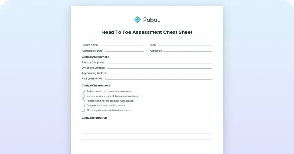
Head to toe assessment cheat sheet: Printable nursing guide
A head to toe assessment cheat sheet gives you one fixed order at the bedside, s...

3
Last Updated: Oct 23, 2026

The Watson test, or scaphoid shift test, detects scapholunate instability. You hold palmar thumb pressure on the scaphoid tubercle while moving the wrist from ulnar to radial deviation.
A positive Watson test produces pain and/or a dorsal clunk as pressure is released, indicating possible scapholunate ligament disruption.
The best accuracy data come from a single arthroscopy-controlled cohort: Sensitivity of 50% and specificity of 78%. A negative test cannot rule out a tear.
Sensitivity swings with the grade of the tear, from 27% for minor tears to 66% for the tears that let the carpus collapse.
Practice management software like Pabau fixes the assessment fields before typing starts, so wrist findings stay comparable across visits and across the team.
Missed scapholunate instability is one of the more consequential diagnostic misses in wrist assessment. A patient presents with vague radial-sided pain, gets a normal scaphoid series, and goes home with a sprain diagnosis. The ligament tear then goes undetected until the joint reaches cartilage damage or fixed deformity.
The Watson test, also known as the scaphoid shift test, is the most widely used clinical screen for catching that pathology early. A positive result is pain, a dorsal clunk, or both, as thumb pressure comes off the scaphoid tubercle.
This guide covers the anatomy, the technique, the interpretation framework, and the accuracy evidence. It also sets out what to document so the next clinician can read your finding.
The Watson test is a clinical provocation test used to assess the integrity of the scapholunate ligament and screen for scapholunate instability. H.K. Watson first described it in 1988.
The test exploits the biomechanical coupling between the scaphoid and the lunate. When the scapholunate ligament is disrupted, the scaphoid can no longer flex and extend in concert with the lunate. That leaves it prone to abnormal dorsal subluxation under examiner pressure.
The test goes by several names: Scaphoid shift test, Watson scaphoid shift test, and scapholunate provocation test. In clinical practice it sits within the broader battery of structured clinical assessment tools used alongside imaging to confirm or refute carpal instability.
The scapholunate ligament (SLL) is a C-shaped intrinsic carpal ligament connecting the scaphoid and lunate bones. It has three components: Dorsal, proximal (membranous), and palmar. The dorsal component bears the greatest tensile load and is the most clinically significant in scapholunate dissociation (SLD).
The scaphoid tubercle is the bony prominence on the palmar-radial surface of the scaphoid, palpable just distal to the distal wrist crease. It serves as the examiner’s contact point during the Watson test.
When the SLL is torn, the scaphoid loses its tether to the lunate. Under radial deviation loading, the scaphoid attempts to flex while the lunate extends. That produces a dorsal intercalated segment instability (DISI) pattern, seen on lateral radiographs as a scapholunate angle above 60 degrees.
Understanding this anatomy explains why the Watson test works: Thumb pressure prevents the scaphoid from flexing during radial deviation. If the SLL is intact, this creates a stable block.
If it is torn, the scaphoid subluxes dorsally over the dorsal rim of the radius, producing the characteristic pain and clunk. Anyone planning a return-to-sport protocol for a wrist injury needs that mechanism to interpret the finding.
Perform the Watson test when the history and the pain location both point at the scapholunate interval. It earns its place after a fall on the outstretched hand, and in any dorso-radial wrist pain that has outlasted the expected recovery.
Hold the test back where loading the carpus could cause harm, or where the result would not be interpretable.
None of these are permanent exclusions. Most are timing decisions, and the test becomes useful again once fracture is excluded or the acute phase passes.
Technique consistency is the biggest determinant of result reliability. Minor variations in thumb position or wrist movement speed change the mechanical load applied to the scaphoid and alter the test’s sensitivity. Follow this sequence precisely.
Seat the patient with their forearm resting on the examination table in full pronation. The elbow is flexed to approximately 90 degrees and supported. This position relaxes the wrist flexors and extensors, reducing muscular guarding that could mask or exaggerate the scaphoid shift.
With thumb pressure maintained on the scaphoid tubercle, passively move the patient’s wrist from full ulnar deviation to full radial deviation. Perform the movement slowly and steadily over approximately two seconds. Do not allow the wrist to flex or extend during the arc.
Always perform the Watson test bilaterally. Compare the symptomatic wrist to the contralateral side, as physiological scaphoid mobility varies between individuals. A response present bilaterally is more likely to represent normal variation than unilateral instability.
A positive Watson test is indicated by one or more of the following responses during or immediately after thumb pressure release:
Pain without a clunk is more common in partial SLL tears. A clunk with minimal pain suggests a more established instability where the joint has adapted to recurrent subluxation. Neither presentation definitively differentiates a partial from a complete tear without imaging.
The Watson test cannot grade instability severity on its own. Mapping the clinical presentation onto the traditional radiographic staging of scapholunate instability does give you a framework for how urgently to escalate. The stages below run from occult laxity through to fixed deformity, and each one carries a different referral timeline.
This radiographic staging is separate from the Garcia-Elias algorithm, which sets out six pathoanatomic stages to guide the choice of surgical reconstruction. The four stages above describe what the imaging shows, not which operation follows.
One clinical point matters most here. In a patient with a history of wrist trauma, a clunk without pain warrants the same urgency as a clunk with pain. Long-standing instability can turn relatively painless as the joint adapts, even as cartilage damage progresses.
The Watson test catches about half of the scapholunate ligament tears that arthroscopy goes on to confirm. The strongest evidence for that figure is a single-center retrospective cohort study published in the Journal of Clinical Medicine in 2022 (PMC9656589).
Its authors reviewed 447 patients who underwent wrist arthroscopy between January 2001 and January 2006. Two features of that study shape how its numbers should be read. It is one cohort from one unit, not a pooled analysis.
No systematic review or meta-analysis of scaphoid shift test accuracy has been published anywhere. Any pooled range quoted for this test has no published review behind it. Every figure below comes from that cohort, scored against arthroscopic findings graded on the Geissler scale.
The study reports no likelihood ratios, so none are quoted here. A post-test probability shift for the Watson test would need a separate source with its own arthroscopic reference standard.
The spread in these figures comes from subgroups inside one study, not from disagreement between studies. Sensitivity climbed to 66% for Geissler grade 3 and 4 tears, and fell to 27% for grade 1 and 2 tears. The chart below sets those subgroups side by side.
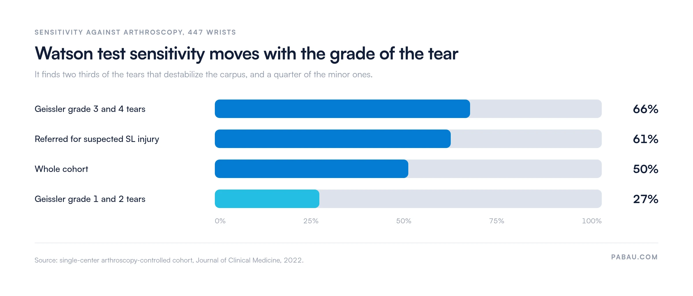
In the 211 patients scoped specifically because scapholunate injury was suspected, sensitivity rose to 61% while specificity dropped to 62%. That pattern tells you what the test is good for. It is a workable screen for the tears that let the carpus collapse, and a weak screen for early partial tears.
It also loses specificity as clinical suspicion rises, because a wrist that already looks unstable is more likely to provoke on testing. One caveat applies to all of it. Every patient in the cohort went on to arthroscopy, so the sample carries far more pathology than a routine outpatient caseload.
Treat these figures as performance in a surgical population, and expect a lower positive predictive value in a community practice.
False-positive Watson test results occur primarily in two populations: Individuals with generalized joint hypermobility, and those with physiologically lax scapholunate ligaments who are asymptomatic. In hypermobile patients, the scaphoid shifts dorsally under examiner pressure without any ligamentous pathology.
Clinicians can reduce false-positive misclassification by comparing findings bilaterally and correlating with the Beighton hypermobility score. A bilateral positive Watson test in an asymptomatic contralateral wrist strongly suggests normal variation rather than pathology. Bilateral comparison is part of the test itself.
Always compare your Watson test findings bilaterally before drawing conclusions. A unilateral positive test in the symptomatic wrist is far more diagnostically significant than bilateral findings. Document both sides in your clinical notes to support downstream clinical decision-making.
What the Watson test cannot tell you shapes how much weight its result should carry. Several structural limitations affect how findings sit in the overall clinical picture.
These limitations explain the consensus position. The American Society for Surgery of the Hand (ASSH) treats the Watson test as a screening tool, and the cohort figures above support that framing. It belongs inside a multimodal assessment battery, not as a standalone diagnostic instrument.
Pairing it with a validated decision rule tightens the picture further, much as the Ottawa ankle rules do for acute ankle injury.
A positive Watson test should be treated as a flag for further investigation, not a stand-alone diagnosis. The imaging pathway depends on the acuity of the injury and the severity of the clinical findings.
The imaging decision pathway reflects instability grade. Pain-only positive tests without a clunk can be investigated with MRI on a semi-urgent basis. A clunk with a dorsal step deformity warrants same-day plain radiographs and urgent hand surgery referral.
The Watson test sits within a broader battery of wrist special tests. Using it alongside complementary tests improves diagnostic precision by ruling in or out pathologies that can mimic scapholunate instability.
The lunotriquetral ballottement test is the most important complement to the Watson test in a standard wrist instability screen. Together they cover the two most common intrinsic carpal instability patterns. A positive Watson test with a negative lunotriquetral test narrows the differential to scapholunate pathology rather than pancarpal or ulnar-sided instability.
Record the character of the response, not only whether it was positive or negative. A note that reads “Watson positive” does not say whether the wrist hurt, clunked, or did both.
A structured wrist assessment template should capture the following every time the test is performed:
That level of specificity keeps serial assessments comparable and supports the handoff to a hand surgeon. It also protects you if the injury is disputed later, which matters most when the first presentation looked minor.
Documentation and coding move together. Once imaging confirms a frank carpal dislocation rather than a ligament sprain, the record carries a different diagnosis. A subsequent encounter on the right side codes as S63.004D. The examination note is what justifies that change to a payer.
The safer clinical notes framework offers practical guidance on structuring musculoskeletal assessment records. Retention rules sit alongside that, and record retention periods cover the timescales that apply to your notes.
Free-text notes drift. Two clinicians in the same practice will record the same Watson test in two different shapes. By the third review, nobody can tell whether the clunk changed. Practice management software like Pabau closes that off by fixing the fields before anyone starts typing.
Pabau’s digital assessment forms let hand therapy and occupational therapy teams build one wrist template with a mandatory field for each special test. An incomplete Watson entry cannot be filed, so the record stays comparable across visits and across the team.

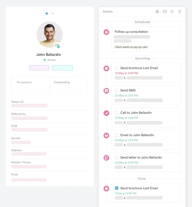
Structured client records then hold the examination findings, the imaging report, and the referral outcome on a single patient timeline. Patients moving between physical therapy, hand surgery, and occupational therapy stop losing their baseline on the way.
For larger physical therapy EMR deployments, you can also report on documentation completeness across the whole team. That turns a clinical audit into a query rather than a file review. It helps whether you are preparing for inspection or checking the physical therapy clinic requirements in your state.
Practices opening a physiotherapy practice or reviewing their physiotherapy clinic management systems get the most from setting these fields up once, before the caseload builds.

Pabau helps hand therapy and physical therapy teams build structured assessment templates and capture findings consistently. Patient timelines stay complete from first assessment through to discharge. See how it works for your practice.

The Watson test earns its place as a screen, not as a verdict. A clunk in a wrist with a trauma history is enough to escalate on the same day. A negative test in a painful wrist is not enough to stop looking.
That asymmetry is the judgment worth carrying. Half the tears in the reference cohort were missed. Treat a negative result as one input among the history, the imaging, and the other carpal tests.
Whichever way the test lands, write down what you felt, not just the label. Book a demo to see how Pabau keeps wrist assessment findings comparable across a hand therapy team.

Building out the rest of your special-test battery? Apley’s test gives you the knee equivalent, with the same procedure and interpretation structure.
Assessing an ankle that gives way rather than a wrist? Peroneal tendon tear test walks through the exam and supplies a recording template.
Need the diagnosis code once imaging confirms a carpal dislocation? S63.004D sets out the documentation a payer expects for a subsequent encounter.
Looking for a structured approach to defensible clinical notes? Safer clinical notes gives a practitioner-focused framework for recording examination findings.
Need to know how long wrist assessment records must be kept? Medical record retention sets out the timescales that apply to examination notes.
The Watson test assesses the integrity of the scapholunate ligament. It screens for scapholunate instability, which is the most common form of carpal instability. It is typically performed by physical therapists, hand therapists, and orthopedic clinicians during a wrist examination following a suspected ligament injury or unexplained wrist pain.
With the patient seated, forearm pronated, apply sustained palmar thumb pressure over the scaphoid tubercle. Passively move the wrist from full ulnar deviation to full radial deviation while maintaining that pressure. Then release the thumb and watch for a dorsal clunk or reproduction of concordant pain.
A positive Watson test indicates likely scapholunate ligament disruption. The response is concordant dorsal-radial wrist pain, a palpable dorsal clunk as the scaphoid subluxes and reduces, or apprehension during radial deviation loading. A positive result should prompt plain radiographs and consideration of MRI arthrography or wrist arthroscopy to confirm the diagnosis and grade instability severity.
Yes. False positives occur most commonly in individuals with generalized joint hypermobility, where the scaphoid shifts dorsally under examiner pressure despite intact ligaments. Always perform the Watson test bilaterally: A bilateral positive finding in an asymptomatic contralateral wrist is strong evidence of normal variation rather than pathology.
Scapholunate dissociation (SLD) is the complete disruption of the scapholunate ligament complex, producing a fixed, visible gap between the scaphoid and lunate on plain radiographs. Dynamic instability is an earlier stage where the gap only appears under stress loading, with normal static films. The Watson test can be positive in both stages, but only imaging can differentiate them reliably.
Start with PA and lateral plain radiographs, including a clenched-fist stress view to assess the scapholunate interval. If radiographs are normal but clinical suspicion remains, MRI arthrography is the preferred next step for partial SLL tears. Wrist arthroscopy is the gold standard for definitive diagnosis and surgical planning when imaging is inconclusive.
Scapholunate instability presents with radial-sided wrist pain and a positive Watson test, while lunotriquetral instability presents with ulnar-sided pain and a positive lunotriquetral ballottement test. Both are forms of carpal instability, but they involve different ligaments and require different imaging protocols and surgical approaches if operative treatment is indicated.
One arthroscopy-controlled cohort of 447 patients reported 50% sensitivity and 78% specificity for the scaphoid shift test. Sensitivity was 66% for Geissler grade 3 and 4 tears, and 27% for grade 1 and 2 tears. No systematic review of the test has been published, so pooled accuracy ranges quoted elsewhere should be treated with caution.