



GLP-1 diet plan
A GLP-1 diet plan is a structured eating framework for patients taking semagluti...

2
Last Updated: Oct 13, 2026

A headache physical exam separates primary headaches from secondary causes that need urgent treatment.
The sequence runs vital signs, general inspection, head and neck assessment, then a full neurological examination.
Red flags such as thunderclap onset, papilledema, neck stiffness, and fever always need urgent investigation.
The SNOOP mnemonic screens for systemic illness, neurologic symptoms, sudden onset, onset after 50, and pattern change.
Practice management software like Pabau lets you build headache templates that prompt every examination domain in order.
Papilledema, meningism, and a new focal deficit are all easy findings to spot. They are also easy to skip when a busy consultation runs on instinct instead of a sequence. That is the risk a structured headache physical exam removes.
This guide walks through the exam in the order you perform it. It starts with the history that shapes your priorities, then vital signs, head and neck assessment, meningismus testing, and the neurological examination. It ends with the red flags that force escalation. Solid patient care management workflows keep each step on the record.
The exam has one job. It tells you whether a headache is primary, meaning migraine, tension-type, or cluster, or secondary to an underlying condition. Primary headaches account for over 90% of presentations. The remaining 10% include conditions that turn fatal when they are missed.
A complete examination draws on history, physical assessment, and neurological evaluation. No single finding is diagnostic on its own. Together they point you toward a working diagnosis and tell you how urgently to investigate.
The American Academy of Neurology recommends pairing a structured examination with guideline-driven neuroimaging decisions. Neither one substitutes for the other.
History is the most powerful tool in headache evaluation. Gather it before you put your hands on the patient, because it decides where the examination goes. The SOCRATES framework structures this consistently. It covers site, onset, character, radiation, associations, timing, exacerbating factors, and severity.
Key history questions that shape the physical exam:
A structured physical exam checklist at the point of consultation stops any of these domains being skipped under time pressure.
The examination follows a set order: vital signs, general inspection, head and neck assessment, then neurological examination. Deviating from that order is how steps get skipped.
Blood pressure is the vital sign that matters most in a headache presentation. Hypertensive emergency is a secondary cause you can identify and act on immediately.
It means a systolic above 180 mmHg or a diastolic above 120 mmHg with end-organ damage. Measure both arms if the reading is unexpectedly high.
Routine blood pressure monitoring between visits gives you a baseline to compare against. Neurological vital signs add pupil response and consciousness level when intracranial pressure is a concern.
Systematic palpation of the head and neck takes under three minutes. It gives you information the history cannot. Pericranial muscle tenderness supports tension-type headache. Temporal artery tenderness or induration in a patient over 50 raises suspicion for giant cell arteritis, which needs urgent ESR, CRP, and clinical correlation.
A full head and neck assessment also picks up the cervical contribution that primary care easily attributes to migraine. Restricted flexion with reproducible pain is common in chiropractic practices and manual therapy caseloads.
Meningismus is neck stiffness caused by meningeal irritation. Two bedside tests give clinical evidence of it: the Kernig sign and the Brudzinski sign. Perform both in any patient where meningitis or subarachnoid hemorrhage cannot be excluded on history alone.
Kernig sign: With the patient supine, flex the hip and knee to 90 degrees. Slowly extend the knee. Resistance or pain in the hamstrings before 135 degrees of extension is a positive result, indicating meningeal irritation.
Brudzinski sign: With the patient supine, passively flex the neck toward the chest. Involuntary flexion of the hips and knees is a positive sign. Both tests have low sensitivity but high specificity in confirmed bacterial meningitis.
Neither a negative Kernig nor a negative Brudzinski sign excludes meningitis. If suspicion stays high, lumbar puncture remains the investigation of choice.
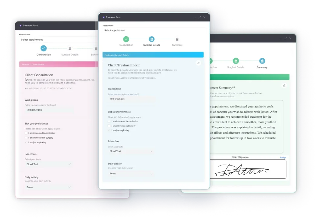
Document what you find, because those records support the escalation decision. Practice management software like Pabau can hold meningism checkboxes inside digital intake forms built for headache consultations.
A thorough neurological examination is the centerpiece of the assessment. It detects the focal deficits that point to structural or vascular pathology. Most primary headache patients examine normally, and that normal result is itself useful information.
Cranial nerve assessment in headache focuses on the oculomotor nerve, CN III. It is compressed by posterior communicating artery aneurysms, which present as a painful third nerve palsy. Assess visual acuity, visual fields, pupillary responses, and extraocular movements as a minimum.
A cranial nerve checklist keeps the order the same for every clinician, which matters when findings are compared across visits.
Fundoscopy to assess the optic disc is an essential part of the examination. Papilledema is swelling of the optic disc caused by raised intracranial pressure, or ICP. It shows as blurred disc margins, loss of the physiological cup, and engorged retinal veins. Its presence mandates urgent neuroimaging before lumbar puncture.
A normal fundoscopic examination does not exclude raised ICP in early or acute presentations. Weigh it against headache character, postural variation, and visual obscurations. The ICHD-3 diagnostic criteria from the International Headache Society then classify what you have found.
Cerebellar testing detects posterior fossa pathology, including cerebellar hematoma, tumor, and infarct. All three can present with headache. The finger-nose test looks for dysmetria and intention tremor. The heel-shin test, where the patient runs a heel from knee to ankle, assesses coordination.
Gait evaluation, including tandem walking, reveals cerebellar ataxia and subtle instability that predates more overt signs. Graded tools such as the modified Ashworth scale keep motor findings comparable between clinicians and between visits.
Red flags are findings or history features that raise the probability of a serious secondary cause. None of them confirms a diagnosis. Each one means the presentation cannot be attributed to a benign primary headache until it is investigated.
The SNOOP mnemonic gives that screening a structure. AAFP’s diagnostic approach to acute headache in adults sets it out, along with the expanded SNNOOP10 version.
Four more examination findings call for immediate action:
Document every red flag finding or its explicit absence. ‘Meningism not detected’ is a meaningful clinical record. ‘Neuro exam NAD’ is not. Specific negative findings show that the clinician actively assessed for and excluded serious pathology, which matters for patient safety and for medicolegal records.
Mapping findings onto likely diagnoses is the interpretation step that follows the examination. Nothing here is pathognomonic on its own, so use the whole clinical picture. The table below covers the most common headache types, ordered for quick bedside reasoning.
Not every headache needs neuroimaging or a specialist. The findings and the history together decide the next step. NICE guideline CG150 indicates neuroimaging for sudden onset, focal neurological signs, or headache after head trauma. It is not routinely indicated for uncomplicated migraine or tension-type headache with a normal examination.
Referral to a neurologist is appropriate when:
For urgent presentations, the pathway is emergency assessment rather than outpatient referral. That covers thunderclap headache, a new focal deficit, fever with neck stiffness, and altered consciousness.
Examination findings never override clinical urgency. Automated referral workflows can hold the escalation pathway steady, but the clinical decision comes first.
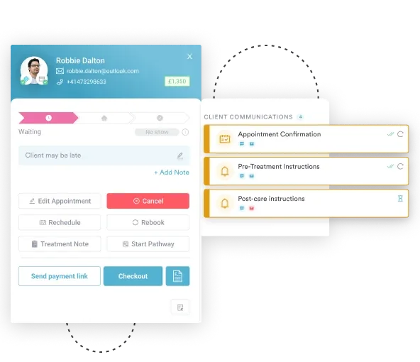
Primary care adds another layer. Clinicians running headache examinations daily need documentation that serves both the internal record and the referral letter. That is what GP practice software is built to do. A multidisciplinary review template helps when a case crosses neurology, physical therapy, and pain management.
After the examination, tell the patient what you found, what the working diagnosis is, and what should bring them back urgently. Then record that conversation.
Write the after-care instructions and the follow-up triggers into the record too. The same discipline behind safer clinical notes applies here, because what you write shapes every later decision about that patient.
A systematic examination is only as useful as the record it leaves behind. When findings are written up differently by each clinician, the pattern that should trigger escalation never surfaces. Consistent templates are what stop that happening.
Pabau lets a practice build a headache consultation template that prompts every domain in turn. Vital signs, meningism assessment, cranial nerve findings, and the red flag checklist all appear in the same order. You configure it once, and every clinician works from the same version.
The structured client record keeps a patient’s headache history, previous findings, and referral decisions in one place. A clinician seeing them for the third time does not rebuild the history from scratch. Worsening frequency and emerging neurological features become visible across visits.
Pabau Scribe, our AI scribe, captures consultation notes as you work, so your attention stays on the patient. In a headache consultation, where a subtle finding decides the outcome, that attention is the priority.
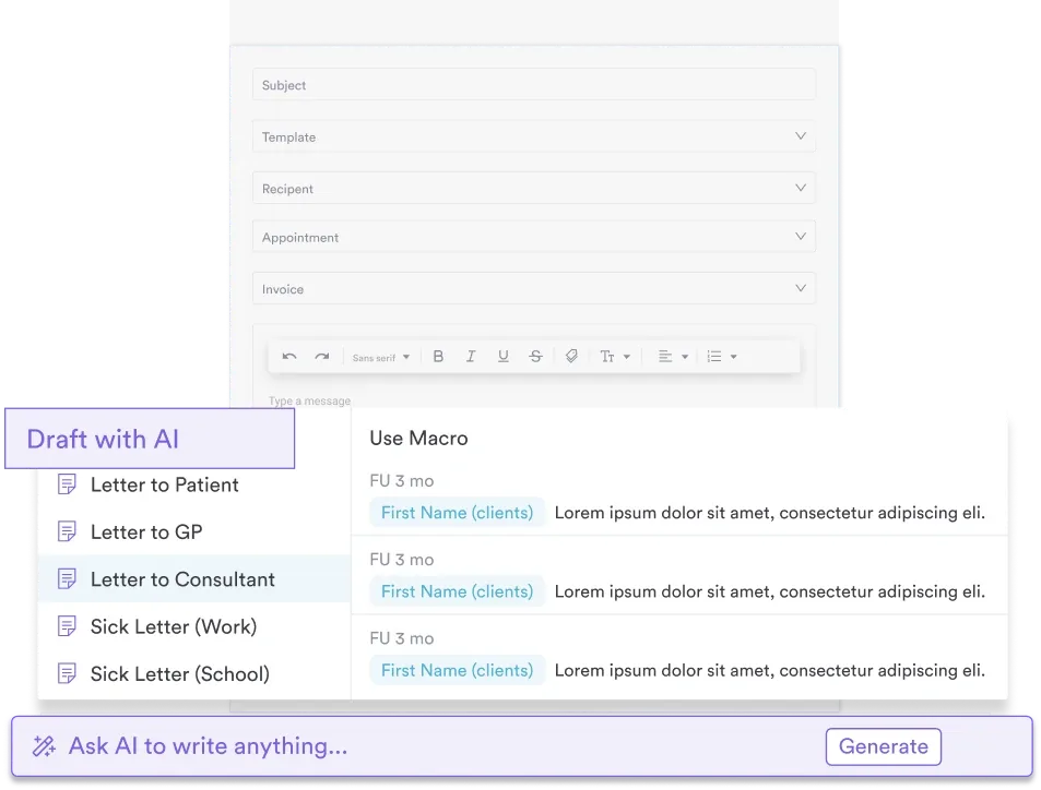

Pabau’s structured templates, digital forms, and AI documentation prompt your team through each examination domain and store the findings. See how practices use Pabau to run safer, better-documented neurological consultations.

Run the same sequence every time: vital signs, head and neck, meningism, then the full neurological examination. A clinician who never varies the order rarely has to wonder what was skipped.
The trade-off worth remembering is that a thorough examination costs minutes, and a missed subarachnoid hemorrhage costs far more. Build the sequence into the template your team already opens, and that cost disappears into the consultation.
The practical next step is to open your headache consultation template and check that every domain above appears in it. Book a demo to see how Pabau prompts every examination domain and keeps the record ready for referral.

Responding to a patient’s hypoglycemic episode? Hypoglycemia nursing diagnosis template structures rapid assessment and intervention when blood glucose drops below 70 mg/dL.
Addressing gaps in a patient’s understanding? Knowledge deficit nursing care plan organizes patient education interventions and documents learning barriers under the NANDA-I diagnosis.
Opting out of Medicare billing? Medicare private contract template lists the 15 requirements in 42 CFR 405.415 and the 10-day affidavit deadline.
Reviewing a patient’s full medication list? Medication review template checks every medicine for safety, dose, and adherence across three review levels.
Need blood pressure reference ranges by age? Normal blood pressure chart gives age-stratified thresholds using the ACC/AHA 2017 guideline classifications.
It covers vital signs, head and neck examination, and a full neurological assessment. Vital signs focus on blood pressure. The head and neck examination includes temporal artery palpation and meningism testing. The neurological assessment covers mental status, cranial nerves, motor and sensory function, cerebellar testing, and gait. The goal is to separate primary headache from secondary causes that need urgent investigation.
The main ones are papilledema on fundoscopy, positive Kernig or Brudzinski signs, and any new focal neurological deficit. Thunderclap onset within 60 seconds, fever with neck stiffness, and tender temporal arteries over age 50 also count. Any of these needs urgent escalation and neuroimaging before you attribute the headache to a primary cause.
SNOOP stands for systemic illness, neurologic symptoms, sudden onset, onset after age 50, and previous headache change. Systemic illness covers fever, weight loss, and immunosuppression. Neurologic symptoms cover focal deficit and altered consciousness. Sudden onset means thunderclap pain peaking within 60 seconds. Each letter flags a category that raises the probability of a serious secondary cause.
Fundoscopy, also called ophthalmoscopy, lets you view the optic disc directly. Look for blurred disc margins, loss of the physiological cup, elevation of the disc, and engorged retinal veins. Papilledema indicates raised intracranial pressure and mandates urgent neuroimaging before any lumbar puncture.
Look for restricted cervical range of motion and reproduction of the headache when you palpate the C2-C3 facet joints. The pattern is typically unilateral, radiating from the occipital region to the forehead. Unlike migraine, it lacks autonomic features and is consistently aggravated by sustained neck postures.
Refer when the examination shows a new focal neurological deficit, or when papilledema is present. Refer also when raised ICP cannot be excluded, when the pattern has changed without explanation, or when frequency is escalating despite primary care management. Thunderclap headache, fever with neck stiffness, and altered consciousness need emergency assessment instead.






