



GLP-1 diet plan
A GLP-1 diet plan is a structured eating framework for patients taking semagluti...

2
Last Updated: Oct 13, 2026

Head and neck assessment is a systematic physical exam covering the scalp, face, eyes, ears, nose, mouth, throat, lymph nodes, and thyroid gland using inspection and palpation.
Normal findings include a normocephalic atraumatic head, PERRLA pupils, patent nares, moist mucous membranes, midline trachea, and non-palpable lymph nodes under 1 cm.
Lymphadenopathy, tracheal deviation, or thyroid enlargement are red-flag findings that always require further clinical investigation.
Practice management software like Pabau provides digital forms and AI-assisted documentation tools that help clinicians capture structured head and neck findings accurately at the point of care.
Missed findings during a head and neck assessment don’t announce themselves. A palpable lymph node that goes undocumented, a subtle tracheal deviation noted but not charted, a thyroid nodule detected without a follow-up plan. Each one erodes the record over time.
The head and neck assessment is one of the most data-dense portions of a physical examination, covering more than a dozen anatomical structures across a small anatomical region.
This guide walks through every region systematically: anatomy, equipment, technique, normal findings, red flags, and documentation, so clinicians can perform and record this assessment with confidence.
A head and neck assessment is a structured physical examination of the anatomical structures from the scalp to the base of the neck. It uses inspection, palpation, percussion, and auscultation (commonly abbreviated as IPPA) to gather objective clinical data, combined with a subjective history of symptoms.
The exam covers the scalp, skull, face, eyes, ears, nose and sinuses, mouth and throat, lymph nodes, thyroid gland, trachea, and carotid arteries.
In nursing and clinical practice, this assessment is performed as part of a comprehensive head-to-toe evaluation, alongside body-system exams like a gastrointestinal assessment, or as a focused exam when a patient presents with head, neck, or upper respiratory complaints. Findings are documented in the patient record and inform differential diagnoses, referral decisions, and treatment planning.
Understanding what you’re examining before you examine it reduces errors and speeds documentation. These are the structures assessed in every complete head and neck examination:
Gather equipment before approaching the patient. A basic head and neck assessment relies on three core items: a penlight, a tongue blade, and gloves. Most clinicians also bring an otoscope, since ear canal and tympanic membrane inspection is part of a complete exam.
Some clinical settings also include a tuning fork (hearing screening), ophthalmoscope (detailed ocular exam), and stethoscope (carotid auscultation). The specific equipment used depends on the clinical context and the level of assessment required. When neck complaints overlap with cardiac or respiratory symptoms, pair this exam with a focused chest exam.
Understanding which tools to use with which skin assessment tools and clinical instruments helps clinicians build thorough, reproducible examinations. Practice management software like Pabau offers digital intake forms that let clinical teams pre-screen relevant patient history before the encounter begins, so the clinician enters knowing which sub-regions need closer attention.

Work systematically from the head downward. Rushing between regions is the most common source of missed findings. The sequence below follows the head and neck assessment checklist taught in nursing programs and used across clinical and medical education resources.
Inspect the scalp for lesions, redness, or trauma. Palpate the skull for tenderness, deformities, or masses, which in a trauma presentation can prompt a work-up for ICD-10 code S02.101B. Note hair distribution and texture. Assess facial symmetry: ask the patient to smile, raise eyebrows, and puff cheeks to screen cranial nerves VII. A normocephalic atraumatic head is the standard normal finding. Document any asymmetry, ptosis, or involuntary movements.
Inspect the conjunctivae for pallor or injection, the sclera for jaundice, and the eyelids for ptosis or edema. Assess pupils using a penlight: document whether they are PERRLA (Pupils Equal, Round, and Reactive to Light and Accommodation), the standard documented normal finding. Note any anisocoria, sluggish reactivity, or abnormal extraocular movements.
Inspect the external ear (pinna) for lesions, redness, or discharge. Pull the pinna gently upward and backward to straighten the canal, then insert the otoscope to visualize the tympanic membrane.
A normal tympanic membrane appears pearly grey, intact, with a visible cone of light. Note any erythema, perforation, or fluid behind the membrane. Screen hearing bilaterally using the whisper test or a tuning fork (Rinne and Weber tests). If the patient reports ear pain, discharge, or hearing loss, a full nursing ear assessment adds a targeted history and clear referral red flags.
Inspect the nares for patency, discharge, and mucosal color. Use the penlight or otoscope to assess the nasal septum for deviation. Normal findings include patent nares bilaterally and a midline septum.
Palpate the maxillary sinuses (below the cheekbones) and frontal sinuses (above the eyebrows) by applying firm upward pressure. Tenderness on palpation is an abnormal finding consistent with sinusitis. A sinuses assessment should note whether tenderness is unilateral or bilateral, as laterality guides diagnosis.
Inspect the lips for color, moisture, and lesions. Examine the gums for bleeding or recession, the teeth for condition and alignment, and the tongue for mobility, color, and coating. Ask the patient to say “ahh” to depress the tongue and inspect the palate, uvula, and posterior pharynx.
Throat assessment normal findings include a midline uvula, moist mucous membranes, and tonsils that are absent or present but not erythematous or exudative.
Abnormal findings to note: white patches (possible candidiasis or leukoplakia), tonsillar enlargement graded I-IV, posterior pharyngeal erythema, or a deviated uvula suggesting unilateral mass effect.
Assess the neck in a logical anterior-to-posterior sequence. Inspect for symmetry, visible masses, and jugular venous distension. Then palpate, auscultate, and document each structure in turn.
Lymph node assessment deserves its own focused treatment because findings here carry significant diagnostic weight. Lymphadenopathy may indicate infection, inflammation, or malignancy. The clinical significance of an enlarged node depends on its characteristics, not just its size.
Palpate each chain bilaterally, noting these four dimensions for every abnormal node found:
The supraclavicular region is particularly significant: supraclavicular lymphadenopathy is almost always pathological and should prompt urgent investigation for intrathoracic or intra-abdominal malignancy.
The table below maps each anatomical region to its expected normal findings and clinically significant abnormal findings. This is the most referenced section of any head and neck assessment nursing guide, and for good reason: findings documentation starts here.
Poor documentation is a clinical and legal liability. A finding noted on exam but absent from the record might as well not have happened. Good clinical note-taking practice means documenting each region systematically, using standardized terminology, distinguishing clearly between normal and abnormal findings, and pairing findings with a signed consent for treatment when the exam supports a procedure or referral.
The American Nurses Association (ANA) and nursing governing bodies including the NMC in the UK emphasize that clinical documentation must be accurate, contemporaneous, and complete. Keeping records up to date is both a professional obligation and a patient safety requirement. For patient care management, assessment findings form the baseline against which all future changes are measured.
A complete head and neck assessment note typically reads as follows:
Head normocephalic, atraumatic. Face symmetric without ptosis or facial droop. PERRLA bilaterally, conjunctivae clear, sclera white.
Tympanic membranes pearly grey, intact. Nares patent, septum midline, no sinus tenderness. Oral mucosa moist, uvula midline, oropharynx without erythema or exudate. No palpable cervical lymphadenopathy. Thyroid non-palpable, non-tender. Trachea midline. No carotid bruits.
This format uses region-by-region structure, specific clinical terms, and no vague language. When an abnormality is present, document its characteristics fully (location, size, tenderness, mobility) rather than just noting “lymph node palpated.” Use structured assessment templates, like this emergency nursing assessment template, to reduce omission errors across complex examinations.
Document findings immediately after the exam, not at the end of a shift. Recall accuracy drops quickly, and delayed documentation increases the risk of missing or misrecording a finding that may be clinically significant.
Most competing resources on head and neck assessment stop at the clinical content. But for practice teams seeing multiple patients daily, capturing a thorough assessment accurately is as much a workflow challenge as a knowledge one.
Pabau’s platform supports clinical documentation workflows across physical therapy practices, GP practices, dermatology settings, and multi-specialty private practices.
The AI-assisted clinical documentation tool (Pabau Scribe) captures spoken notes during or after a consultation and structures them into the patient record, reducing the time spent typing templated findings.
For head and neck assessments specifically, this means a clinician can verbalize findings region-by-region during the exam and have them formatted correctly in the EHR without post-consultation transcription burden. Teams weighing documentation platforms can review the DrChrono pros and cons to see how EHR-first tools compare. This also applies to virtual visits: the telemedicine physical exam covers how to adapt these techniques for video consultations.
Beyond AI scribing, digital medical forms in Pabau allow practices to deploy pre-consultation intake forms that capture relevant symptoms, patient history, and any previous head and neck findings before the patient enters the consultation room.
This means the clinician focuses examination time on active assessment rather than history-taking. The paperless practice workflow also reduces the administrative burden on reception teams while keeping clinical records complete and retrievable.
For practices managing ongoing patient relationships, structured client records within Pabau allow clinicians to compare current findings against previous assessments, making it easier to detect new lymphadenopathy, thyroid changes, or progressive abnormalities over time. Compliance management tools within the platform also help practices meet documentation standards required by governing bodies including the NMC and HIPAA, depending on jurisdiction.
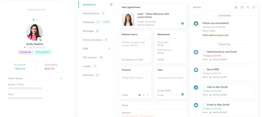
Keeping every assessment in one client record means the next clinician opens the full head and neck history at a glance, so a new lymph node or thyroid change stands out against the previous baseline instead of being reconstructed from scattered notes.

From AI-assisted note capture to structured digital forms, Pabau helps practice teams record head and neck assessment findings accurately, every time. Book a demo to see it in action.

Use this checklist as a quick-reference guide before, during, or after the exam. It covers every region in assessment order and flags the key normal findings to verify at each step.
A thorough head and neck assessment is only as useful as its documentation. Missed findings and vague charting undermine continuity of care. For clinical teams managing high patient volumes, the real challenge is capturing every finding accurately before the next patient arrives.
Pabau’s structured client records and AI-assisted documentation tools help practice teams move from exam to complete, retrievable notes without added administrative burden. Structured templates, like our nursing shift report template, show how Pabau replaces paper-based records across a practice. To explore the full platform, book a demo with the Pabau team.

Documenting a patient’s food allergies? Food allergy form template documents allergies clearly so every team member can respond appropriately in an emergency.
Screening a senior for memory decline? Memory test for seniors compares the SAGE, MoCA, and SLUMS screens along with their score cutoffs.
Admitting a patient into hospice care? Hospice admission note template structures a CMS-aligned admission note that covers advance directives and care planning.
A head and neck assessment is a systematic physical examination of the structures from the scalp to the base of the neck, including the eyes, ears, nose, sinuses, mouth, throat, lymph nodes, thyroid, trachea, and carotid arteries, using inspection, palpation, percussion, and auscultation to collect objective clinical data.
Normal findings include a normocephalic atraumatic head, symmetric facial features, PERRLA pupils, clear conjunctivae, intact tympanic membranes, patent nares with a midline septum, moist mucous membranes, a midline uvula, non-palpable lymph nodes under 1 cm, a non-palpable thyroid, midline trachea, and no carotid bruits.
Abnormal neck assessment findings include lymph nodes greater than 1 cm that are tender, fixed, or matted; thyroid enlargement, nodularity, or tenderness; tracheal deviation from midline; and carotid bruits on auscultation. Supraclavicular lymphadenopathy is particularly concerning and warrants urgent investigation.
Palpate bilaterally using the fingerpads in a systematic chain sequence: preauricular, postauricular (mastoid), occipital, submental, submandibular, tonsillar, anterior cervical, posterior cervical, and supraclavicular. For each node found, document size, tenderness, mobility, and consistency. Non-palpable or small, soft, mobile, non-tender nodes are normal findings.
The standard equipment includes a penlight, otoscope, tongue blade, and gloves. Some assessments also require a stethoscope for carotid auscultation, a tuning fork for hearing screening, and an ophthalmoscope for a detailed ocular examination.
Document each region in sequence using standardized clinical terminology. Record normal findings explicitly (e.g., “PERRLA, conjunctivae clear”) rather than simply noting “normal.” For abnormal findings, describe location, size, tenderness, and mobility. Document immediately after the exam to preserve accuracy.






