Nitrates medication list
A nitrates medication list is a point-of-care reference that sets out the main c...

1
Last Updated: Oct 6, 2026
The randot test is a clinical stereoacuity assessment that measures depth perception using polarized random-dot targets. The patient wears polarized glasses, so each eye receives a slightly different image.
Results are recorded in seconds of arc, and a lower number means sharper stereoacuity. Adults with intact binocular vision typically reach 20 to 40 seconds of arc.
Its random-dot design is what separates it from contour-based tests such as the Titmus fly, which a patient can sometimes pass using monocular shape cues. According to the National Institutes of Health (PMC), the standard near test measures stereo acuity from 20 to 400 seconds of arc. Three variants cover different ages and fixation distances, and picking the wrong one skews the result.

The randot test measures stereoacuity in seconds of arc using polarized random-dot vectographs, which removes the monocular cues that contour-based tests can leave open.
Three variants are in clinical use: standard near Randot, Randot Preschool, and Distance Randot. Randot Preschool uses its own random-dot picture shapes, not LEA Symbols.
A result worse than 40 seconds of arc in adults, or outside age-normed thresholds in children, warrants clinical attention and possible referral.
Practice management software like Pabau stores serial stereoacuity scores against a baseline, so a change between visits shows up at the next appointment.
The randot test is a binocular depth perception assessment. It uses polarized vectographic plates printed with random-dot patterns to measure stereoacuity in seconds of arc. Contour-based tests leave visible shapes that a patient can sometimes recognize with one eye. The randot plates leave no such cue, so a correct answer requires binocular fusion.
Developed by the Stereo Optical Company, the test presents a booklet of polarized plates. The patient wears polarized glasses so that each eye receives a slightly different image. The visual cortex fuses these disparate inputs into perceived depth.
The test records the finest disparity at which the patient can reliably identify the target, expressed in seconds of arc. A lower number indicates sharper stereoacuity. That single figure is what clinicians compare between visits and against age-normed data.
Clinicians who see only near-vision patients sometimes overlook the distance variant, but near testing does not always predict distance stereoacuity deficits. A patient can pass a near stereopsis test and still fail at distance fixation, particularly after strabismus surgery or in early amblyopia. Keeping both scenarios in view prevents missed diagnoses.
Random-dot stereograms work by embedding a hidden shape within a field of dots that looks identical to both eyes when viewed separately. The shape becomes visible only when the brain combines the two slightly offset images through binocular disparity processing. Because the pattern contains no monocular depth cues, a patient cannot identify the target by looking with one eye alone.
Contour-based tests such as the Titmus fly present recognizable shapes. A patient with poor stereopsis but intact monocular form recognition can sometimes identify the correct answer from the contour alone, producing a false-normal result. The randot test’s random-dot approach closes that loophole, which makes it more sensitive where subtle suppression or reduced binocularity is suspected.
Two types of stereopsis are relevant here. Global stereopsis relies entirely on binocular disparity, with no monocular shape information, and is what random-dot tests like the Randot measure. Local (contour) stereopsis involves targets with visible edges that provide some monocular cues. Assessing both types gives a more complete picture of binocular function.
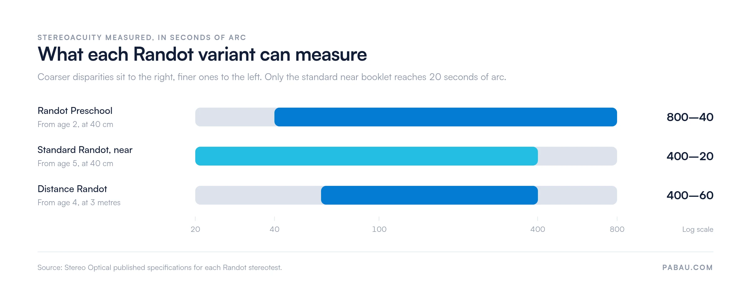
Three variants of the test are used in clinical practice, all of them made by Stereo Optical. Which one you reach for depends on the patient’s age and on the fixation distance you need to assess. Choosing on age alone is where protocol errors usually start.
Randot Preschool presents its own random-dot picture shapes, so a pre-verbal child can point to a matching card instead of describing what they see. Stereo Optical designs it for patients as young as 2 years, though reliable results come more consistently from around age 3.
A separate product, the Randot Stereotest Including LEA Symbols, uses LEA Symbols for gross screening at 500 and 250 seconds of arc. The two are routinely confused in clinical write-ups, and they do different jobs. Confirm which booklet is on the shelf before you record a Preschool result.
Plotted on one scale, the three ranges overlap less than the table suggests. The variant you pick for the patient’s age also caps how fine a deficit you can detect.
Consistent administration is where practices introduce the variability that undermines the test’s diagnostic value. The protocol below follows standard clinical practice as described in peer-reviewed optometry literature and the manufacturer’s instructions.
Flag any result where the patient consistently responds faster on contour targets than random-dot targets at the same disparity level. This asymmetry sometimes indicates the patient is using monocular form cues on the contour plates rather than stereopsis. Re-administer with one eye occluded to check whether the contour response degrades appropriately.
Stereoacuity thresholds improve rapidly during childhood and stabilize in early adulthood. A result that would be clinically normal for a 3-year-old represents a significant deficit in a 10-year-old. Using age-appropriate normative data is therefore non-negotiable when interpreting any stereopsis test result.
These ranges come from published normative data, including the Distance Randot Stereotest normative study (PMC) and developmental optometry literature. They are reference ranges, not absolute diagnostic thresholds. Referral decisions belong in the context of the full eye examination and the clinical history.
The American Academy of Ophthalmology (AAO) amblyopia guidance carries the current evidence-based thresholds for clinical action. Check it before you set a referral threshold for your own protocol.
The table describes single visits. A patient who passes at 40 arcsec today may retest at 60 arcsec six months later, and that change is clinically meaningful. Serial records scored against the same normative baseline make it visible. Visits interpreted in isolation do not.
The randot test earns its place as a screening and monitoring tool inside a broader binocular vision workup. On its own it diagnoses nothing. Stereopsis testing sits alongside the rest of the eye physical examination, and the result is read against the other findings.
The five most common indications are outlined below, drawing on American Optometric Association (AOA) guidance on binocular vision assessment.
A practice manager running a busy optometry or ophthalmology service faces a retrieval problem here. Stereoacuity results from six visits ago need to be on screen in seconds at the next consultation. That is the job patient record management is there to do.

Choosing between the available stereotests is a practical clinical decision. Each test has a different sensitivity profile, monocular cue risk, and age floor. The comparison below covers the tests most commonly used in optometry and ophthalmology settings.
The Titmus fly’s higher monocular cue risk is its main clinical limitation. A child who sees the fly’s wings rise up may be responding to the contour of the image rather than to depth.
That matters most when you need a confident negative result, such as clearing a child from amblyopia suspicion. In those cases the randot random-dot subtest gives the more defensible finding.
The Lang stereotest needs no polarized glasses, which makes it practical for infants. Its coarser disparity levels mean it cannot detect the subtle adult-level deficits the Randot picks up.
The near and distance variants are not interchangeable. Near and distance stereoacuity can dissociate, especially in patients with convergence excess esotropia, post-surgical strabismus, or early binocular dysfunction. A patient who achieves 40 arcsec at 40 cm may perform far worse at 3 metres, and a difference that size is clinically actionable.
The Distance Randot Stereotest was developed specifically to capture that dissociation. Administered at 3 metres using polarized vectographic targets, it has shown good test-retest reliability in peer-reviewed validation studies.
Some patients drive, play sports, or work in roles that demand distance depth perception. For them the distance variant is the right choice, even when the near result looks normal.
A practical guide for choosing between them:
Stereoacuity data loses most of its clinical value if it sits in a paper record that nobody reviews between visits. The test’s strength is longitudinal comparison. A single result is a data point, while six results plotted against age-normed thresholds guide treatment decisions and referral timing.
Effective documentation captures the variant used, the testing distance, and the date. It also records the patient’s habitual correction, each subtest score in seconds of arc, and any compliance issues. A structured stereopsis test template pre-populates those fields, which cuts transcription errors and makes serial comparison straightforward. Nobody has to reconstruct the context from free-text notes.
Practice managers overseeing multi-practitioner optometry or ophthalmology teams face one more layer. A result entered by one clinician has to read the same way to a colleague at the next visit.
A standard form is what makes that happen. Paired with automated clinical workflows, practices can trigger recall appointments from a threshold crossing instead of relying on someone to remember.

Build a stereoacuity check into your amblyopia recall protocol. Set a review at 6 weeks after treatment starts, then at 3 months and 6 months. At each visit, record the randot test result against the patient’s age-normed baseline. Flag any increase of more than one disparity level as a discussion item, so slow regression does not go unnoticed between annual exams.
Most practices type a stereoacuity result into a free-text consultation note. Finding it again means opening the last three visits and reading through them. In a full clinic day, serial comparison quietly gets skipped.
Practice management software like Pabau stores the result as a tracked measurement instead. Pabau’s measurement tracking for clinicians holds each stereoacuity value against the patient’s own baseline, so the trend is on screen at the next appointment. Structured records keep the variant, the distance, and the correction worn alongside the score.
That pays off in amblyopia monitoring and post-surgical follow-up, where one disparity level decides whether treatment continues. The reviewing clinician reads the history instead of rebuilding it, and the recall for the next check can be scheduled from the same record.

Pabau helps eye care practices track serial clinical measurements, automate follow-up scheduling, and keep structured patient records. Every stereoacuity result is where you need it, when you need it.

A stereoacuity score is only as useful as the consistency behind it. Match the variant to the patient’s age and to the distance you need to assess. Run the same protocol every time, and score against age-normed thresholds. Those three habits decide whether the number means anything six months later.
The trade-off worth remembering is sensitivity against cooperation. Contour tests are quicker and reach younger children, but they let a patient answer from shape alone. When you need a confident negative, the random-dot subtest is the finding that will hold up.
If serial results are the point, the record has to make them easy to compare. Book a demo to see how Pabau keeps stereoacuity data, recalls, and patient records together for eye care teams.

Separating refractive from pathological acuity loss? Pinhole test explains a quick chairside check that narrows the cause of reduced acuity.
Assessing ocular motility in children? Eye movement test covers the pursuit and saccade checks that pair with stereopsis testing.
Looking for the neurological side of the exam? Neurological eye exam walks through cranial nerve and pupil testing in a clinical sequence.
The randot test measures stereoacuity, the finest binocular depth disparity a patient can reliably detect, expressed in seconds of arc. It uses polarized random-dot vectographic plates that remove monocular form cues. Lower scores, meaning fewer seconds of arc, indicate sharper stereoscopic vision.
Normal thresholds vary by age. Children aged 5 to 7 years typically achieve 40 to 100 seconds of arc. From age 8, results should be at or near the adult norm of 20 to 40 seconds of arc. Adults with intact binocular vision typically score 20 to 40 arcsec, and 20 is optimal. Always interpret a result against a published normative reference for the variant used.
Stereo Optical designs the Randot Preschool Stereoacuity Test for patients as young as 2 years. It measures from 800 to 40 seconds of arc using random-dot picture shapes that the child matches to a card. Reliable results come more consistently from around age 3 in clinical practice.
The randot test is a sensitive screening tool for amblyopia rather than a standalone diagnostic instrument. Reduced stereoacuity is a reliable marker, particularly where monocular visual acuity alone looks near-normal. A result outside age-normed thresholds should prompt a full binocular vision workup.
The randot test uses random-dot patterns with no monocular shape cues, which makes it more sensitive to stereopsis deficits. The Titmus fly is contour-based, so a patient may identify the correct answer from shape recognition alone. The Titmus is faster and suits younger children, but it carries a higher risk of false-normal results.
Repeat testing is indicated at key clinical milestones. Those are before and after strabismus surgery, and at intervals during amblyopia treatment (typically 6 weeks, 3 months, and 6 months). Annual testing suits any patient with known binocular dysfunction. A change of more than one disparity level between visits warrants clinical review, as does a result that crosses a normative threshold.