



GLP-1 diet plan
A GLP-1 diet plan is a structured eating framework for patients taking semagluti...

2
Last Updated: Oct 13, 2026
Pleural fluid analysis interpretation is the systematic reading of a thoracentesis sample to work out why fluid has collected in the pleural space. You classify the fluid as a transudate or an exudate using Light’s criteria. Then you read glucose, pH, the cell differential, cytology, and microbiology as a pattern. That pattern points to a cause: heart failure, infection, malignancy, tuberculosis, or one of roughly 60 other conditions.
Pleural effusion reaches an estimated 1.5 million patients annually in the United States, according to StatPearls. Reading the fluid correctly decides whether a patient needs a chest drain today or treatment for the underlying condition. This guide walks the workup in order, from what the fluid looks like through to the special tests.

Pleural fluid analysis interpretation classifies fluid as a transudate or an exudate using Light’s criteria, which narrows a differential of roughly 60 conditions.
Light’s criteria misclassify up to 25% of transudates in patients on diuretics, so read the numbers alongside the clinical picture.
pH below 7.2, glucose below 60 mg/dL, and neutrophil predominance together point to a complicated parapneumonic effusion that needs drainage.
Cytology detects malignancy in roughly 60% of first samples, so a negative result never rules out malignant pleural disease.
Pleural fluid analysis interpretation begins the moment fluid is aspirated at thoracentesis. The workup runs in a fixed order. Macroscopic appearance comes first, then biochemical classification, then cells and microbiology. Specialist tests follow where the clinical picture calls for them.
Skipping a stage, or acting on one result alone, causes most of the diagnostic error in this workup. Each analyte answers a different question. The combined pattern narrows the differential far better than any single test, and the five stages below show what each one decides.
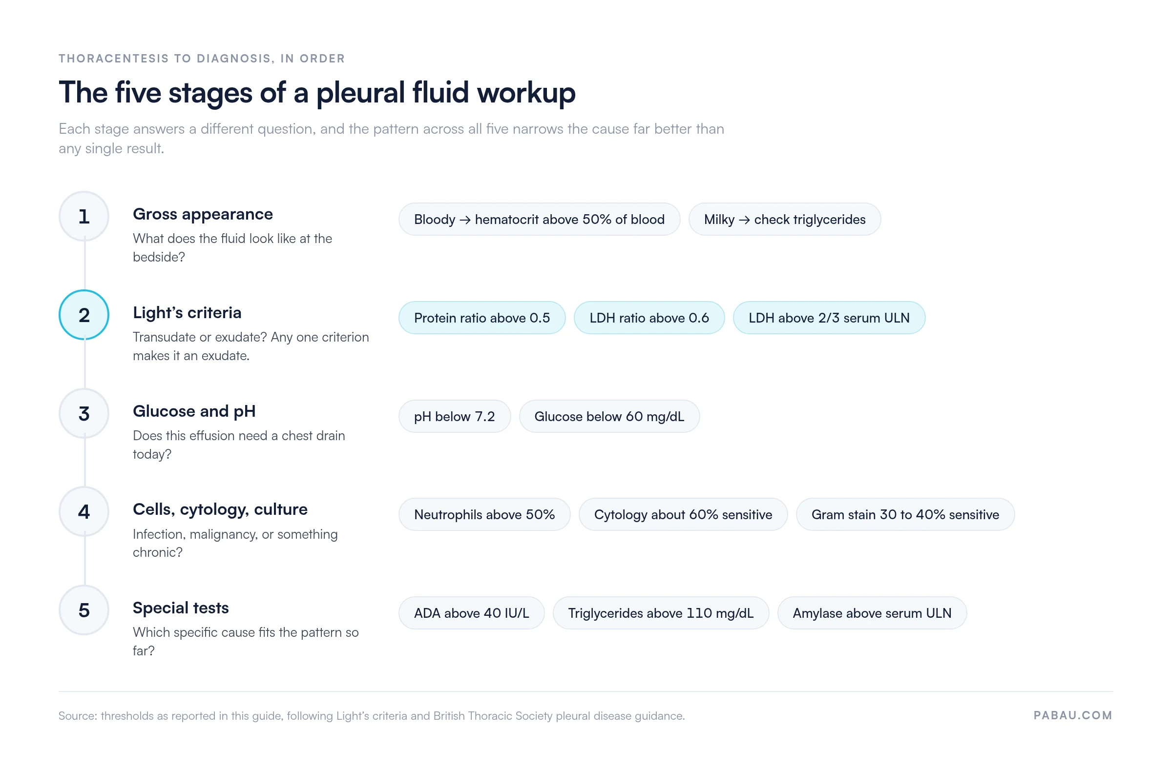
Recording those five stages consistently matters as much as running them. Respiratory assessment forms built in patient intake software capture every analyte in one place, so no result is left off the chart. Practices running high diagnostic volumes attach results to the record through medical lab software, which removes the manual transcription step where errors creep in.

Macroscopic appearance gives you diagnostic clues before any laboratory result returns. Straw-colored, clear fluid is the most common finding and is non-specific. Turbid or cloudy fluid raises suspicion for infection or a high cellular count. Bloody fluid calls for a pleural fluid hematocrit, and a ratio above 50% of the peripheral blood hematocrit confirms hemothorax. Milky or opalescent fluid points toward chylothorax, though a pleural lipoma or empyema can look similar and only triglycerides separate them.
The transudate-exudate distinction is the first binary decision in any pleural fluid workup. Transudates form when hydrostatic or oncotic pressure changes push fluid across an intact pleural membrane. Heart failure, hepatic hydrothorax, and nephrotic syndrome are the usual culprits. Exudates come from local pleural inflammation or disruption that raises capillary permeability, which produces protein-rich fluid.
The distinction decides how far the workup goes. A transudate rarely needs pleural investigation beyond treating the underlying systemic condition. An exudate needs the full analytical panel.
Light’s criteria classify pleural fluid as an exudate when any one of three conditions is met. That makes them highly sensitive for exudates, at roughly 98%, with specificity around 72% to 83%. Fluid meeting none of the three is a transudate. Richard Light and colleagues set the thresholds, and decades of clinical practice have validated them.
The two-test rule is a validated alternative described by Life in the Fast Lane. It uses only the protein and LDH ratios and drops the absolute LDH criterion. Sensitivity falls slightly and specificity improves. Some guidelines prefer it for exactly that reason, since it misclassifies fewer patients on diuretics.
Protein and pleural fluid LDH carry diagnostic weight beyond their roles in Light’s criteria. Absolute pleural fluid protein above 3 g/dL (30 g/L) fits an exudate, while values below 2.5 g/dL (25 g/L) suggest a transudate. A pleural LDH more than three times the upper limit of normal suggests empyema, malignancy, or rheumatoid pleurisy. A rising LDH on serial taps can flag a developing exudative process, even within the transudate range.
Reference ranges vary between laboratories. A value sitting near a Light’s criteria threshold is read against the clinical picture, not treated as a hard boundary. Flagging borderline analytes for review inside medical records management stops a threshold result being acted on without corroborating evidence. Sequential documentation also surfaces the trends a single sample hides.
Glucose and pH narrow the differential inside the exudate category and drive time-sensitive management. Normal pleural fluid glucose mirrors serum glucose. A pleural fluid glucose below 60 mg/dL (3.3 mmol/L) is shared by a short list of conditions. Those are complicated parapneumonic effusion or empyema, rheumatoid pleurisy, malignant effusion, tuberculous pleurisy, and esophageal rupture. pH is a parallel marker read against the same differential.
The British Thoracic Society pleural disease guidelines set pH below 7.2 as the threshold supporting chest tube drainage in parapneumonic effusion. No single value overrides clinical assessment. pH also has to be measured on a blood-gas analyzer. Sending fluid in a standard specimen tube lets CO2 escape and falsely raises the reading.
The white cell count and differential push the interpretation toward an acute or a chronic process, and toward infection, malignancy, or inflammation. A total white cell count above 10,000 cells per microliter in an exudate points strongly to a parapneumonic effusion or empyema. The differential pattern is usually more informative than the total count alone.
Send cytology whenever malignancy is in the differential for an exudative effusion. According to data published in NIH PubMed Central, cytological examination detects malignant cells in roughly 60% of first samples. Four in ten malignant effusions therefore return a false negative on the first look. A second sample improves the yield modestly, then the curve flattens.
A negative cytology result does not exclude malignant pleural disease. When suspicion stays high after two negative samples, image-guided or thoracoscopic pleural biopsy is the next step. Adenocarcinoma gives the highest cytology yield. Mesothelioma has low cytological sensitivity and usually needs biopsy for histological subtyping.
Microbiological testing is warranted when infection is suspected clinically, or when the fluid is an exudate with neutrophil predominance. Gram stain has low sensitivity in parapneumonic effusion, around 30% to 40%, but it is fast and a positive result guides antimicrobial choice immediately. Send bacterial culture in blood culture bottles, inoculated at the bedside, which raises yield compared with laboratory inoculation from a specimen pot.
Prior antibiotic therapy cuts culture yield substantially. In a treated patient with suspected empyema, a negative culture does not change the management decision, which rests on pH, glucose, and macroscopic appearance. Standardized medical forms that prompt for microbiological test selection reduce the risk of omitting a culture that would have confirmed the diagnosis.
Special tests are ordered on specific clinical suspicion, never as a routine panel. Three are worth knowing by threshold and indication.
ADA testing earns its place where tuberculosis remains prevalent. In low-prevalence settings, a positive ADA needs corroboration from cytology, culture, or biopsy before anti-TB therapy starts. Pre-test probability always modifies how a threshold result should be acted on, and the ADA cutoff is no exception.
Five errors cause most of the misclassification and delayed diagnosis in pleural workups. Each one is avoidable, and each one turns on reading the pattern rather than a single number.
Several of these are documentation failures as much as clinical ones. A result flagged for follow-up and then lost between encounters ends the same way as a result nobody ordered. Clinical notes software that prompts for the next action at the point of entry stops the second cytology.
Each effusion type produces a recognizable pattern across the panel. The table below consolidates the characteristic findings for the five most clinically important types.
The table below puts the whole panel on one page: every analyte, its threshold, and what crossing it means. Check your local laboratory reference ranges too, since thresholds shift slightly between institutions.
A table like this one only helps if the reading reaches the record intact. Practice management software like Pabau writes the interpretation into structured fields instead of free text, so the next clinician can retrieve it. That matters most on sequential taps, where the trend carries as much weight as any single value.
Always draw serum protein and LDH at the same time as the thoracentesis. Light’s criteria ratios lose accuracy when the two samples are hours apart. Biological variation can shift a borderline result across the exudate threshold and misdirect the whole workup.
In most practices a pleural workup is spread across three places. The thoracentesis note sits in the chart, the laboratory results arrive by portal or fax, and the follow-up plan lives in somebody’s head. Reconciling the three usually takes a phone call.
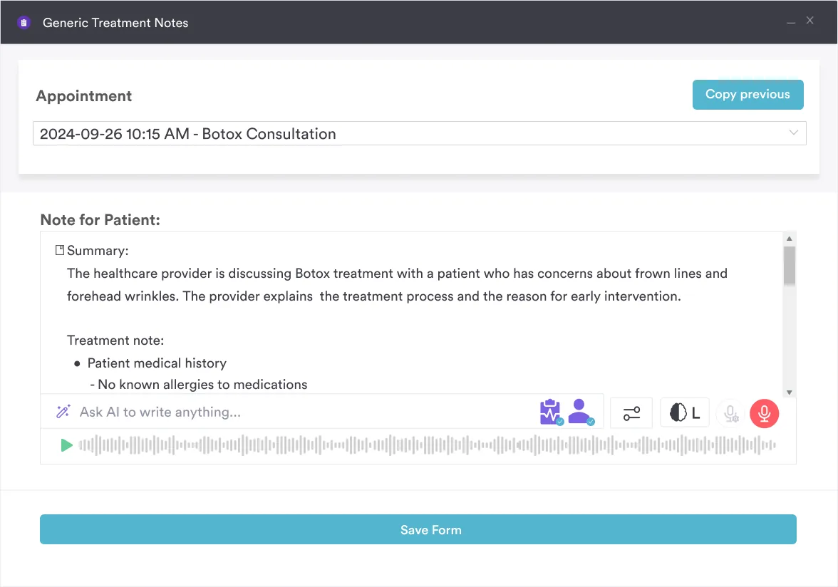
Pabau holds the whole sequence on the patient record instead. Custom assessment forms capture appearance, Light’s criteria, glucose, pH, and the differential as discrete fields rather than prose. Laboratory results attach to the same record, and Pabau Scribe drafts your interpretation straight into the treatment note.
The outcome is a record that answers the next clinician’s question without a search. A borderline protein ratio from the first tap sits beside the second, and the drainage decision keeps its evidence attached. None of that sits behind a higher tier, because every Pabau subscription includes every feature.

Pabau gives respiratory and acute teams structured assessment forms, attached lab results, and clinical notes that hold a workup together from thoracentesis to diagnosis. See how it fits your practice.

Read the panel, not the number. Light’s criteria classify the effusion, glucose and pH decide whether it drains today, and cytology, microbiology, and special tests name the cause. Where one result disagrees with the rest, the rest usually wins.
Three habits prevent most of the errors above, and none of them costs anything. Draw serum at the same time as the tap. Treat a diuretic-treated transudate as a transudate until the protein gradient says otherwise. Send a second cytology whenever suspicion stays high after a negative first sample.
Book a demo to see how Pabau keeps a pleural workup, its results, and its follow-up actions on one patient record.

Comparing systems for clinical documentation? Clinical documentation software walks through what to look for when notes, results and forms have to live in one place.
Want a ready-made progress note structure? Clinical progress notes template gives you a format for recording sequential findings across repeat encounters.
Documenting a patient with a large effusion? Respiratory failure nursing care plan template covers assessment, goals and interventions for deteriorating respiratory function.
Need a care plan for the oxygenation problem underneath? Impaired gas exchange nursing care plan sets out the monitoring and interventions that follow a compromised pleural space.
Pleural fluid analysis interpretation is the process of reading biochemical, cellular, and microbiological results from a thoracentesis sample to identify the cause of an effusion. Clinicians read the analyte pattern as a whole, including Light’s criteria, glucose, pH, the cell differential, cytology, and special tests. That pattern narrows the differential toward infection, malignancy, heart failure, tuberculosis, or another cause.
Light’s criteria are the gold-standard method for classifying pleural fluid as a transudate or an exudate. A fluid is an exudate if it meets any one of three thresholds. Those are a protein ratio above 0.5, an LDH ratio above 0.6, or an absolute LDH above two thirds of the serum upper limit. Their main limitation is misclassifying up to 25% of transudates in patients on diuretics, where protein and LDH concentrate. The serum-to-pleural protein gradient helps resolve those borderline cases.
Tuberculous pleurisy typically produces a lymphocyte-predominant exudate with adenosine deaminase (ADA) above 40 IU/L, few mesothelial cells, and low or normal glucose. Mycobacterial culture is the gold standard, but it takes four to eight weeks. An ADA above the threshold in a lymphocyte-predominant exudate has high sensitivity where TB is prevalent.
Pleural fluid pH below 7.2 is the decision threshold for chest tube drainage in parapneumonic effusion, per British Thoracic Society guidelines. Alongside low glucose and neutrophil predominance, it indicates a complicated effusion or early empyema. pH must be measured on a blood-gas analyzer with the sample kept on ice. Standard specimen-pot collection lets CO2 escape and produces falsely elevated readings.






