Nitrates medication list
A nitrates medication list is a point-of-care reference that sets out the main c...

Last Updated: Oct 6, 2026

Impaired gas exchange is the NANDA-I diagnosis, code 00030, for an excess or deficit in oxygenation or carbon dioxide elimination at the alveolar-capillary membrane.
A healthy adult breathes 12 to 20 times a minute, so any rate above 20 counts as tachypnea.
Normal SpO2 for a healthy adult is 95 to 100%, while 94 to 98% is an oxygen therapy target range.
Write the related factor as the mechanism, then support it with data you have already charted.
Core interventions are supplemental oxygen, upright positioning, and pursed-lip breathing, each recorded with its rationale.
A fillable plan with space for your assessment findings, the three-part NANDA-I diagnosis statement, short and long-term goals, interventions with rationale, and evaluation criteria. Worked examples for COPD, pneumonia, and pulmonary edema are included.
Download templateA patient’s SpO2 reads 91%, their respiratory rate is 26, and they are pulling on their neck muscles to breathe. You know what you are looking at. Writing it into a plan that a clinical instructor or a chart auditor will accept is the harder part.
Impaired gas exchange is the NANDA-I diagnosis for a patient whose alveoli cannot move oxygen and carbon dioxide properly. It is one of the most common diagnoses in acute care, and one of the easiest to document badly.
Three problems come up again and again. The related factor is really a medical diagnosis. The goal carries no measurable target. And the supporting data never made it into the chart.
A good impaired gas exchange nursing care plan follows the five-step nursing process, and the template below is built around it. You fill it in as you assess, rather than reconstructing the shift from memory afterward.
NANDA-I defines impaired gas exchange as an excess or deficit in oxygenation, carbon dioxide elimination, or both, at the alveolar-capillary membrane. It carries the code 00030 and sits in Domain 3, elimination and exchange, under respiratory function.
The distinction that matters at the bedside is where the problem sits. The airway can be wide open and the patient can still be hypoxemic, because the lung tissue itself cannot move gas across the membrane.
Why this matters clinically: the diagnosis flags respiratory failure that is already underway. Chronic obstructive pulmonary disease (COPD), pneumonia, pulmonary edema, pulmonary fibrosis, and acute respiratory distress syndrome all produce it.
Hypoxemia. Catching the diagnosis early lets you prioritize oxygen, positioning, and monitoring before the patient decompensates.
Start with a full minute of counting, not a 15-second estimate. A healthy adult breathes 12 to 20 times a minute. Anything above 20 is tachypnea, and in a hypoxemic patient it is usually the first sign to appear.
Then look at the work of breathing. Retractions, accessory muscle use, and nasal flaring all point the same way. The patient is spending energy to breathe. Note skin color, cyanosis, chest pain, and any complaint of shortness of breath.
Check oxygen saturation with pulse oximetry. For a healthy adult, normal SpO2 sits between 95 and 100%. The 94 to 98% figure you often see quoted is a treatment target for oxygen therapy rather than a baseline normal.
Patients with chronic COPD are the exception. Many sit at a baseline of 88 to 92%, and over-oxygenating them risks carbon dioxide retention. Document their usual range, not the textbook one.
An arterial blood gas draw, known as an ABG, gives you pH, PaO2, PaCO2, and HCO3. That is the test that shows carbon dioxide retention, which oximetry alone cannot. Finish with auscultation, and let the sound point you toward the cause.
The template follows the five steps of the nursing process. Here is what belongs in each one.
The three parts do different jobs, and mixing them up is the most common reason a plan comes back.
Put together, the statement reads like this. Impaired gas exchange related to ventilation-perfusion mismatch, as evidenced by SpO2 88% on room air and a respiratory rate of 26.
The label stays the same. The related factor, the target numbers, and the interventions all change with the underlying condition.
Where the picture tips into failure of oxygenation or ventilation, the respiratory failure care plan takes over. Asthma follows a different pattern again.
The difference sits in where the obstruction is. Ineffective airway clearance means something is in the way, such as mucus, swelling, a foreign body, or a lost cough reflex. Impaired gas exchange means air reaches the alveoli and still cannot cross into the blood.
Plenty of patients have both. A pneumonia patient with thick secretions plugging the small airways is a good example. The secretions cause the clearance problem, and the inflamed alveoli cause the diffusion problem.
Writing both keeps both sets of interventions on the plan. Suctioning and cough exercises address the airway. Oxygen and positioning address the exchange. Drop one diagnosis and half your interventions lose their justification.
These five turn up in almost every set of student plans, and in plenty of professional charts too.
Evaluate at the end of every shift, and again before any transfer or discharge. Compare each goal to what happened, then write met, partially met, or not met against it.
When a goal is not met, the reason matters more than the label. Has the underlying condition worsened? Is the patient struggling with positioning or the breathing technique? Is the oxygen order no longer enough?
Record the barrier, then revise the plan with the team, including respiratory therapy, the physician. Many respiratory patients carry a second active problem.
Chart SpO2 and ABG results as a trend, not as isolated numbers. If saturation plateaus while oxygen delivery keeps rising, the underlying cause is probably worsening. That is a reason to escalate, rather than a reason to turn the flow up again.
Run through this short check before the plan leaves your hands.
Nursing students use it for coursework and clinical practicum, where the marking usually rewards a clean three-part statement and measurable goals.
Registered nurses in acute care, intensive care, and respiratory units use it as a working document rather than an assignment. Primary care teams following patients with chronic respiratory disease use it to track baselines between visits.
Nurse practitioners and clinical nurse specialists managing complex pulmonary patients adapt it for their own caseload. Educators use it to teach the nursing process without writing a fresh handout every term.
Clinical accuracy. A fixed set of fields means SpO2, respiratory rate, breath sounds, and ABG values get recorded every time. Anything left blank is obvious on the page.
Continuity. The layout follows NANDA-I structure and American Nurses Association guidance, so any nurse can read the plan at speed. That is what makes a nursing shift report quick instead of a reconstruction.
Time. Starting from a structured plan and customizing it is faster than composing one from nothing. On a busy unit, that difference is often the reason the plan gets written at all.
Patient safety. Bringing patients and families into goal setting improves adherence to positioning and breathing techniques. Pair it with a fall risk assessment if the patient is repositioned often. Store completed plans somewhere HIPAA compliant, and share them only with the team looking after that patient.
In most units the care plan lives in one place and the assessment data lives in another. Vital signs go on a flow sheet. The plan sits in a binder or a separate module. The two only meet when somebody updates both by hand.
Practice management software like Pabau keeps the whole picture in one client record. Assessment findings, treatment notes, and the active plan sit against the same patient.
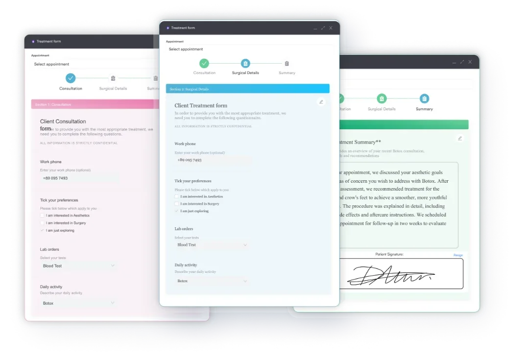
Digital forms carry the structure of this template, so a nurse fills in the same fields every time.
The outcome is a plan that reflects the last shift, rather than what somebody remembered to copy over. That is the difference between a document nurses trust and one they work around.

Pabau stores assessment findings, treatment notes, and care plans in one client record, so the next nurse on shift sees the current version. Nobody re-keys data or hunts for a paper folder.

Spotting impaired gas exchange is usually the easy part. Writing it down so it survives a chart review, and tells the next nurse what to do, takes more care.
Fill the template in while you are still at the bedside. Keep every “as evidenced by” item traceable to your own charting. Give each goal a number and a deadline. Do that, and the plan stops being coursework and starts working as a handover tool.
If your team still keeps care plans separate from the patient record, that copying is where versions drift apart. Book a demo to see how Pabau keeps assessment findings and care plans together in one client record.

Need to move fast when a patient desaturates? Emergency nursing assessment gives you a rapid head-to-toe sequence for an unstable patient.
Managing a chronic respiratory caseload? Cystic fibrosis ATI template covers airway clearance, nutrition, and infection control in one plan.
Referring a patient out for dental care? Dental referral form carries the tooth number, imaging, and history a specialist needs to schedule care.
Worried about a clot reaching the lungs? DVT nursing care plan sets out the prevention and monitoring steps behind pulmonary embolism risk.
Planning a discharge on home oxygen? Caregiver care plan helps you document what the family needs to manage at home.
The main signs are hypoxemia, hypercapnia, abnormal arterial pH, and tachypnea above 20 breaths a minute. Look also for accessory muscle use, nasal flaring, cyanosis, confusion, and restlessness. Patients usually report shortness of breath or chest tightness.
They describe different problems. Ineffective breathing pattern, code 00032, covers inspiration or expiration that does not move enough air. Impaired gas exchange covers a diffusion problem at the alveolar-capillary membrane. A patient can breathe at a normal rate and depth and still be hypoxemic.
Hypoxemia means low oxygen in the arterial blood, which is what your oximeter and ABG measure. Hypoxia means the tissue itself is not getting enough oxygen. Hypoxemia usually causes hypoxia, but a patient in shock can be hypoxic with normal saturation.
Airway comes before breathing, so ineffective airway clearance is usually the priority. Clear the secretions and the gas exchange problem often eases on its own. Keep both diagnoses active, and record interventions against each.
Not directly. Pediatric respiratory rates are banded by age and run much higher than the adult 12 to 20 range. Use your unit’s age-specific reference chart, and treat any drift from the child’s own baseline as significant.