



GLP-1 diet plan
A GLP-1 diet plan is a structured eating framework for patients taking semagluti...

2
Last Updated: Oct 13, 2026
A dilated eye exam is a clinical examination in which eye drops widen the pupil.
That opening lets an eye doctor inspect the retina, optic nerve, macula, and blood vessels at the back of the eye. According to the National Eye Institute (NEI), it is the gold-standard method for detecting sight-threatening disease before vision loss starts.
Without dilation, the pupil stays too small for a full view of those structures. Early glaucoma, diabetic retinopathy, and macular degeneration can go undetected as a result. This guide covers what happens during the exam, who needs one, how often, and what to expect afterward.

A dilated eye exam uses drops to widen the pupil, so the eye doctor can see the retina and optic nerve clearly.
Six conditions, including glaucoma, diabetic retinopathy, and macular degeneration, can be caught on dilation before any symptoms appear.
Type 2 diabetes calls for a dilated exam at diagnosis, then annually. Type 1 calls for one within five years of diagnosis, then annually.
Adults over 60 and African American adults over 40 should be examined every 1 to 2 years, per NEI guidance.
Dilation usually lasts 4 to 6 hours, so bring sunglasses and arrange a driver where you can.
A dilated eye exam runs in two parts. First, a clinician places mydriatic drops in each eye to widen the pupil. Once the pupil is large enough, they examine the interior of the eye with a slit lamp, indirect ophthalmoscope, or fundus lens.
The undilated pupil measures roughly 4 to 8 mm across, even in a dim room. That opening is too narrow for a complete view of the peripheral retina and optic disc. Dilation widens it further and gives the clinician a clear line of sight to the structures that show disease first.
The distinction matters clinically. A standard refraction check tests visual acuity, meaning how well you read the chart. A dilated eye exam evaluates the health of the tissue producing that vision. Only the second question catches disease early enough to prevent irreversible damage. A home nearsighted test with a printed Snellen chart checks acuity only.
Dilation is one component of a wider comprehensive eye exam, which also covers acuity, eye pressure, and eye movement. Clinicians decide at each visit whether the dilated portion is needed.
The dilated eye exam is the most reliable tool for detecting six major eye and systemic conditions at a stage when treatment can preserve vision.
The BrightFocus Foundation notes that dilation is especially critical for glaucoma and AMD because both progress silently. By the time a patient notices symptoms, meaningful nerve or macular tissue is already lost.
Patients monitoring AMD at home often track their central vision with an Amsler grid chart between visits. That is a self-check for new distortion, not a substitute for the dilated exam.
Not every patient needs annual dilation, but specific groups face enough risk to warrant regular dilated exams. Optometry and ophthalmology practices typically flag these groups at intake.
Dilated eye exam frequency depends on age and risk factors, and the interval tightens sharply as risk accumulates. The table below summarizes guidance from the NEI, the American Academy of Ophthalmology (AAO), and the ADA.
The two bodies slice the low-risk population differently. The AAO tiers healthy adults by age band, while the NEI groups everyone over 60 together. Where the two overlap, the shorter interval is the safer default. Side by side, the intervals collapse from a decade to a few months.
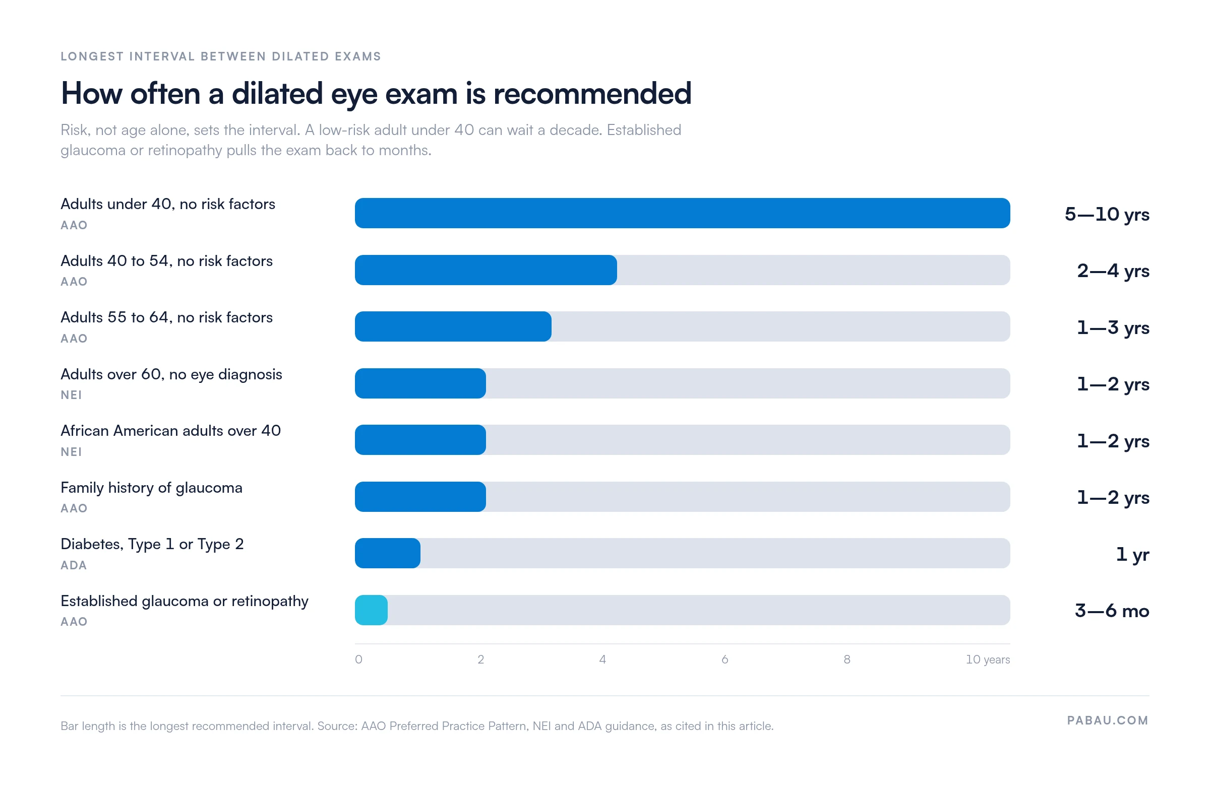
These are population-level guidelines. Individual frequency should always be set by the treating clinician, based on the patient’s full medical history.
A dilated eye exam typically runs 45 to 90 minutes from check-in to discharge. Here is what happens at each stage.
Practices that collect medical history and consent digitally before the appointment cut chair time and start the clinical conversation sooner. Practice management software like Pabau lets patients complete those forms from a phone, so the record is ready when they walk in.

The choice of mydriatic agent affects onset time, depth of dilation, and how long the effects last. Most eye care practices use a combination of two agents. According to the American Academy of Ophthalmology (AAO), the two main pharmacological classes are sympathomimetics, such as phenylephrine, and anticholinergics, such as tropicamide and cyclopentolate.
Who should avoid dilating drops? Patients with known or suspected narrow-angle glaucoma face a risk of acute angle-closure crisis from mydriatics. The AAO recommends screening for narrow angles with a penlight test or gonioscopy before the drops go in. Patients should also disclose any prior angle-closure episode to their eye doctor. Phenylephrine at 10% carries a separate cardiovascular caution for patients with uncontrolled hypertension or known cardiac disease.
Ask your eye doctor which drops they plan to use before your appointment. If you have cardiovascular disease, narrow angles, or a prior angle-closure reaction, say so ahead of time. That lets the clinician choose a safer combination or adjust the concentration.
Dilated eye exam side effects are predictable and temporary for most patients. The two most common are light sensitivity, known as photophobia, and blurry near vision from the drop’s cycloplegic effect on the ciliary muscle.
Systemic absorption of phenylephrine can cause a short-lived rise in blood pressure and heart rate, particularly at the 10% concentration. Most practices now use the 2.5% formulation to minimize that risk.
Eye dilation typically lasts 4 to 6 hours for most adults, though this varies by the agent used and by the individual. Cyclopentolate, used in pediatric exams, can keep pupils dilated for 24 hours or longer.
Eye color also plays a role. Blue, green, and hazel eyes tend to respond faster to mydriatic drops than brown eyes do. Brown irises carry more pigment, which absorbs more of the medication. Some patients with dark irises need a second set of drops or a stronger concentration.
Light sensitivity and near-vision blur ease gradually over those 4 to 6 hours. Avoid driving, detailed close-up work, and bright sun without sunglasses during that window. Most patients are back to normal by early evening after a morning appointment.
Most eye care providers recommend arranging a driver for after a dilated eye exam. The decision depends on the individual’s sensitivity, the drops used, and the light outside. Dilation impairs near vision and increases glare sensitivity. Distance vision and reaction time are less affected.
There is no universal legal prohibition on driving after dilation in the United States or United Kingdom. Bright midday sun does amplify the discomfort, which makes an afternoon drive home harder than an early morning one. Polarized sunglasses reduce glare substantially.
Patients who choose to drive should wait at least 30 minutes after leaving the practice. Stick to a familiar route, and avoid highway or high-speed driving until the light sensitivity settles. For older adults, patients with prior vision problems, and anyone given cyclopentolate, a driver is the safer call.
A dilated eye exam for diabetics follows a different cadence than for the general population. The ADA recommends a dilated retinal exam at diagnosis for Type 2 diabetes, then annually. For Type 1 diabetes, the first exam falls within five years of diagnosis, and annually from there. Neither schedule loosens because blood sugar is well controlled.
Diabetic retinopathy develops in stages. Early non-proliferative retinopathy shows microaneurysms and small hemorrhages on the retinal surface. As it progresses, retinal blood vessels leak fluid into the macula, causing the blurred central vision of diabetic macular edema.
In advanced proliferative retinopathy, abnormal new vessels grow across the retina and into the vitreous. That risks traction retinal detachment and sudden vision loss. A dilated exam identifies each of these stages.
Treatment at the non-proliferative stage is far more effective than waiting for symptoms. Options at that point include blood sugar optimization, intravitreal anti-VEGF injections, and laser photocoagulation.
Many patients skip the eye exam when their vision feels stable. The exam earns its place by finding damage they cannot yet notice. Practices running annual screening at scale often standardize what gets recorded with a diabetic eye exam form. Every visit then captures the same retinal findings, so the next clinician can see the trend.
Non-dilated exams and dilated eye exams answer different clinical questions. The table below compares what each can and cannot detect.
Digital retinal photography, also called fundus photography, can capture wide-field retinal images without dilation. Some teleretinal screening programs for diabetic retinopathy work this way. The AAO cautions that it does not fully replace a dilated exam for every indication, particularly peripheral retinal disease and detailed optic nerve evaluation. Read it as a supplement, or as a screening tool where dilation is not feasible.
Preparation is straightforward, but a few practical steps make the appointment easier.
The dilated eye exam creates specific workflow problems for an eye care practice. Patients need longer slots, and the dilation wait has to be built into the day. Recall intervals differ by risk group, and pre-appointment instructions decide whether patients arrive ready. Most practices track those intervals in a spreadsheet or in someone’s head.
Pabau’s recall automation for practices sets a recall interval per patient risk category instead. A patient with diabetes is flagged for a 12-month recall from their last dilated exam. A low-risk adult over 40 gets a 24-month recall. When the interval passes, the recall goes out by SMS or email with no prompting from the front desk.
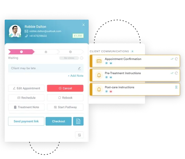
Slot length is the other half of the problem. A dilated exam booked into a 20-minute refraction slot runs late and pushes the rest of the clinic back. Pabau’s scheduling tools for practices support service-specific slot lengths, so the dilation wait is already in the calendar before the patient books.
The outcome is fewer no-shows, better recall compliance, and fewer rescheduled appointments caused by patients who arrived unprepared. Screening rates for diabetic retinopathy and glaucoma follow the recall rate closely, so the admin work pays back clinically.

Pabau lets eye care practices set risk-based recall intervals, automate pre-exam instructions, and build the dilation wait into appointment slot lengths. See how it works for optometry and ophthalmology practices.

In a higher-risk group, the dilated exam is the one test that reliably finds glaucoma, diabetic retinopathy, AMD, and retinal tears before vision goes. All four stay quiet until the damage is done, which is why the interval matters more than the symptoms.
Weigh an afternoon of blurred near vision against what it buys. Book the appointment in the morning, bring sunglasses, and line up a ride if the drops have hit you hard before.
For practices, the harder job is keeping every at-risk patient on their interval year after year. Book a demo to see how Pabau runs risk-based recall for optometry and ophthalmology teams.

Want the full comprehensive exam sequence? Eye physical examination walks through every component of an eye exam, dilation included.
Need to document annual diabetic screening? Diabetes eye exam template gives you a ready-made form for recording retinal findings at each visit.
Checking pupils and eye movement too? PERRLA eye exam form template covers the pupil reaction findings clinicians record before the drops go in.
Looking for the neurological side of an eye exam? Neurological eye exam explains what cranial nerve testing adds to the picture.
Wondering when a pinhole test is used? Pinhole test shows how clinicians separate refractive blur from retinal or nerve problems.
A dilated eye exam is a clinical examination in which mydriatic eye drops widen the pupil. That lets a clinician inspect the retina, optic nerve, macula, and retinal blood vessels at the back of the eye. It is the gold-standard method for detecting glaucoma, diabetic retinopathy, and macular degeneration before symptoms develop.
Dilation typically lasts 4 to 6 hours for most adults using standard tropicamide or phenylephrine drops. Cyclopentolate, used in pediatric exams, can last 24 hours or more. Patients with lighter irises tend to dilate faster than those with dark brown irises.
Most providers recommend arranging a driver, particularly for afternoon appointments in bright sunlight. There is no universal legal prohibition, but dilation impairs near vision and increases glare sensitivity for several hours. Polarized sunglasses help, and patients who do drive should wait at least 30 minutes after leaving the practice.
Dilated exams are recommended regularly for people with diabetes, adults over 60, and anyone with a family history of glaucoma. African American adults over 40 are in that group too. The NEI also recommends one for anyone who has not had a comprehensive eye exam in more than five years, whatever their risk factors.
Type 2 diabetes calls for one at diagnosis and annually after that, while Type 1 calls for one within five years of diagnosis, then annually. Adults over 60 with no eye diagnosis should have one every 1 to 2 years. Low-risk adults under 40 can go every 5 to 10 years, per AAO guidance.
No, but it is recommended at regular intervals for at-risk groups. A standard vision check or refraction does not examine the back of the eye. Whether dilation is needed at a given visit depends on age, health history, and the last dilation date. Clinicians assess that at every appointment.






