Newborn head circumference chart
A newborn head circumference chart plots a baby’s head measurement against...

1
Last Updated: Oct 14, 2026

Endocrine system assessment combines subjective history-taking, physical examination, and laboratory investigation to identify hormonal dysfunction.
TSH is the first thyroid test to order, and HbA1c or fasting glucose is the standard diabetes screen.
Inspection and palpation of the thyroid are the core physical techniques, and abnormal findings include goiter, nodules, and bruits.
Pabau’s digital intake forms and lab result tracking help private practices keep endocrine documentation consistent between visits.
Most endocrine conditions go undetected for years. The American Thyroid Association estimates that 20 million Americans have some form of thyroid disease. Up to 60 percent of them do not know it. In private practice, where patients arrive with vague, overlapping complaints, a structured endocrine system assessment is what separates an early diagnosis from a missed one.
This guide covers the whole process. It walks through subjective history-taking, objective physical examination, the key laboratory panels, and imaging. It also marks where a nurse’s scope ends and a physician’s order is required, which matters most for dynamic adrenal testing.
An endocrine system assessment is a systematic clinical evaluation designed to detect hormonal imbalance and glandular dysfunction. It follows a fixed sequence. Gather the patient’s history, perform a targeted physical examination, then order the confirmatory laboratory or imaging investigation the findings point to.
The endocrine system regulates metabolism, growth, reproduction, and homeostasis by releasing hormones directly into the bloodstream. When one gland produces too much or too little hormone, the effects show up across several organ systems at once. That is why pattern recognition matters more here than any single result.
The conditions most commonly identified through endocrine assessment include hypothyroidism, hyperthyroidism, type 2 diabetes, Cushing’s syndrome, and adrenal insufficiency. Each has a distinct clinical signature. Many also share overlapping symptoms such as fatigue, weight change, and mood disturbance.
Before assessing the endocrine system, you need a working map of which gland makes which hormone and what happens when it fails. The table below summarizes the major glands, their primary hormones, and the signs of dysfunction.
The pituitary gland acts as the master regulator. It releases trophic hormones that stimulate the peripheral glands. That hierarchy means a failing thyroid produces a different laboratory pattern than pituitary failure does.
The clinical history is where most endocrine diagnoses begin. Patients rarely arrive saying they think they have a thyroid problem. They describe fatigue, unexplained weight change, hair thinning, cold intolerance, or mood shifts. Your job is to recognize the hormonal pattern underneath those complaints.
Structure the history around four areas. Start with the presenting complaint, then run a targeted review of systems, then medical and family history, then current medication. A health assessment form collects all four before the appointment, so the consultation starts from a written record.
Ask the patient to describe their main symptom, when it started, how long it has lasted, and how severe it is. Then run a review of systems focused on endocrine presentations. A nursing review of systems template keeps that sweep consistent between clinicians and between visits.
Record medical history, including previous thyroid disease, diabetes, and autoimmune conditions. Record family history too, covering thyroid cancer, type 1 or type 2 diabetes, and MEN syndromes. Then list current medication, especially anything that affects thyroid function or cortisol, such as corticosteroids, lithium, and amiodarone.
The physical examination remains a cornerstone of endocrine disease evaluation, even as laboratory testing has grown more sophisticated. Careful inspection and palpation, read alongside systemic signs, narrows the differential before a single test is ordered.
Begin with general appearance. Observe body habitus, affect, skin tone, and any obvious dysmorphic features. Then record vital signs. Hypertension points toward Cushing’s or pheochromocytoma, tachycardia toward hyperthyroidism, and bradycardia toward hypothyroidism.
Thyroid assessment is the most detailed part of the endocrine physical examination. The steps run in the same order as a complete physical examination, narrowed to one gland. Follow this sequence.
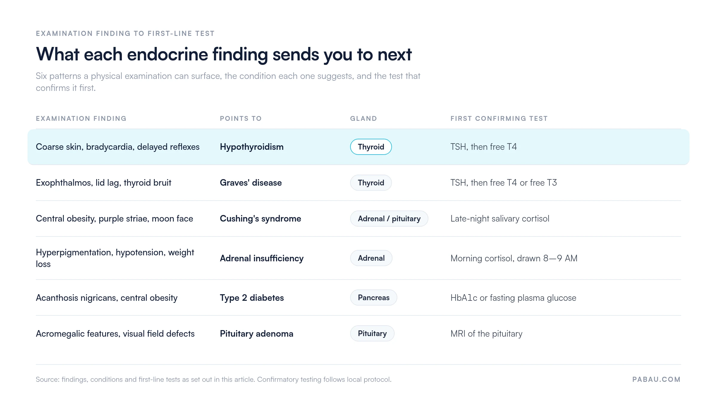
Neither the adrenal nor the pituitary gland is directly palpable. Assessment relies on recognizing characteristic clinical constellations. The table below maps key examination findings to their likely endocrine source.
A finding only earns its place when it changes what you order next. The map below carries each of those patterns through to the test that confirms it first.
Also assess the skin for thinning, easy bruising, and acanthosis nigricans at the neck and axillae, which suggests insulin resistance. Check hair distribution and reflexes, since a delayed relaxation phase is a classic hypothyroid sign. Document each finding in the patient records with a timestamp and the examiner’s name.
Standardize your endocrine examination sequence before every appointment. Clinicians who follow a fixed inspection, palpation, and vital signs order miss fewer low-prevalence signs, such as a thyroid bruit or acanthosis nigricans.
Laboratory investigation confirms or rules out the impression formed during history and examination. Ordering the right panel for each suspected condition reduces patient burden and speeds diagnosis. Read every result against the clinical picture rather than treating a reference range as an absolute threshold.
According to the American Thyroid Association, measuring TSH is the best way to test thyroid function initially. A single elevated or suppressed TSH then triggers free T4 and free T3 testing, which characterizes the disorder’s nature and severity.
Reference ranges vary between laboratories. Always interpret a result against that laboratory’s own interval, and correlate it with what you found clinically. A TSH at the upper end of normal can still matter in a symptomatic patient.
Adrenal function is assessed mainly through morning serum cortisol, with the ACTH stimulation test reserved for suspected insufficiency. Both are established tools for evaluating adrenal reserve. The panels below cover the adrenal and metabolic side of an endocrine work-up.
Imaging confirms structural abnormalities found on examination or suggested by laboratory results. It supplements clinical findings rather than replacing them. Work out which modality answers your clinical question before you refer.
A systematic endocrine assessment reliably surfaces a core group of conditions. Recognizing their characteristic presentation patterns makes referral or treatment initiation faster and more confident.
Practices running endocrine assessments on paper lose clinical detail between intake forms, separate lab portals, and handwritten examination notes. The history sits in one place and the results in another, so nobody sees the trend that matters.
Practice management software like Pabau brings those pieces back together. Our patient intake software lets you build endocrine-specific pre-assessment questionnaires covering symptom checklists, medication history, and family history of hormonal conditions. Patients complete them before they arrive, so you start the consultation with the subjective data already captured and structured.
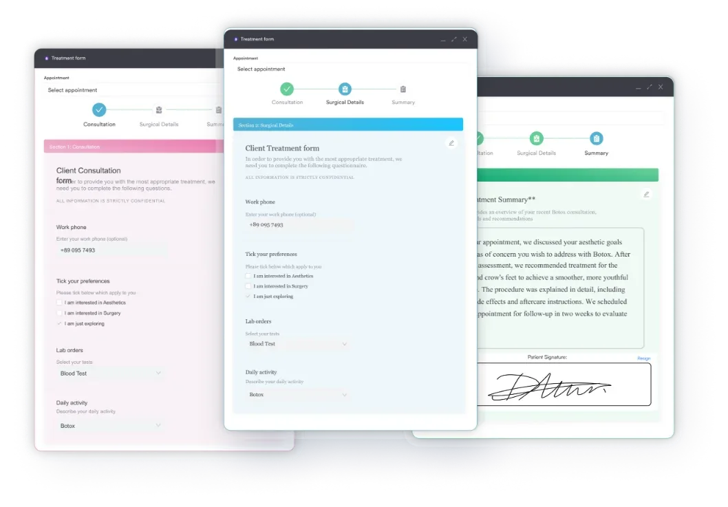
Examination findings and clinical notes go straight into the patient record using customizable templates. Pabau’s lab result tracking logs TSH, free T3, free T4, HbA1c, and cortisol against each patient’s timeline. Monitoring a trend no longer means switching systems.
Endocrine conditions run across many appointments, so Pabau also handles recall scheduling, automated follow-up messages, and an audit-ready record of who documented what. Pabau Scribe, our AI medical scribe, drafts the note at the point of care. Examination findings and clinical reasoning reach the record without breaking the consultation.
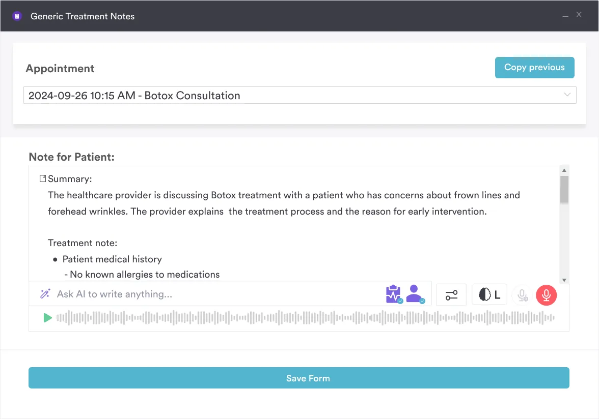

Pabau’s intake forms, note templates, and lab result tracking capture endocrine history, examination findings, and panel results in one patient record. See how it fits your practice.

Endocrine dysfunction is common, multisystem, and easy to miss without a deliberate approach. The sequence is what protects you. Take the history first, examine second, then order the test the finding actually points to.
Standardize that sequence and the documentation follows it. Digital forms, structured note templates, and lab tracking keep each visit comparable to the last. That comparison is the whole point in a condition that moves slowly. Book a demo to see how Pabau holds an endocrine assessment together across visits.

Assessing another body system with the same structure? Gastrointestinal assessment walks the same history, examination, and investigation sequence.
Need the symptom sweep in a reusable form? Nursing review of systems gives you a head-to-toe checklist you can fill in during the consultation.
Working in urgent or unscheduled care? Emergency nursing assessment covers the rapid version of the same assessment steps.
Want one form that carries both history and examination? History and physical form keeps subjective and objective findings on a single sheet.
Documentation getting picked apart in audit? Nursing documentation explains what a defensible clinical note has to contain.
An endocrine system assessment is a structured clinical evaluation that detects hormonal imbalance and glandular dysfunction. It combines history-taking, physical examination, laboratory investigation, and imaging. It identifies conditions such as thyroid disorders, diabetes mellitus, Cushing’s syndrome, and adrenal insufficiency.
A complete assessment has four parts. The clinical history covers symptoms, medication, and family history. The physical examination targets the thyroid, skin, hair, and vital signs, plus systemic signs of hormonal excess or deficiency. Laboratory panels include TSH, free T3, free T4, cortisol, HbA1c, and fasting glucose. Imaging such as thyroid ultrasound or pituitary MRI is added where indicated.
The endocrine system maintains homeostasis through hormone-mediated negative feedback loops. When a hormone level deviates from its set point, the hypothalamic-pituitary axis responds by increasing or suppressing trophic hormone release to restore balance. Disruption of these feedback pathways is what clinicians detect during endocrine system assessment.
Key techniques include inspection of the neck for thyroid enlargement and swallowing movement. Next come anterior and posterior palpation of the gland, then auscultation for a thyroid bruit. Systemic signs matter too, including skin texture, hair distribution, tendon reflexes, blood pressure, and heart rate. Each finding maps to specific glandular dysfunction patterns.